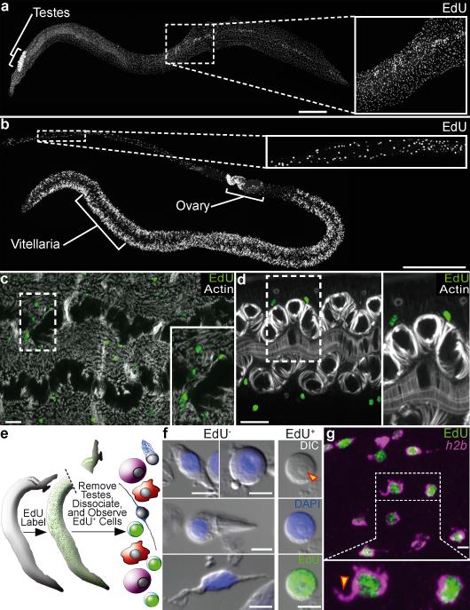
Figure 1. Proliferation of somatic cells in adult schistosomes.
a–b, EdU labeling in (a) male and (b) female parasites.
c–d, Distribution of mesenchymal PSCs in (c) male and (d) female parasites. Phalloidin staining for actin shows male enteric and dorso-ventral muscles and female enteric and uterine muscles.
e, Strategy to characterize PSC morphology.
f, The morphology of EdU− and EdU+ cells. Arrowhead indicates a nucleolus.
g, FISH for histone h2b with EdU labeling. Arrowhead indicates a cytoplasmic projection.
(a–d, g) are confocal projections; (a–b) are derived from tiled stacks. Scale bars: (a–b) 500 μm, (c–d) 20 μm, (f–g) 5 μm.

