Abstract
Allostery is well documented for proteins but less recognized for DNA-protein interactions. Here we report that specific binding of a protein on DNA is substantially stabilized or destabilized by another protein bound nearby. The ternary complex's free energy oscillates as a function of the separation between the two proteins with a periodicity of ~10 base pairs, the helical pitch of B-form DNA, and a decay length of ~15 base pairs. The binding affinity of a protein near a DNA hairpin is similarly dependent on their separation, which—together with molecular dynamics simulations—suggests that deformation of the double-helical structure is the origin of DNA allostery. The physiological relevance of this phenomenon is illustrated by its effect on gene expression in live bacteria and on a transcription factor's affinity near nucleosomes.
Upon binding of a ligand, a macromolecule often undergoes conformational changes that modify the binding affinity of a second ligand at a distant site. This phenomenon, known as “allostery”, is responsible for dynamic regulation of biological functions. Although extensive studies have been done on allostery in proteins or enzymes (1, 2), less is known for that through DNA, which is normally considered as a mere template providing binding sites. In fact, multiple proteins, such as transcription factors and RNA or DNA polymerases, bind close to each other on genomic DNA to carry out their cellular functions in concert. Such allostery through DNA has been implicated in previous studies (3–10) but has not been quantitatively characterized or mechanistically understood.
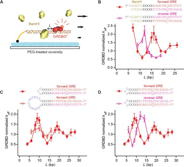
We performed a single-molecule study of allostery through DNA by measuring the dissociation rate constant (koff) of a DNA-bound protein affected by the binding of another protein nearby. In the assay, DNA duplexes (dsDNA), tethered on the passivated surface of a flow cell, contained two specific protein binding sites separated by a linker sequence of L base pairs (bp) (Fig. 1A and figs. S1 and S2) (11). One of the proteins was fluorescently labeled, and many individual protein-DNA complexes were monitored in a large field of view with a total internal reflection fluorescence microscope. Once the labeled protein molecules were bound to DNA, the second protein at a certain concentration was flowed in. Stochastic dissociation times of hundreds of labeled protein molecules were then recorded, the average of which yields the koff (fig. S3) (12).
Fig. 1.
Allostery through DNA affecting koff of GRDBD near BamHI or near a hairpin loop. (A) Schematic for the single-molecule assay in a flow cell. The structural model is for L = 11 with GRDBD from Protein Data Bank (PDB) ID 1R4R (13) and BamHI from PDB ID 2BAM (32). (B) Oscillation in the koff of GRDBD for the forward (red solid circles) and reverse (magenta open circles) GRE sequences, normalized to that measured in the absence of BamHI (±SEM). DNA sequences are shown with the linker DNA (L = 5). The central base of GRE, which makes the sequence non-palindromic, is underlined. (C) Protein binding affinity affected by a nearby DNA hairpin loop, 3 bp and 15 bp (±SEM). (D) Effect of 3-bp loop on the forward and reverse GRE (±SEM). The DNA sequence is shown for L = 5. koff is normalized to that measured on DNA without a hairpin loop.
We first present a protein pair that does not substantially bend DNA, namely a Cy3B-labeled DNA-binding domain of glucocorticoid receptor (GRDBD), a eukaryotic transcription factor, together with BamHI, a type II endonuclease (Fig. 1A) (13, 14). To prevent the endonuclease activity of BamHI, we used buffer containing Ca2+ instead of Mg2+. At a saturating concentration of BamHI, the koff of GRDBD was found to oscillate as a function of L with significant amplitude spanning a factor of 4 and a periodicity of 10 bp, which intriguingly coincides with the helical pitch of B-form DNA (Fig. 1B, red).
When we reversed the DNA sequence of the nonpalindromic GRDBD binding site (GRE) with respect to that of BamHI, the koff of GRDBD oscillated with a phase shift of 4 bp, nearly 180° relative to that of the forward GRE (Fig. 1B). On the other hand, the binding sequence of BamHI is palindromic; therefore, its reversion is not expected to cause any phase shift.
Similar oscillatory modulation in koff was observed with other protein pairs, such as lac repressor (LacR) and EcoRV, or LacR and T7 RNA polymerase (T7 RNAp) (figs. S5 and S6). These proteins differ in size, shape, surface charge distribution, and DNA binding affinity (15–18). In fact, the oscillation was independent of ionic strength (fig. S7), suggesting that the electrostatic interaction between the two proteins is not the origin of the allosteric phenomenon. However, the presence of a nick, mismatched bases, or GC-rich sequences in the linker region attenuated the oscillation (figs. S8 to S10), implying that the allostery is largely dependent on the mechanical properties of the linker DNA.
To prove this hypothesis, we replaced the BamHI binding site with a DNA hairpin loop (Fig. 1C), which allows examination of the effect of DNA distortion alone. When the length of linker DNA between the hairpin loop and GRE (L) was varied, we observed a similar oscillation in the koff of GRDBD (Fig. 1C). A larger hairpin loop decreased the amplitude of the oscillation, likely because of a smaller distortion induced by the larger hairpin (Fig. 1C). Again when GRE was reversed, the oscillation showed a 4-bp phase shift (Fig. 1D).
The oscillation dampens out with a characteristic decay length of ~15 bp (Fig. 1 and fig. S12) (12), which is much shorter than either the bending persistence length (~150 bp) (19) or the twisting persistence length (~300 bp) of DNA (20). On the other hand, recognizing that proteins primarily interact with the DNA major groove (21, 22), we hypothesized that allostery through DNA results mainly from distortion of the major groove.
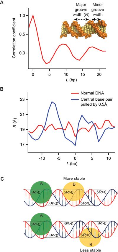
We carried out molecular dynamics (MD) simulations first on free dsDNA in aqueous solutions at room temperature (12). We evaluated the spatial correlation between the major groove widths (R) (Fig. 2A inset) at two positions (base pairs i and i+L) as a function of their separation, averaged over time t. We observed that the correlation coefficient has a clear oscillation with a periodicity of ~10 bp and dampens within a few helical turns (Fig. 2A). A similar yet slightly weaker oscillation was also observed for the correlation of the minor groove widths. We attribute the oscillation in Fig. 2A to thermally excited low-frequency vibrational modes of dsDNA, which are dictated by the double helical structure of DNA.
Fig. 2.
Allostery through DNA induced by distortion of the major groove. (A) MD simulation at room temperature reveals the spatial correlation between the major groove widths (inset, defined as the distance between C3 atoms of the ith and i+7th nucleotide sugar-rings) at two positions as a function of their separation L, averaged over time t, [<δR(i;t)δR(i+L;t)>t, i=5]. The correlation oscillates with a periodicity of ~10 bp and is attributed to thermally excited low-frequency vibrational modes of dsDNA. (B) Upon breaking the symmetry by pulling apart a base pair in the middle of the dsDNA (defined as L=0) by 0.5Å (12), the time-averaged R (blue) deviates from that of a free DNA (red) and oscillates as a function of the distance (L) from the perturbed base pair with a periodicity of ~10 bp. (C) Oscillation of R(L) causes the variation of the allosteric coupling between two DNA-binding proteins A and B. If protein B widens R, it would energetically favor binding at positions where R is already widened (δR>0) by protein A (top), but disfavor where R is narrowed (δR<0) (bottom).
Such spatial correlation as well as the time-averaged R (Fig. 2B, red curve) are translationally invariant across a free DNA unless the symmetry is broken, as in the case of hairpin formation or protein binding. We simulated such an effect by applying a harmonic potential to pull a base pair apart in the middle of the strand. Under this condition, we observed that the time-averaged R (Fig. 2B, blue curve) deviates from that of a free DNA (Fig. 2B, red curve) and oscillates as a function of the distance (L) from the perturbed base pair with a periodicity of ~10 bp. In contrast, no such oscillation was observed for the inter-helical distance.
Such deviation from the free DNA, δR(L), is expected to cause variation in the binding of the second DNA-binding protein at a distance L bases away. For example, in Fig. 2C, if protein B widens R, its binding would be energetically favored at positions where R is already widened (δR>0) by the hairpin or protein A (Fig. 2C, top), but disfavored where R is narrowed (δR<0) (Fig. 2C, bottom). Consequently, reversing a nonpalindromic binding sequence would invert the binding preference of the protein, explaining the phase shift in Figure 1. This model is also well supported by the observation that the koff of LacR oscillates with an opposite phase in the presence of BamHI or EcoRV (fig. S6), which is consistent with the fact that BamHI widens whereas EcoRV narrows the major groove (14, 15).
Next, to investigate the effect of DNA allostery on transcription regulation, we studied modulation of RNA polymerase binding affinity when a protein binds near the promoter both in vitro and in vivo. The protein pair we chose is LacR and T7 RNAp, both of which, unlike GRDBD and BamHI, bend DNA (17, 23, 24) but nevertheless exhibit a similar allosteric effect.
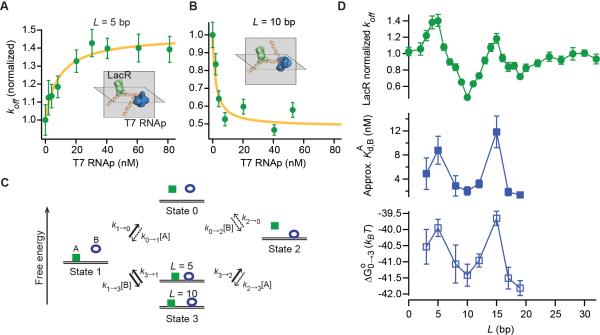
In the in vitro assay, we measured the binding affinity of unlabeled T7 RNAp on its promoter by titrating koff of labeled LacR on lac operator O1 (lacO1) with T7 RNAp. koff exhibited hyperbolic T7 RNAp concentration dependence (Michaelis-Menten—like kinetics) (Fig. 3, A and B, and fig. S4), as can be rigorously derived from the kinetic scheme for LacR (Protein A) and T7 RNAp (Protein B) in Fig. 3C (12):
| (1) |
where ki→j is the rate constant from state i to j and [B] is the concentration of T7 RNAp. The plateau value in the titration curve is k3→2. We observed that k3→2 oscillates as a function of L with the periodicity and amplitude similar to those of GRDBD and BamHI (Fig. 3D, top).
Fig. 3.
Allostery through DNA between LacR and T7 RNAp in vitro. (A and B) Titration curves, where koff values were normalized to those measured in the absence of T7 RNAp on the given template (±SEM). The hyperbolic fit (yellow) is based on Eq. 1. Structural models illustrate the ternary complex of LacR [PDB ID 2PE5 (33)] and T7 RNAp [PDB ID 3E2E (18)]. (C) Kinetic model for the binding of proteins A (LacR) and B (T7 RNAp). Our experiments start with state 1 and proceed to the dissociation of LacR to state 0 or state 2 (via state 3), as shown with solid arrows. Dashed arrows indicate reactions that are not considered in our derivation of Eq.1. (D) The maximum koff of LacR (k3→2), Kd of T7 RNAp in the presence of LacR , and the free energy of the ternary complex , as function of L, oscillating with a periodicity of 10 bp. Error bars reflect SEM for k3→2 and 1 SD of the χ2 fit for and (12).
According to Eq. 1, the dissociation constant of B on the A-bound DNA, , can be measured from the value of [B] at which koff reaches half of the plateau value in the titration curves (Fig. 3, A and B) (12). We found that oscillates as a function of L (Fig. 3D, middle) in phase with k3→2 [that is, ] (Fig. 3D, top). Therefore, the cooperativity in DNA binding, if present, exhibits either simultaneous stabilization or destabilization between the two proteins. This is a consequence of the fact that free energy is a path-independent thermodynamic state function (Fig. 3C) (12):
| (2) |
where Kd,A and Kd,B are the dissociation constant of a protein in the absence of the other, kB is the Boltzmann constant, and T is temperature.
Based on the second line of Eq. 2, the free energy of the ternary complex, , was found to oscillate with a periodicity of ~10 bp and an amplitude of ~2 kBT (Fig. 3D, bottom).
In general, for a ternary complex formed with DNA and Proteins A and B, the free energy of the overall system is , where and are the binding free energies of the two individual proteins on DNA, respectively. , small relative to or , is the energetic coupling involving in the linker DNA, given by the sum of two terms—the variation of protein A binding caused by protein B and the variation of protein B binding caused by protein A:
| (3) |
In each δR, or the distortion of the major groove widths, the subscripts indicate where the distortion occurs (binding site of protein A or B) and the superscripts indicate the protein that causes the DNA distortion (12). According to our proposed mechanism, and propagate periodically (Fig. 2B), yielding a damped oscillation in . This explains the oscillations of the coupling energy for LacR and T7 RNAp (Fig. 3D, bottom and fig. S14A) and for GRDBD and BamHI (fig. S14B).
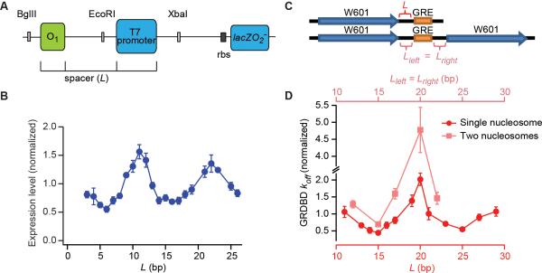
The allosteric coupling between LacR and T7 RNAp is likely to affect transcription in vivo because the efficiency of transcription initiation correlates with the binding affinity of T7 RNAp (25). We therefore inserted DNA templates used in vitro (fig. S15) into the chromosome of Escherichia coli and measured the expression level of lacZ using the Miller assay (Fig. 4A) (26). Indeed, the gene expression level oscillates as a function of L with a periodicity of ~10 bp (Fig. 4B). Similar oscillations of T7 RNAp activity were observed on plasmids in E. coli cells by using a yellow fluorescent protein as a reporter (fig. S16). The oscillation of gene expression levels with a 10-bp periodicity was also seen in a classic experiment on lac operon with a DNA loop formed by two operators (27). However, our T7 RNAp result illustrates that DNA allostery results in such an oscillatory phenomenon even without a DNA loop, which is consistent with a recent study in which E. coli RNA polymerase was used (10).
Fig. 4.
Physiological relevance of DNA allostery. (A) E. coli strains constructed to examine cooperativity between LacR and T7 RNAp on the bacterial chromosome. (B) The expression level of lacZ (normalized to the average expression levels of all Ls) oscillates as a function of L with a periodicity of 10 bp, similar to the corresponding in vitro data (fig. S15). Error bars reflect SEM (n = 3). (C) Schematic for the DNA sequences used in the GRDBD-nucleosome experiment. W601 is the Widom 601 nucleosome positioning sequence (34). (D) Oscillation of the koff of GRDBD as a function of L (±SEM). Data was normalized to koff of GRDBD in the absence of histone (fig. S17).
Pertinent to eukaryotic gene expression, DNA allostery may affect the binding affinity of transcription factors near nucleosomes that are closely positioned (28, 29). We placed GRE downstream of a nucleosome (Fig. 4C) and observed a similar DNA allosteric effect in the koff of GRDBD (Fig. 4D, and fig. S17). To evaluate DNA allostery in an internucleosomal space, we used two nucleosomes to flank a GRE (Fig. 4C). At the same separation L, GRDBD resides on GRE for a relatively longer time with a single nucleosome nearby than it does with a pair of nucleosomes on both sides of GRE (Fig. 4D). Nonetheless, the fold change between the maximal and minimal koff is larger for GRDBD with two nucleosomes (approximately sevenfold). This indicates moderately large cooperativity between the two flanking nucleosomes in modifying the binding affinity of GRDBD, which is in line with previous in vivo experiments (30, 31). The fact that histones modify a neighboring transcription factor's binding suggests that allostery through DNA might be physiologically important in affecting gene regulation.
Supplementary Material
Acknowledgements
We thank K. Wood for his early involvement and J. Hynes, A. Szabo, C. Bustamante and J. Gelles for helpful discussions. This work is supported by NIH Director's Pioneer Award and NIH grant GM096450 to X.S.X., Peking University for BIOPIC, Thousand Youth Talents Program for Y.S., as well as the Major State Basic Research Development Program (2011CB809100), National Natural Science Foundation of China (31170710, 31271423, 21125311).
References and Notes
- 1.Monod J, Wyman J, Changeux JP. J. Mol. Biol. 1965;12:88. doi: 10.1016/s0022-2836(65)80285-6. [DOI] [PubMed] [Google Scholar]
- 2.Koshland DE, Némethy G, Filmer D. Biochemistry. 1966;5:365. doi: 10.1021/bi00865a047. [DOI] [PubMed] [Google Scholar]
- 3.Pohl FM, Jovin TM, Baehr W, Holbrook JJ. Proc. Natl. Acad. Sci. U.S.A. 1972;69:3805. doi: 10.1073/pnas.69.12.3805. [DOI] [PMC free article] [PubMed] [Google Scholar]
- 4.Hogan M, Dattagupta N, Crothers DM. Nature. 1979;278:521. doi: 10.1038/278521a0. [DOI] [PubMed] [Google Scholar]
- 5.Parekh BS, Hatfield GW. Proc. Natl. Acad. Sci. U.S.A. 1996;93:1173. doi: 10.1073/pnas.93.3.1173. [DOI] [PMC free article] [PubMed] [Google Scholar]
- 6.Rudnick J, Bruinsma R. Biophys. J. 1999;76:1725. doi: 10.1016/S0006-3495(99)77334-0. [DOI] [PMC free article] [PubMed] [Google Scholar]
- 7.Panne D, Maniatis T, Harrison SC. Cell. 2007;129:1111. doi: 10.1016/j.cell.2007.05.019. [DOI] [PMC free article] [PubMed] [Google Scholar]
- 8.Moretti R, et al. ACS Chem. Biol. 2008;3:220. doi: 10.1021/cb700258r. [DOI] [PMC free article] [PubMed] [Google Scholar]
- 9.Koslover EF, Spakowitz AJ. Phys. Rev. Lett. 2009;102:178102. doi: 10.1103/PhysRevLett.102.178102. [DOI] [PubMed] [Google Scholar]
- 10.Garcia Hernan G., et al. Cell Reports. 2012;2:150. doi: 10.1016/j.celrep.2012.06.004. [DOI] [PMC free article] [PubMed] [Google Scholar]
- 11.Kim S, Blainey PC, Schroeder CM, Xie XS. Nat. Methods. 2007;4:397. doi: 10.1038/nmeth1037. [DOI] [PubMed] [Google Scholar]
- 12. Materials and methods are available as supplementary materials on Science online.
- 13.Luisi BF, et al. Nature. 1991;352:497. doi: 10.1038/352497a0. [DOI] [PubMed] [Google Scholar]
- 14.Newman M, Strzelecka T, Dorner L, Schildkraut I, Aggarwal A. Science. 1995;269:656. doi: 10.1126/science.7624794. [DOI] [PubMed] [Google Scholar]
- 15.Winkler FK, et al. EMBO J. 1993;12:1781. doi: 10.2210/pdb4rve/pdb. [DOI] [PMC free article] [PubMed] [Google Scholar]
- 16.Lewis M, et al. Science. 1996;271:1247. doi: 10.1126/science.271.5253.1247. [DOI] [PubMed] [Google Scholar]
- 17.Kalodimos CG, et al. EMBO J. 2002;21:2866. doi: 10.1093/emboj/cdf318. [DOI] [PMC free article] [PubMed] [Google Scholar]
- 18.Durniak KJ, Bailey S, Steitz TA. Science. 2008;322:553. doi: 10.1126/science.1163433. [DOI] [PMC free article] [PubMed] [Google Scholar]
- 19.Smith S, Finzi L, Bustamante C. Science. 1992;258:1122. doi: 10.1126/science.1439819. [DOI] [PubMed] [Google Scholar]
- 20.Bryant Z, et al. Nature. 2003;424:338. doi: 10.1038/nature01810. [DOI] [PubMed] [Google Scholar]
- 21.Travers AA. Annu. Rev. Biochem. 1989;58:427. doi: 10.1146/annurev.bi.58.070189.002235. [DOI] [PubMed] [Google Scholar]
- 22.Rohs R, et al. Annu. Rev. Biochem. 2010;79:233. doi: 10.1146/annurev-biochem-060408-091030. [DOI] [PMC free article] [PubMed] [Google Scholar]
- 23.Újvári A, Martin CT. J. Mol. Biol. 2000;295:1173. doi: 10.1006/jmbi.1999.3418. [DOI] [PubMed] [Google Scholar]
- 24.Tang G-Q, Patel SS. Biochemistry. 2006;45:4936. doi: 10.1021/bi0522910. [DOI] [PubMed] [Google Scholar]
- 25.Jia Y, Kumar A, Patel SS. J. Biol. Chem. 1996;271:30451. doi: 10.1074/jbc.271.48.30451. [DOI] [PubMed] [Google Scholar]
- 26.Miller JH. Experiments in molecular genetics. Cold Spring Harbor Laboratory; 1972. [Google Scholar]
- 27.Müller J, Oehler S, Müller-Hill B. J. Mol. Biol. 1996;257:21. doi: 10.1006/jmbi.1996.0143. [DOI] [PubMed] [Google Scholar]
- 28.Lee W, et al. Nat. Genet. 2007;39:1235. doi: 10.1038/ng2117. [DOI] [PubMed] [Google Scholar]
- 29.Sharon E, et al. Nat. Biotech. 2012;30:521. doi: 10.1038/nbt.2205. [DOI] [PMC free article] [PubMed] [Google Scholar]
- 30.John S, et al. Nat. Genet. 2011;43:264. doi: 10.1038/ng.759. [DOI] [PMC free article] [PubMed] [Google Scholar]
- 31.Meijsing SH, et al. Science. 2009;324:407. doi: 10.1126/science.1164265. [DOI] [PMC free article] [PubMed] [Google Scholar]
- 32.Viadiu H, Aggarwal AK. Nat. Struct. Biol. 1998;5:910. doi: 10.1038/2352. [DOI] [PubMed] [Google Scholar]
- 33.Daber R, Stayrook S, Rosenberg A, Lewis M. J. Mol. Biol. 2007;370:609. doi: 10.1016/j.jmb.2007.04.028. [DOI] [PMC free article] [PubMed] [Google Scholar]
- 34.Lowary PT, Widom J. J. Mol. Biol. 1998;276:19. doi: 10.1006/jmbi.1997.1494. [DOI] [PubMed] [Google Scholar]
- 35.Strähle U, Klock G, Schütz G. Proc. Natl. Acad. Sci. U.S.A. 1987;84:7871. doi: 10.1073/pnas.84.22.7871. [DOI] [PMC free article] [PubMed] [Google Scholar]
- 36.Tang G-Q, Bandwar RP, Patel SS. J. Biol. Chem. 2005;280:40707. doi: 10.1074/jbc.M508013200. [DOI] [PubMed] [Google Scholar]
- 37.Joo C, Ha T. In: Single molecule techniques: a laboratory manual. Selvin PR, Ha T, editors. Cold Spring Harbor Laboratory Press; Cold Spring Harbor: 2008. pp. 3–35. [Google Scholar]
- 38.Eismann ER, Müller-Hill B. J. Mol. Biol. 1990;213:763. doi: 10.1016/S0022-2836(05)80262-1. [DOI] [PubMed] [Google Scholar]
- 39.Elf J, Li G-W, Xie XS. Science. 2007;316:1191. doi: 10.1126/science.1141967. [DOI] [PMC free article] [PubMed] [Google Scholar]
- 40.Chen J, Matthews KS. J. Biol. Chem. 1992;267:13843. [PubMed] [Google Scholar]
- 41.Gottesfeld JM, et al. J. Mol. Biol. 2001;309:615. doi: 10.1006/jmbi.2001.4694. [DOI] [PubMed] [Google Scholar]
- 42.Schroeder CM, Blainey PC, Kim S, Xie XS. In: Single molecule techniques: a laboratory manual. Selvin PR, Ha T, editors. Cold Spring Harbor Laboratory Press; Cold Spring Harbor: 2008. pp. 461–492. [Google Scholar]
- 43.Blainey PC, van Oijen AM, Banerjee A, Verdine GL, Xie XS. Proc. Natl. Acad. Sci. U.S.A. 2006;103:5752. doi: 10.1073/pnas.0509723103. [DOI] [PMC free article] [PubMed] [Google Scholar]
- 44.Luo G, Wang M, Konigsberg WH, Xie XS. Proc. Natl. Acad. Sci. U.S.A. 2007;104:12610. doi: 10.1073/pnas.0700920104. [DOI] [PMC free article] [PubMed] [Google Scholar]
- 45.Gilbert W, Müller-Hill B. Proc. Natl. Acad. Sci. U.S.A. 1966;56:1891. doi: 10.1073/pnas.56.6.1891. [DOI] [PMC free article] [PubMed] [Google Scholar]
- 46.Datsenko KA, Wanner BL. Proc. Natl. Acad. Sci. U.S.A. 2000;97:6640. doi: 10.1073/pnas.120163297. [DOI] [PMC free article] [PubMed] [Google Scholar]
- 47.Lee EC, et al. Genomics. 2001;73:56. doi: 10.1006/geno.2000.6451. [DOI] [PubMed] [Google Scholar]
- 48.Oehler S, Eismann ER, Krämer H, Müller-Hill B. EMBO J. 1990;9:973. doi: 10.1002/j.1460-2075.1990.tb08199.x. [DOI] [PMC free article] [PubMed] [Google Scholar]
- 49.Munteanu MG, Vlahovicek K, Parthasarathy S, Simon I, Pongor S. Trends Biochem. Sci. 1998;23:341. doi: 10.1016/s0968-0004(98)01265-1. [DOI] [PubMed] [Google Scholar]
- 50.Emsley P, Cowtan K. Acta Crystallogr. D. 2004;60:2126. doi: 10.1107/S0907444904019158. [DOI] [PubMed] [Google Scholar]
- 51.Cheetham GMT, Jeruzalmi D, Steitz TA. Nature. 1999;399:80. doi: 10.1038/19999. [DOI] [PubMed] [Google Scholar]
- 52.Dolinsky TJ, Nielsen JE, McCammon JA, Baker NA. Nucl. Acids Res. 2004;32:W665. doi: 10.1093/nar/gkh381. [DOI] [PMC free article] [PubMed] [Google Scholar]
- 53.Li H, Robertson AD, Jensen JH. Proteins. 2005;61:704. doi: 10.1002/prot.20660. [DOI] [PubMed] [Google Scholar]
- 54.Baker NA, Sept D, Joseph S, Holst MJ, McCammon JA. Proc. Natl. Acad. Sci. U.S.A. 2001;98:10037. doi: 10.1073/pnas.181342398. [DOI] [PMC free article] [PubMed] [Google Scholar]
- 55.Case D, et al. AMBER 11. University of California; San Francisco: 2010. [Google Scholar]
- 56.Berendsen HJC, Grigera JR, Straatsma TP. J. Phys. Chem. 1987;91:6269. 1987/11/01. [Google Scholar]
- 57.Pérez A, et al. Biophys. J. 2007;92:3817. doi: 10.1529/biophysj.106.097782. [DOI] [PMC free article] [PubMed] [Google Scholar]
- 58.Ryckaert J-P, Ciccotti G, Berendsen HJC. J. Comput. Phys. 1977;23:327. [Google Scholar]
- 59.Darden T, York D, Pedersen L. J. Chem. Phys. 1993;98:10089. [Google Scholar]
- 60.Berendsen HJC, Postma JPM, Gunsteren W. F. v., DiNola A, Haak JR. J. Chem. Phys. 1984;81:3684. [Google Scholar]
- 61.Falcon CM, Matthews KS. Biochemistry. 2000;39:11074. doi: 10.1021/bi000924z. [DOI] [PubMed] [Google Scholar]
- 62.Romanuka J, et al. J. Mol. Biol. 2009;390:478. doi: 10.1016/j.jmb.2009.05.022. [DOI] [PubMed] [Google Scholar]
- 63.Perona JJ. Methods. 2002;28:353. doi: 10.1016/s1046-2023(02)00242-6. [DOI] [PubMed] [Google Scholar]
- 64.Vipond IB, Baldwin GS, Halford SE. Biochemistry. 1995;34:697. doi: 10.1021/bi00002a037. [DOI] [PubMed] [Google Scholar]
- 65.Horton NC, Perona JJ. Proc. Natl. Acad. Sci. U.S.A. 2000;97:5729. doi: 10.1073/pnas.090370797. [DOI] [PMC free article] [PubMed] [Google Scholar]
- 66.Israelachvili J. Intermolecular & Surface Forces. 2nd Academic Press; 1991. [Google Scholar]
- 67.Baumann CG, Smith SB, Bloomfield VA, Bustamante C. Proc. Natl. Acad. Sci. U.S.A. 1997;94:6185. doi: 10.1073/pnas.94.12.6185. [DOI] [PMC free article] [PubMed] [Google Scholar]
- 68.Wenner JR, Williams MC, Rouzina I, Bloomfield VA. Biophys. J. 2002;82:3160. doi: 10.1016/S0006-3495(02)75658-0. [DOI] [PMC free article] [PubMed] [Google Scholar]
- 69.Gore J, et al. Nature. 2006;439:100. doi: 10.1038/nature04319. [DOI] [PMC free article] [PubMed] [Google Scholar]
- 70.Hormeño S, et al. Biophys. J. 2011;100:1996. doi: 10.1016/j.bpj.2011.02.051. [DOI] [PMC free article] [PubMed] [Google Scholar]
- 71.Yin YW, Steitz TA. Science. 2002;298:1387. doi: 10.1126/science.1077464. [DOI] [PubMed] [Google Scholar]
- 72.Holmes VF, Cozzarelli NR. Proc. Natl. Acad. Sci. U.S.A. 2000;97:1322. doi: 10.1073/pnas.040576797. [DOI] [PMC free article] [PubMed] [Google Scholar]
- 73.Dillon SC, Dorman CJ. Nat. Rev. Microbiol. 2010;8:185. doi: 10.1038/nrmicro2261. [DOI] [PubMed] [Google Scholar]
- 74.Jain A, et al. Nature. 2011;473:484. doi: 10.1038/nature10016. [DOI] [PMC free article] [PubMed] [Google Scholar]
- 75.Vasudevan D, Chua EYD, Davey CA. J. Mol. Biol. 2010;403:1. doi: 10.1016/j.jmb.2010.08.039. [DOI] [PubMed] [Google Scholar]
Associated Data
This section collects any data citations, data availability statements, or supplementary materials included in this article.


