Otosclerosis is a disease of the human otic capsule1. Otosclerosis is well known to the otologist, as it is the most common cause for progressive conductive hearing loss in early adulthood. As hearing loss progresses, a sensorineural component is sometimes seen, resulting in a mixed hearing loss. In 1% of cases the disease is considered purely cochlear, presenting with a sensorineural hearing loss only, as the otosclerosis involves areas other than the footplate1.
CASE REPORT
A 66 year old Caucasian male with no family history of otosclerosis first noted hearing loss in his early twenties, and assumed it was caused by military and occupational noise exposure, and possibly also frequent ear infections as a child. No other major health issues were reported. He started wearing hearing aids in his twenties. Hearing loss progressed during the fourth and fifth decades of life, until he was profoundly hearing impaired by his mid-forties. Audiometry at age 54 showed no responses at equipment limits to either air or bone conduction stimuli; tympanometry was normal. Aided thresholds did not allow for any speech recognition, and he communicated through lip reading and writing. At this time he was accepted for cochlear implantation in his right ear. A preoperative CT-scan of the temporal bones revealed no structural abnormalities. The internal and external auditory canals, and middle and inner ear structures including the oval windows were visualized bilaterally. At surgery, entrance through the round window was obstructed by a bony occlusion; however, the electrode was successfully inserted into the cochlear duct during a modified approach several months later.
HISTOPATHOLOGY
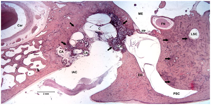
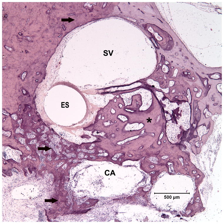
The external and middle ears were basically normal, excluding the stapedes. The stapedes were relatively spared, with maintained anatomy, although both footplates were thickened, and slightly displaced, appearing fixed anteriorly (Fig 1a). There was bilateral extensive otosclerotic remodeling of the bony labyrinth, extending throughout the complete temporal bone specimens, from the genu area of the facial nerve to beneath the round window niche. All stages of otosclerosis were seen: active and inactive areas with interconnecting vascular channels, devitalized, and degenerate areas. The otosclerotic foci extended through the endosteum and large areas of cavitation were seen, both anterior to the apical turn and posterior to the basal turn (Fig 1a). Foci were seen within the interscalar septa, the modiolus, and the ostia of the cribrose area. In the basal turn of the right cochlear duct, adjacent to the electrode site, there was new lamellar bone formation (Fig 1b) within the scala tympani even extending into the scala vestibuli. In the left ear there were fibrous strands in the scala tympani. There was severe atrophy of the organ of Corti, with extensive loss of inner and outer hair cells, and slight to moderate hydrops in all turns. The stria vascularis was atrophied with hyalinization. There was widespread loss of spiral ganglion cells. There were foci adjacent to all six semicircular canals, surrounding the labyrinth. The endolymphatic ducts appeared patent, and unaffected by otosclerosis, but the endolymphatic sacs were filled with macrophages and hyaline bodies.
Fig 1.
Fig 1a. A histopathologic section of the right ear, at the level of the oval window, showing extensive otosclerosis (thick arrows) of the otic capsule with multiple cavitations (CA). The stapes footplate (FP) is relatively unaffected. Hydrops of the middle turn is shown with a thin arrow. H&E
Fig 1b. Detail of 1a, showing the basal turn of the cochlear duct, with ossification (*) of the scala tympani, medial to the electrode site (ES). H&E
Abbreviations used in figures:
C, cochlea; Car, carotid artery; ES, electrode site; FN, facial nerve; IAC, internal auditory canal; LSC, lateral semicircular canal; ME, middle ear; PSC, posterior semicircular canal; SSC, superior semicircular canal; SV, scala vestibule; Utr, utricle. H&E, Hematoxylin and eosin.
DISCUSSION
In this case, the extensive otosclerosis was apparently not recognized during the patient’s lifetime. There was no mention of conductive hearing loss or the word otosclerosis in the medical charts, audiometric data or even at surgery, where massive new “bone” formation was encountered in the round window area. The CT scans, both before and five years after surgery, were evaluated as normal. The implant functioned in a stable fashion up until fourteen years after surgery, i.e. one year prior to his death. This is in accordance with the findings of Green et al 2, who showed that ossification in the scala tympani did not affect the function of the implant.
Diagnosis of otosclerosis remains enigmatic, as this case demonstrates. It is essential to consider cochlear otosclerosis in cases of progressive sensorineural hearing loss in young adults to optimize timely rehabilitation.
Acknowledgments
The authors wish to thank Monika Schachern for technical assistance.
References
- 1.Schuknecht HF, Kirchner JC. Cochlear otosclerosis: fact or fantasy. The Laryngoscope. 1974 May;84(5):766–782. doi: 10.1288/00005537-197405000-00008. [DOI] [PubMed] [Google Scholar]
- 2.Green JD, Jr, Marion MS, Hinojosa R. Labyrinthitis ossificans: histopathologic consideration for cochlear implantation. Otolaryngology--head and neck surgery : official journal of American Academy of Otolaryngology-Head and Neck Surgery. 1991 Mar;104(3):320–326. doi: 10.1177/019459989110400306. [DOI] [PubMed] [Google Scholar]


