

Abstract
We aimed to investigate whether increased consumption of fructose is linked to the increased prevalence of fatty liver. The prevalence of nonalcoholic steatohepatitis (NASH) is 3% and 20% in nonobese and obese subjects, respectively. Obesity is a low-grade chronic inflammatory condition and obesity-related cytokines such as interleukin-6, adiponectin, leptin, and tumor necrosis factor-α may play important roles in the development of nonalcoholic fatty liver disease (NAFLD). Additionally, the prevalence of NASH associated with both cirrhosis and hepatocellular carcinoma was reported to be high among patients with type 2 diabetes with or without obesity. Our research group previously showed that consumption of fructose is associated with adverse alterations of plasma lipid profiles and metabolic changes in mice, the American Lifestyle-Induced Obesity Syndrome model, which included consumption of a high-fructose corn syrup in amounts relevant to that consumed by some Americans. The observation reinforces the concerns about the role of fructose in the obesity epidemic. Increased availability of fructose (e.g., high-fructose corn syrup) increases not only abnormal glucose flux but also fructose metabolism in the hepatocyte. Thus, the anatomic position of the liver places it in a strategic buffering position for absorbed carbohydrates and amino acids. Fructose was previously accepted as a beneficial dietary component because it does not stimulate insulin secretion. However, since insulin signaling plays an important role in central mechanisms of NAFLD, this property of fructose may be undesirable. Fructose has a selective hepatic metabolism, and provokes a hepatic stress response involving activation of c-Jun N-terminal kinases and subsequent reduced hepatic insulin signaling. As high fat diet alone produces obesity, insulin resistance, and some degree of fatty liver with minimal inflammation and no fibrosis, the fast food diet which includes fructose and fats produces a gene expression signature of increased hepatic fibrosis, inflammation, endoplasmic reticulum stress and lipoapoptosis. Hepatic de novo lipogenesis (fatty acid and triglyceride synthesis) is increased in patients with NAFLD. Stable-isotope studies showed that increased de novo lipogenesis (DNL) in patients with NAFLD contributed to fat accumulation in the liver and the development of NAFLD. Specifically, DNL was responsible for 26% of accumulated hepatic triglycerides and 15%-23% of secreted very low-density lipoprotein triglycerides in patients with NAFLD compared to an estimated less than 5% DNL in healthy subjects and 10% DNL in obese people with hyperinsulinemia. In conclusion, understanding the underlying causes of NAFLD forms the basis for rational preventive and treatment strategies of this major form of chronic liver disease.
Keywords: Nonalcoholic, Fatty liver, Diabetes, Insulin resistance, Cytokines, Obesity, Fructose
INTRODUCTION
Excessive accumulation of triglycerides in hepatocytes in the absence of significant alcohol consumption occurs in about 20%-30% of adults[1-5]. Excessive fat in the liver, called nonalcoholic fatty liver disease (NAFLD), predisposes to the development of nonalcoholic steatohepatitis (NASH). NASH constitutes the subset of NAFLD that is most worrisome because it is a significant risk factor for developing cirrhosis and its complications, including hepatocellular carcinoma (HCC)[6-9]. Because the accumulation of excess fat in the liver is a prerequisite for the development of NASH, understanding the underlying causes of NAFLD forms the basis for rational preventive and treatment strategies of this major form of chronic liver disease.
Obesity is a low-grade chronic inflammatory condition and obesity-related cytokines such as interleukin-6 (IL-6), adiponectin, leptin, and tumor necrosis factor (TNF) α may play important roles in the development of NAFLD. The prevalence of NASH is 3% and 20% in nonobese and obese subjects, respectively. Additionally, the prevalence of NASH associated with both cirrhosis and HCC was reported to be high among patients with type-2 diabetes with or without obesity.
OBESITY EPIDEMIC
A balance exists between energy demand and intake in the human body. Obesity is one of the major abnormalities of this well preserved equilibrium. Obesity, and its consequences such as insulin resistance and the metabolic syndrome, is a growing threat to the health of people in developed nations[10]. A diet based on high cholesterol, high saturated fat, and high fructose (cafeteria or fast food type) recapitulates features of the metabolic syndrome and NASH with progressive fibrosis (Figure 1).
Figure 1.
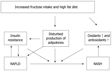
Diet based on high cholesterol, high saturated fat, and high fructose (cafeteria or fast food type) recapitulates features of the metabolic syndrome and nonalcoholic fatty liver disease and nonalcoholic steatohepatitis with progressive fibrosis in human and mice. NAFLD: Nonalcoholic fatty liver disease; NASH: Nonalcoholic steatohepatitis.
“FAST FOOD” OR “CAFETERIA” TYPE DIET COMPOSED OF HIGH SATURATED FATS, CHOLESTEROL, AND FRUCTOSE
The basis of the composition of “fast food” or “cafeteria” style food is high saturated fats, cholesterol, and fructose[11]. As the high fat diet produces obesity, insulin resistance, and some hepatic steatosis with minimal inflammation and no fibrosis, the fast food diet produces a gene expression signature of increased hepatic fibrosis, inflammation, endoplasmic reticulum stress and lipoapoptosis (Figure 2). Our research group previously showed that consumption of fructose is associated with adverse alterations of plasma lipid profiles and metabolic changes in mice, the American Lifestyle-Induced Obesity Syndrome (ALIOS) model, which included consumption of a high-fructose corn syrup (HFCS) in amounts relevant to that consumed by some Americans[11]. The observation that the ALIOS mice indeed consumed a greater quantity of food beyond the additional calories consumed from the HFCS when fed HFCS compared with control water supports this observation and reinforces the concerns about the role of fructose in the obesity epidemic[12-15]. In adolescents, higher fructose consumption is associated with multiple markers of cardiometabolic risk, but it appears that these relationships are mediated by visceral obesity.
Figure 2.
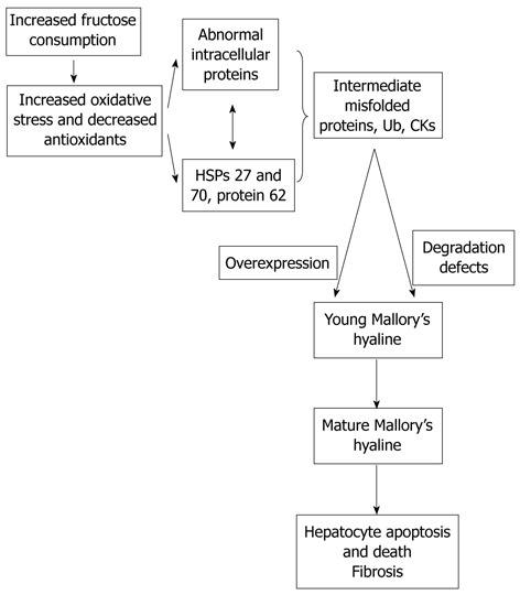
As the high fat diet produces obesity, insulin resistance, and some hepatic steatosis with minimal inflammation with no fibrosis, the fast food diet produces a gene expression signature of increased hepatic fibrosis, inflammation, and endoplasmic reticulum stress and lipoapoptosis. HSP: Heat shock proteins.
The most commonly used HFCS in soft drinks and other carbohydrate-sweetened beverages is a blend composed of 55% fructose, 41% glucose, and 4% complex polysaccharides. Fructose has increasingly been used as a sweetener since the introduction of high-fructose corn syrups in the 1960s[10-13,16] and is now an abundant source of dietary carbohydrate in the United States. The annual per capita consumption of extrinsic or added fructose was approximately 0.2 kg in 1970 to approximately 28 kg in 1997. This increased consumption has been linked to the increased prevalence of obesity, type 2 diabetes and fatty liver in the United States.
The liver is exquisitely sensitive to changes in nutrient delivery and is uniquely suited to metabolize ingested simple sugars, such as fructose and glucose[13,14]. Stress-activated protein kinases, principally the c-Jun N-terminal kinases (JNK), are activated by cell stress-inducing stimuli. Increased fructose supply provokes a hepatic stress response involving activation of JNK and subsequent reduced hepatic insulin signaling.
UNIQUE METABOLISM OF FRUCTOSE
Fructose, glucose, and galactose are the 3 major dietary monosaccharides. Sucrose (glucose-fructose), lactose (glucose-galactose), and maltose (glucose-glucose) are the major disaccharides. Dietary fructose occurs in 2 forms: mono- or disaccharide. The rate of fructose absorption appears to be between that of mannose and glucose[12-15]. Fructose is absorbed by carrier-mediated facilitated diffusion, an energy-dependent process. The fructose carrier is a member of the glucose transport family and is referred to as glucose transporter 5. Sucrose is cleaved to glucose and fructose by sucrase, an enzyme located in the brush border of small intestine enterocytes.
Fructose was previously accepted as a beneficial dietary component because it does not stimulate insulin secretion. However, since insulin signaling plays an important role in the central mechanisms of NAFLD, this property of fructose may be undesirable[13-15]. Additionally, fructose may prevent suppression of ghrelin secretion, resulting in impaired satiety mechanisms[14]. In large quantities, fructose can also stress the liver by depleting hepatic energy supplies. Normal subjects and patients with NASH exhibited a similar depletion of hepatic ATP levels after an injection of fructose, but recovery of ATP levels after depletion was slower in NASH patients compared with healthy controls. A mixture of fructose and glucose might induce metabolic abnormalities that differ from sucrose, a disaccharide cleaved to fructose and glucose in the small intestine.
Phosphorylation of glucose by glucokinase is a rate-determining step in hepatic glucose metabolism. In contrast to glucose, phosphorylation of fructose in the liver occurs via the enzyme fructokinase. In addition, the metabolism of fructose 1-phosphate in the liver occurs independently of phosphofructokinase, a second rate-determining step in glucose metabolism[13-15]. As a result, the liver is the primary site of fructose extraction and metabolism, with extraction approaching 50% to 70% of fructose delivery. Therefore, increased availability of fructose (e.g., high-fructose corn syrup) will increase not only abnormal glucose flux but also fructose metabolism in the hepatocyte. Thus, the anatomic position of the liver places it in a strategic buffering position for absorbed carbohydrates and amino acids.
Fructose extraction and metabolism by the liver are exceptionally high compared to glucose due both to the extensive amount of fructokinase that phosphorylates fructose to fructose 1-phosphate in the liver and to the subsequent metabolism of fructose 1-phosphate at the triose phosphate level, which bypasses flux control at phosphofructokinase[13-16]. Previous studies comparing the metabolism of fructose and glucose in postabsorptive humans over short intervals have shown that fructose is used faster than glucose and that more is converted to liver glycogen. Fructose oxidation represented a significant portion of fructose metabolism, accounting for 56% to 59% of the ingested fructose and approximately 33% of the infused fructose. It is likely that extrahepatic lactate oxidation subsequent to hepatic fructolysis contributed significantly to the estimated rate of fructose oxidation. Thus, increments in fructose after infusion produced immediate changes in hepatic and extrahepatic substrate metabolism, but did not induce changes in overall glucose production. An immediate fructose infusion in humans induced both hepatic and extrahepatic insulin resistance. These data are consistent with the notion that high concentrations of fructose elicit adaptations in the liver that include metabolic intermediates, gene expression, and insulin action.
SYSTEMIC AND HEPATIC INSULIN RESISTANCE IN NAFLD
While insulin receptor defects cause severe insulin resistance, most patients with insulin resistance have impaired post-receptor intracellular insulin signaling. Insulin binds α-subunits of its receptor, which is a cell surface receptor on the major insulin sensitive cells such as skeletal muscle, adipocytes, and hepatocytes, leading to autophosphorylation of the cytoplasmic domains (β-subunits) of the receptor[2-5,17]. The insulin receptor has intrinsic tyrosine kinase activity activated by insulin binding and the autophosphorylated receptor activates its substrates that include insulin receptor substrate (IRS)-1, IRS-2, Src homology collagen, and adaptor protein with a pleckstrin homology and Src homology 2 domain by tyrosine phosphorylation. These phosphorylated docking proteins bind and activate several downstream components of the insulin signaling pathways. Activated IRS-1 associates with phosphatidyl inositol 3-kinase, which then activates Akt. These events and insulin-dependent inhibition of hepatic glucose output maintain glucose homeostasis. Insulin also affects glucose homeostasis indirectly by its regulatory effect on lipid metabolism. Any interference in this insulin signaling pathway causes glucotoxicity, insulin resistance and, when islet beta cells are capable of responding, compensatory hyperinsulinemia.
Hepatic expression of insulin receptor protein in humans and the levels of both IRS-1 and IRS-2 in animals were decreased in chronic hyperinsulinemic states[11]. IRS-1 was more closely linked to glucose homeostasis with the regulation of glucokinase expression while IRS-2 was more closely linked to lipogenesis with the regulation of lipogenic enzymes sterol regulatory element-binding protein-1c (SREBP-1c) and fatty acid synthase[18,19]. Additional physiological roles of insulin include regulating the metabolism of macronutrients and stimulating cellular growth. Insulin activates synthesis and inhibits catabolism of lipids while shutting off the synthesis of glucose in the liver.
Adipose tissue is one of the major insulin sensitive organs in the human body and the process of differentiation of preadipocytes to adipocytes is induced by insulin[17,18]. Within the adipose tissue, insulin stimulates triglyceride synthesis and inhibits lipolysis by upregulating lipoprotein lipase activity which is the most sensitive pathway in insulin action, facilitating free fatty acid uptake and glucose transport, inhibiting hormone sensitive lipase, and increasing gene expression of lipogenic enzymes.
PROINFLAMMATORY SIGNALING IN INSULIN RESISTANCE
Protein kinase C theta (PKCtheta;) and inhibitor κB kinase β (IKK-β) are two proinflammatory kinases involved in insulin downstream signaling[17,18]. They are activated by lipid metabolites such as high plasma free fatty acid concentrations and there is a positive relationship between the activation of PKCtheta; and the concentration of intermediate fatty acid products. PKCtheta; activates both IKK-β and JNK, leading to increased Ser 307 phosphorylation of IRS-1 and insulin resistance. Activation or overexpression of IKK-β diminishes insulin signaling and causes insulin resistance whereas inhibition of IKK-β improves insulin sensitivity. Inhibition of IKK-β activity prevented insulin resistance due to TNF-α in cultured cells. IKK-β phosphorylates the inhibitor of nuclear factor kappa B (NF-κB), leading to the activation of NF-κB by the translocation of NF-κB to the nucleus. NF-κB is an inducible transcription factor and promotes specific gene expression in the nucleus. For example, NF-κB regulates the production of multiple inflammatory mediators, such as TNF-α and IL-6. TNF-α and reactive oxygen species could also activate NF-κB[19-22]. In contrast, antioxidants inhibit this activation. NF-κB has both apoptotic and anti-apoptotic effects. The finding that NF-κB deficient mice were protected from high-fat diet-induced insulin resistance suggests that NF-κB directly participates in processes that impair insulin signaling. High-dose salicylates also inhibit NF-κB and subsequently improve insulin sensitivity. These subsequently promote hepatic and systemic insulin resistance. The study group also showed that these results were reversed by curcumin which inhibits NF-κB activity. Curcumin also has the ability to induce antioxidant enzymes and scavenge ROS.
Suppressors of cytokine signaling (SOCS) and inducible nitric oxide synthase are two inflammatory mediators recently recognized to play a role in insulin signaling[23-25]. Induction of SOCS proteins (SOCS 1-7 and cytokine-inducible src homology 2 domain-containing protein) by proinflammatory cytokines might contribute to the cytokine-mediated insulin resistance in obese subjects[26-30]. In fact, the isoforms of SOCS are the members of a negative feedback loop of cytokine signaling, regulated by both phosphorylation and transcription events. SOCS-1, and particularly SOCS-3, are involved in the inhibition of insulin signaling either by interfering with IRS-1 and IRS-2 tyrosine phosphorylation or by the degradation of their substrates. SOCS-3 might also regulate central leptin action and play a role in the leptin resistance of obese human subjects. SOCS might be a link between leptin and insulin resistance because insulin levels are increased in leptin resistant conditions due to the diminished insulin suppression effect of leptin because of insufficient leptin levels. Moreover, SOCS proteins might involve insulin/insulin like growth factor-1 signaling. SOCS-1 knockout mice showed low glucose concentrations and increased insulin sensitivity. SREBP-1c is one of the key mediators of lipid synthesis from glucose and other precursors (de novo lipogenesis) in the liver. Indeed, SOCS proteins markedly induce de novo fatty acid synthesis in the liver by both the up-regulation of SREBP-1c and persistent insulin resistance with hyperinsulinemia which stimulates SREBP-1c-mediated gene expression. Liver is the insulin clearance organ. Thus, decreased insulin clearance in patients with NAFLD further elevates insulin levels in the circulation and de novo lipogenesis in the liver. SOCS-1 and SOCS-3 may exert these effects by inhibiting signal transduction and activator of transcription proteins (STAT), particularly STAT-3, via binding Janus tyrosine Kinase (JAK) tyrosine kinase because this binding diminishes the phosphorylation ability of JAK kinase to STAT-3. STAT-3 inhibits the activation of SREBP-1c. Specific STAT-3 knockout mice showed markedly increased expression of SREBP-1c and subsequently increased fat content in the liver. Conversely, inhibition of SOCS proteins, particularly SOCS-3, improved both insulin sensitivity and the activation of SREBP-1c which eventually reduced liver steatosis and hypertriglyceridemia in db/db mice.
Nitric oxide synthase-2 (NOS2) or inducible nitric oxide synthase (iNOS) production are also induced by proinflammatory cytokines[31]. A high-fat diet in rats causes up-regulation of iNOS mRNA expression and increases iNOS protein activity. Increased production of NOS2 might reduce insulin action in both muscle and pancreas and decreased iNOS activity protects muscles from the high-fat diet induced insulin resistance. It was also shown that leptin deficient ob/ob mice without iNOS were more insulin sensitive than ob wild-type mice. Thus, the production of nitric oxide may be one link between inflammation and insulin resistance.
SOURCES OF LIVER FAT
Accumulation of triglycerides as fat droplets within the cytoplasm of hepatocytes is a prerequisite for subsequent events of NASH. Accumulation of excess triglyceride in hepatocytes is generally the result of increased delivery of non-esterified fatty acids (NEFAs), increased synthesis of NEFAs, impaired intracellular catabolism of NEFAs, impaired secretion as triglyceride, or a combination of these abnormalities[32]. Recent techniques, such as isotope methodologies, multiple-stable-isotope approach and gas chromatography/mass spectrometry, provided valuable information regarding the fate of fatty acids during both fasting and fed states[33] such as the relative contribution of three fatty acid sources to the accumulated fat in NAFLD: adipose tissue, de novo lipogenesis, and dietary fat. Additionally, these studies reported that the plasma NEFA pool is the main contributor of both hepatic triglycerides in the fasting state and very low-density lipoproteins (VLDL)-triglycerides in both fasting and fed states.
DYSREGULATED PERIPHERAL LIPOLYSIS
A study showed that adipose tissue makes a major contribution to the plasma NEFA pool, contributing 81.7% in the fasted state and 61.7% in the fed state[33]. Additionally, the contribution of dietary lipids to the plasma NEFA pool was found to be only 26.2% and 10.4% in fed and fasted states, respectively, in the same study. Finally, the contribution of newly made fatty acids (originating from the adipose tissue and liver) to the plasma NEFA pool was 7.0% and 9.4% for the fasted and fed states, respectively.
The liver takes up free fatty acids from the circulating NEFA pool and the rate of uptake depends only on the plasma free fatty acid concentrations. Hepatic NEFA uptake continues despite increased hepatic content of fatty acids and triglycerides[34]. The concentration of free fatty acids is increased in the portal circulation rapidly when lipolysis occurs in visceral adipose tissue. These products directly flux to the liver via the splanchnic circulation and contribute to hepatic triglyceride synthesis, NAFLD, and hepatic insulin resistance.
HEPATIC DE NOVO LIPOGENESIS
Hepatic de novo lipogenesis (fatty acid and triglyceride synthesis) is increased in patients with NAFLD[35-39]. Stable-isotope studies showed that increased de novo lipogenesis (DNL) in patients with NAFLD contributed to fat accumulation in the liver and the development of NAFLD[33]. Specifically, DNL was responsible for 26% of accumulated hepatic triglycerides and 15%-23% of secreted VLDL triglycerides in patients with NAFLD compared to an estimated less than 5% DNL in healthy subjects and 10% DNL in obese people with hyperinsulinemia. Interestingly, Donnelly and colleagues demonstrated the similarity between VLDL-triglycerides and hepatic-triglycerides regarding contributions of fatty acid sources (62% vs 59% for NEFA contribution, respectively; 23% vs 26% for DNL, respectively; and 15% vs 15% for dietary fatty acids, respectively) in NAFLD patients. Substrates used for the synthesis of newly made fatty acids by DNL are primarily glucose, fructose, and amino acids; oleic acid (18:1, a ω-6 monounsaturated fatty acid, which is relatively resistant to peroxidation) is the major end product of de novo fatty acid synthesis[40-42]. Moreover, simple sugars have the ability to stimulate lipogenesis[33]. Ingested carbohydrates are a major stimulus for hepatic delayed neuronal loss and are thus more likely to directly contribute to NAFLD than dietary fat intake[43-46].
In conclusion, fructose has increasingly been used as a sweetener since the introduction of high-fructose corn syrups in the 1960s and is now an abundant source of dietary carbohydrate in the United States[47-50]. The most commonly used HFCS in soft drinks and other carbohydrate-sweetened beverages is a blend composed of 55% fructose, 41% glucose, and 4% complex polysaccharides[51-55]. This increased consumption has been linked to the increased prevalence of obesity and type 2 diabetes and fatty liver in the United States by increased fructose supply, which provokes a hepatic stress response involving activation of JNK and subsequent reduced hepatic insulin signaling[56-59]. Understanding the underlying causes of NAFLD forms the basis for rational preventive and treatment strategies of this major form of chronic liver disease.
Footnotes
P- Reviewers Koutsilieris M, Lee SY S- Editor Gou SX L- Editor O’Neill M E- Editor Zhang DN
References
- 1.Matteoni CA, Younossi ZM, Gramlich T, Boparai N, Liu YC, McCullough AJ. Nonalcoholic fatty liver disease: a spectrum of clinical and pathological severity. Gastroenterology. 1999;116:1413–1419. doi: 10.1016/s0016-5085(99)70506-8. [DOI] [PubMed] [Google Scholar]
- 2.Marchesini G, Brizi M, Morselli-Labate AM, Bianchi G, Bugianesi E, McCullough AJ, Forlani G, Melchionda N. Association of nonalcoholic fatty liver disease with insulin resistance. Am J Med. 1999;107:450–455. doi: 10.1016/s0002-9343(99)00271-5. [DOI] [PubMed] [Google Scholar]
- 3.Marchesini G, Brizi M, Bianchi G, Tomassetti S, Bugianesi E, Lenzi M, McCullough AJ, Natale S, Forlani G, Melchionda N. Nonalcoholic fatty liver disease: a feature of the metabolic syndrome. Diabetes. 2001;50:1844–1850. doi: 10.2337/diabetes.50.8.1844. [DOI] [PubMed] [Google Scholar]
- 4.Seppälä-Lindroos A, Vehkavaara S, Häkkinen AM, Goto T, Westerbacka J, Sovijärvi A, Halavaara J, Yki-Järvinen H. Fat accumulation in the liver is associated with defects in insulin suppression of glucose production and serum free fatty acids independent of obesity in normal men. J Clin Endocrinol Metab. 2002;87:3023–3028. doi: 10.1210/jcem.87.7.8638. [DOI] [PubMed] [Google Scholar]
- 5.Pagano G, Pacini G, Musso G, Gambino R, Mecca F, Depetris N, Cassader M, David E, Cavallo-Perin P, Rizzetto M. Nonalcoholic steatohepatitis, insulin resistance, and metabolic syndrome: further evidence for an etiologic association. Hepatology. 2002;35:367–372. doi: 10.1053/jhep.2002.30690. [DOI] [PubMed] [Google Scholar]
- 6.Marchesini G, Bugianesi E, Forlani G, Cerrelli F, Lenzi M, Manini R, Natale S, Vanni E, Villanova N, Melchionda N, et al. Nonalcoholic fatty liver, steatohepatitis, and the metabolic syndrome. Hepatology. 2003;37:917–923. doi: 10.1053/jhep.2003.50161. [DOI] [PubMed] [Google Scholar]
- 7.Caldwell SH, Oelsner DH, Iezzoni JC, Hespenheide EE, Battle EH, Driscoll CJ. Cryptogenic cirrhosis: clinical characterization and risk factors for underlying disease. Hepatology. 1999;29:664–669. doi: 10.1002/hep.510290347. [DOI] [PubMed] [Google Scholar]
- 8.Poonawala A, Nair SP, Thuluvath PJ. Prevalence of obesity and diabetes in patients with cryptogenic cirrhosis: a case-control study. Hepatology. 2000;32:689–692. doi: 10.1053/jhep.2000.17894. [DOI] [PubMed] [Google Scholar]
- 9.Charlton M, Kasparova P, Weston S, Lindor K, Maor-Kendler Y, Wiesner RH, Rosen CB, Batts KP. Frequency of nonalcoholic steatohepatitis as a cause of advanced liver disease. Liver Transpl. 2001;7:608–614. doi: 10.1053/jlts.2001.25453. [DOI] [PubMed] [Google Scholar]
- 10.Keaney JF, Larson MG, Vasan RS, Wilson PW, Lipinska I, Corey D, Massaro JM, Sutherland P, Vita JA, Benjamin EJ. Obesity and systemic oxidative stress: clinical correlates of oxidative stress in the Framingham Study. Arterioscler Thromb Vasc Biol. 2003;23:434–439. doi: 10.1161/01.ATV.0000058402.34138.11. [DOI] [PubMed] [Google Scholar]
- 11.Tetri LH, Basaranoglu M, Brunt EM, Yerian LM, Neuschwander-Tetri BA. Severe NAFLD with hepatic necroinflammatory changes in mice fed trans fats and a high-fructose corn syrup equivalent. Am J Physiol Gastrointest Liver Physiol. 2008;295:G987–G995. doi: 10.1152/ajpgi.90272.2008. [DOI] [PMC free article] [PubMed] [Google Scholar]
- 12.Elliott SS, Keim NL, Stern JS, Teff K, Havel PJ. Fructose, weight gain, and the insulin resistance syndrome. Am J Clin Nutr. 2002;76:911–922. doi: 10.1093/ajcn/76.5.911. [DOI] [PubMed] [Google Scholar]
- 13.James J, Thomas P, Cavan D, Kerr D. Preventing childhood obesity by reducing consumption of carbonated drinks: cluster randomised controlled trial. BMJ. 2004;328:1237. doi: 10.1136/bmj.38077.458438.EE. [DOI] [PMC free article] [PubMed] [Google Scholar]
- 14.Teff KL, Elliott SS, Tschöp M, Kieffer TJ, Rader D, Heiman M, Townsend RR, Keim NL, D'Alessio D, Havel PJ. Dietary fructose reduces circulating insulin and leptin, attenuates postprandial suppression of ghrelin, and increases triglycerides in women. J Clin Endocrinol Metab. 2004;89:2963–2972. doi: 10.1210/jc.2003-031855. [DOI] [PubMed] [Google Scholar]
- 15.Faeh D, Minehira K, Schwarz JM, Periasamy R, Park S, Tappy L. Effect of fructose overfeeding and fish oil administration on hepatic de novo lipogenesis and insulin sensitivity in healthy men. Diabetes. 2005;54:1907–1913. doi: 10.2337/diabetes.54.7.1907. [DOI] [PubMed] [Google Scholar]
- 16.Dhingra R, Sullivan L, Jacques PF, Wang TJ, Fox CS, Meigs JB, D'Agostino RB, Gaziano JM, Vasan RS. Soft drink consumption and risk of developing cardiometabolic risk factors and the metabolic syndrome in middle-aged adults in the community. Circulation. 2007;116:480–488. doi: 10.1161/CIRCULATIONAHA.107.689935. [DOI] [PubMed] [Google Scholar]
- 17.Weisberg SP, McCann D, Desai M, Rosenbaum M, Leibel RL, Ferrante AW. Obesity is associated with macrophage accumulation in adipose tissue. J Clin Invest. 2003;112:1796–1808. doi: 10.1172/JCI19246. [DOI] [PMC free article] [PubMed] [Google Scholar]
- 18.Xu H, Barnes GT, Yang Q, Tan G, Yang D, Chou CJ, Sole J, Nichols A, Ross JS, Tartaglia LA, et al. Chronic inflammation in fat plays a crucial role in the development of obesity-related insulin resistance. J Clin Invest. 2003;112:1821–1830. doi: 10.1172/JCI19451. [DOI] [PMC free article] [PubMed] [Google Scholar]
- 19.Hansel B, Giral P, Nobecourt E, Chantepie S, Bruckert E, Chapman MJ, Kontush A. Metabolic syndrome is associated with elevated oxidative stress and dysfunctional dense high-density lipoprotein particles displaying impaired antioxidative activity. J Clin Endocrinol Metab. 2004;89:4963–4971. doi: 10.1210/jc.2004-0305. [DOI] [PubMed] [Google Scholar]
- 20.Furukawa S, Fujita T, Shimabukuro M, Iwaki M, Yamada Y, Nakajima Y, Nakayama O, Makishima M, Matsuda M, Shimomura I. Increased oxidative stress in obesity and its impact on metabolic syndrome. J Clin Invest. 2004;114:1752–1761. doi: 10.1172/JCI21625. [DOI] [PMC free article] [PubMed] [Google Scholar]
- 21.Ratziu V, Bonyhay L, Di Martino V, Charlotte F, Cavallaro L, Sayegh-Tainturier MH, Giral P, Grimaldi A, Opolon P, Poynard T. Survival, liver failure, and hepatocellular carcinoma in obesity-related cryptogenic cirrhosis. Hepatology. 2002;35:1485–1493. doi: 10.1053/jhep.2002.33324. [DOI] [PubMed] [Google Scholar]
- 22.Ratziu V, Giral P, Charlotte F, Bruckert E, Thibault V, Theodorou I, Khalil L, Turpin G, Opolon P, Poynard T. Liver fibrosis in overweight patients. Gastroenterology. 2000;118:1117–1123. doi: 10.1016/s0016-5085(00)70364-7. [DOI] [PubMed] [Google Scholar]
- 23.Stein CJ, Colditz GA. The epidemic of obesity. J Clin Endocrinol Metab. 2004;89:2522–2525. doi: 10.1210/jc.2004-0288. [DOI] [PubMed] [Google Scholar]
- 24.Hundal RS, Petersen KF, Mayerson AB, Randhawa PS, Inzucchi S, Shoelson SE, Shulman GI. Mechanism by which high-dose aspirin improves glucose metabolism in type 2 diabetes. J Clin Invest. 2002;109:1321–1326. doi: 10.1172/JCI14955. [DOI] [PMC free article] [PubMed] [Google Scholar]
- 25.Seki S, Kitada T, Sakaguchi H. Clinicopathological significance of oxidative cellular damage in non-alcoholic fatty liver diseases. Hepatol Res. 2005;33:132–134. doi: 10.1016/j.hepres.2005.09.020. [DOI] [PubMed] [Google Scholar]
- 26.Marchesini G, Ridolfi V, Nepoti V. Hepatotoxicity of fast food? Gut. 2008;57:568–570. doi: 10.1136/gut.2007.143958. [DOI] [PubMed] [Google Scholar]
- 27.Milagro FI, Campión J, Martínez JA. Weight gain induced by high-fat feeding involves increased liver oxidative stress. Obesity (Silver Spring) 2006;14:1118–1123. doi: 10.1038/oby.2006.128. [DOI] [PubMed] [Google Scholar]
- 28.Mozaffarian D, Katan MB, Ascherio A, Stampfer MJ, Willett WC. Trans fatty acids and cardiovascular disease. N Engl J Med. 2006;354:1601–1613. doi: 10.1056/NEJMra054035. [DOI] [PubMed] [Google Scholar]
- 29.Feldstein AE, Werneburg NW, Canbay A, Guicciardi ME, Bronk SF, Rydzewski R, Burgart LJ, Gores GJ. Free fatty acids promote hepatic lipotoxicity by stimulating TNF-alpha expression via a lysosomal pathway. Hepatology. 2004;40:185–194. doi: 10.1002/hep.20283. [DOI] [PubMed] [Google Scholar]
- 30.Bradbury MW, Berk PD. Lipid metabolism in hepatic steatosis. Clin Liver Dis. 2004;8:639–671. doi: 10.1016/j.cld.2004.04.005. [DOI] [PubMed] [Google Scholar]
- 31.Wellen KE, Hotamisligil GS. Inflammation, stress, and diabetes. J Clin Invest. 2005;115:1111–1119. doi: 10.1172/JCI25102. [DOI] [PMC free article] [PubMed] [Google Scholar]
- 32.Neuschwander-Tetri BA, Caldwell SH. Nonalcoholic steatohepatitis: summary of an AASLD Single Topic Conference. Hepatology. 2003;37:1202–1219. doi: 10.1053/jhep.2003.50193. [DOI] [PubMed] [Google Scholar]
- 33.Donnelly KL, Smith CI, Schwarzenberg SJ, Jessurun J, Boldt MD, Parks EJ. Sources of fatty acids stored in liver and secreted via lipoproteins in patients with nonalcoholic fatty liver disease. J Clin Invest. 2005;115:1343–1351. doi: 10.1172/JCI23621. [DOI] [PMC free article] [PubMed] [Google Scholar]
- 34.Tamura S, Shimomura I. Contribution of adipose tissue and de novo lipogenesis to nonalcoholic fatty liver disease. J Clin Invest. 2005;115:1139–1142. doi: 10.1172/JCI24930. [DOI] [PMC free article] [PubMed] [Google Scholar]
- 35.Neuschwander-Tetri BA, Ford DA, Acharya S, Gilkey G, Basaranoglu M, Tetri LH, Brunt EM. Dietary trans-fatty acid induced NASH is normalized following loss of trans-fatty acids from hepatic lipid pools. Lipids. 2012;47:941–950. doi: 10.1007/s11745-012-3709-7. [DOI] [PMC free article] [PubMed] [Google Scholar]
- 36.Henkel J, Frede K, Schanze N, Vogel H, Schürmann A, Spruss A, Bergheim I, Püschel GP. Stimulation of fat accumulation in hepatocytes by PGE₂-dependent repression of hepatic lipolysis, β-oxidation and VLDL-synthesis. Lab Invest. 2012;92:1597–1606. doi: 10.1038/labinvest.2012.128. [DOI] [PubMed] [Google Scholar]
- 37.Arai T, Kim HJ, Hirako S, Nakasatomi M, Chiba H, Matsumoto A. Effects of dietary fat energy restriction and fish oil feeding on hepatic metabolic abnormalities and insulin resistance in KK mice with high-fat diet-induced obesity. J Nutr Biochem. 2013;24:267–273. doi: 10.1016/j.jnutbio.2012.06.002. [DOI] [PubMed] [Google Scholar]
- 38.Flannery C, Dufour S, Rabøl R, Shulman GI, Petersen KF. Skeletal muscle insulin resistance promotes increased hepatic de novo lipogenesis, hyperlipidemia, and hepatic steatosis in the elderly. Diabetes. 2012;61:2711–2717. doi: 10.2337/db12-0206. [DOI] [PMC free article] [PubMed] [Google Scholar]
- 39.Tappy L, Lê KA. Does fructose consumption contribute to non-alcoholic fatty liver disease? Clin Res Hepatol Gastroenterol. 2012;36:554–560. doi: 10.1016/j.clinre.2012.06.005. [DOI] [PubMed] [Google Scholar]
- 40.Sashidhara KV, Kumar M, Sonkar R, Singh BS, Khanna AK, Bhatia G. Indole-based fibrates as potential hypolipidemic and antiobesity agents. J Med Chem. 2012;55:2769–2779. doi: 10.1021/jm201697v. [DOI] [PubMed] [Google Scholar]
- 41.Kok BP, Kienesberger PC, Dyck JR, Brindley DN. Relationship of glucose and oleate metabolism to cardiac function in lipin-1 deficient (fld) mice. J Lipid Res. 2012;53:105–118. doi: 10.1194/jlr.M019430. [DOI] [PMC free article] [PubMed] [Google Scholar]
- 42.Herrema H, Meissner M, van Dijk TH, Brufau G, Boverhof R, Oosterveer MH, Reijngoud DJ, Müller M, Stellaard F, Groen AK, et al. Bile salt sequestration induces hepatic de novo lipogenesis through farnesoid X receptor- and liver X receptor alpha-controlled metabolic pathways in mice. Hepatology. 2010;51:806–816. doi: 10.1002/hep.23408. [DOI] [PubMed] [Google Scholar]
- 43.Sevastianova K, Santos A, Kotronen A, Hakkarainen A, Makkonen J, Silander K, Peltonen M, Romeo S, Lundbom J, Lundbom N, et al. Effect of short-term carbohydrate overfeeding and long-term weight loss on liver fat in overweight humans. Am J Clin Nutr. 2012;96:727–734. doi: 10.3945/ajcn.112.038695. [DOI] [PubMed] [Google Scholar]
- 44.Carvalhana S, Machado MV, Cortez-Pinto H. Improving dietary patterns in patients with nonalcoholic fatty liver disease. Curr Opin Clin Nutr Metab Care. 2012;15:468–473. doi: 10.1097/MCO.0b013e3283566614. [DOI] [PubMed] [Google Scholar]
- 45.Tsuchiya H, Ebata Y, Sakabe T, Hama S, Kogure K, Shiota G. High-fat, high-fructose diet induces hepatic iron overload via a hepcidin-independent mechanism prior to the onset of liver steatosis and insulin resistance in mice. Metabolism. 2013;62:62–69. doi: 10.1016/j.metabol.2012.06.008. [DOI] [PubMed] [Google Scholar]
- 46.Utzschneider KM, Bayer-Carter JL, Arbuckle MD, Tidwell JM, Richards TL, Craft S. Beneficial effect of a weight-stable, low-fat/low-saturated fat/low-glycaemic index diet to reduce liver fat in older subjects. Br J Nutr. 2012:1–9. doi: 10.1017/S0007114512002966. [DOI] [PMC free article] [PubMed] [Google Scholar]
- 47.Johnson RJ, Lanaspa MA, Roncal-Jimenez C, Sanchez-Lozada LG. Effects of excessive fructose intake on health. Ann Intern Med. 2012;156:905; author reply 905–906. doi: 10.7326/0003-4819-156-12-201206190-00024. [DOI] [PubMed] [Google Scholar]
- 48.Li M, Feng F, Cheng L. Expression patterns of genes involved in sugar metabolism and accumulation during apple fruit development. PLoS One. 2012;7:e33055. doi: 10.1371/journal.pone.0033055. [DOI] [PMC free article] [PubMed] [Google Scholar]
- 49.Mellouk Z, Zhang Y, Bulur N, Louchami K, Sener A, Ait Yahia D, Malaisse WJ. The metabolic syndrome of fructose-fed rats: effects of long-chain polyunsaturated ω3 and ω6 fatty acids. IV. D-glucose metabolism by isolated pancreatic islets. Int J Mol Med. 2012;29:291–293. doi: 10.3892/ijmm.2011.824. [DOI] [PubMed] [Google Scholar]
- 50.Liu J, Litt L, Segal MR, Kelly MJ, Pelton JG, Kim M. Metabolomics of oxidative stress in recent studies of endogenous and exogenously administered intermediate metabolites. Int J Mol Sci. 2011;12:6469–6501. doi: 10.3390/ijms12106469. [DOI] [PMC free article] [PubMed] [Google Scholar]
- 51.Bray GA. Fructose and risk of cardiometabolic disease. Curr Atheroscler Rep. 2012;14:570–578. doi: 10.1007/s11883-012-0276-6. [DOI] [PMC free article] [PubMed] [Google Scholar]
- 52.Caporaso N, Morisco F, Camera S, Graziani G, Donnarumma L, Ritieni A. Dietary approach in the prevention and treatment of NAFLD. Front Biosci. 2012;17:2259–2268. doi: 10.2741/4049. [DOI] [PubMed] [Google Scholar]
- 53.Zelber-Sagi S, Ratziu V, Oren R. Nutrition and physical activity in NAFLD: an overview of the epidemiological evidence. World J Gastroenterol. 2011;17:3377–3389. doi: 10.3748/wjg.v17.i29.3377. [DOI] [PMC free article] [PubMed] [Google Scholar]
- 54.Nseir W, Nassar F, Assy N. Soft drinks consumption and nonalcoholic fatty liver disease. World J Gastroenterol. 2010;16:2579–2588. doi: 10.3748/wjg.v16.i21.2579. [DOI] [PMC free article] [PubMed] [Google Scholar]
- 55.Alegret M, Laguna JC. Opposite fates of fructose in the development of metabolic syndrome. World J Gastroenterol. 2012;18:4478–4480. doi: 10.3748/wjg.v18.i33.4478. [DOI] [PMC free article] [PubMed] [Google Scholar]
- 56.Sahebkar A. Potential efficacy of ginger as a natural supplement for nonalcoholic fatty liver disease. World J Gastroenterol. 2011;17:271–272. doi: 10.3748/wjg.v17.i2.271. [DOI] [PMC free article] [PubMed] [Google Scholar]
- 57.Mathes AM. Hepatoprotective actions of melatonin: possible mediation by melatonin receptors. World J Gastroenterol. 2010;16:6087–6097. doi: 10.3748/wjg.v16.i48.6087. [DOI] [PMC free article] [PubMed] [Google Scholar]
- 58.Ha HL, Shin HJ, Feitelson MA, Yu DY. Oxidative stress and antioxidants in hepatic pathogenesis. World J Gastroenterol. 2010;16:6035–6043. doi: 10.3748/wjg.v16.i48.6035. [DOI] [PMC free article] [PubMed] [Google Scholar]
- 59.Grattagliano I, Bonfrate L, Diogo CV, Wang HH, Wang DQ, Portincasa P. Biochemical mechanisms in drug-induced liver injury: certainties and doubts. World J Gastroenterol. 2009;15:4865–4876. doi: 10.3748/wjg.15.4865. [DOI] [PMC free article] [PubMed] [Google Scholar]