Abstract
Background
Radiography is the most widely available imaging modality. Precise evaluations of wrist x-ray can help diagnosis and evaluate the prognosis of many wrist disorders.
Methods
We measured length, angles and indices in 150 posteroanterior and lateral wrist x-rays to determine normal dimensions and variations according to age and sex. All x-rays were made with standard exposure, with the wrist and forearm in a neutral position.
Results
The average carpal height ratio was 0.52±0.03 with the Youm method and 1.5±0.09 with the Nattrass method. Mean ulnar variance was +0.99±1.6 mm and mean radial inclination was 25±4 degrees. The average radial tilt was 10±5.1 degrees. Mean scapholunate angle was 50±8.4 degrees (normal range 40-60).
Conclusion
Carpal height, third metacarpal and capitate length were smaller in women than in men. There was a significant positive relationship between all dimensions. Our data base may be used to follow-up in conditions such as carpal instability, osteoarthritis and osteonecrosis, as well as for clinical research.
Keywords: Wrist measurements, Wrist radiography, Wrist indices
Introduction
Despite the availability of various imaging modalities, plain radiography is used most frequently to evaluate wrist bony anatomy. To define normal wrist anatomy and quantify abnormalities, distances from bone landmarks in posteroanterior (PA) and lateral x-ray have been measured, and used to calculate carpal indices. These indices are used to diagnose and treat wrist disorders. For example, radial inclination, radial tilt, radial height and ulnar variance can be used to diagnose and evaluate distal radius fractures. An abnormal scapholunate angle in lateral view can demonstrate intercarpal ligament injuries. Carpal height ratio may be used to assess carpal collapse in lunate osteonecrosis and osteoarthritis. The ulnar-carpal ratio and radial-carpal ratio can provide information about carpal translocation and malalignment as seen in wrist arthritis.
Precise measurements from x-rays have allowed physicians to determine subtle angulations and abnormal bone and joint relationships. However, because of the variability between the right and left wrist, for radial inclination and radial tilt of distal radius and for the ulnar variance, the normal side also not provides a better reference than normal values obtained from data bases (1).
The aim of this report was to prepare normal data from plain x-rays of Iranian individuals.
Methods
The study measurements consisted of 150 normal posteroanterior and lateral wrist X-Rays. Our inclusion criteria were age between 20 and 60 years and either sex. The participants were patients who come to our hospital for contralateral wrist trauma. Our exclusion criteria were included history of disease, trauma, operation or congenital anomaly of the upper limb. We allowed radiography of normal side for patients.
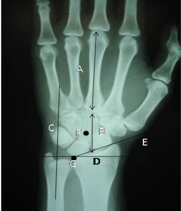
Linear measurements, including third metacarpal length, carpal height, capitate length, radial length and lunate width were made in millimeters (Figs 1 and 2). Angles were measured in degrees, and included radial inclination, distal radioulnar joint angle in posteroanterior view, radial tilt and scapholunate angle in lateral view (Figs. 1 and 3).
Fig. 1.
Line A, axis of the third metacarpal, used to measure length .Line B, continuation of the third metacarpal axis used to measure carpal height. Line C, longitudinal axis of ulnar shaft. Line D, horizontal line from the radiocarpal junction to distal radio-ulnar joint (point G). Line E, from radial styloid process to point G. Radial inclination is the angle between lines D and E. Point F, center of rotation of the carpus in the capitate head.
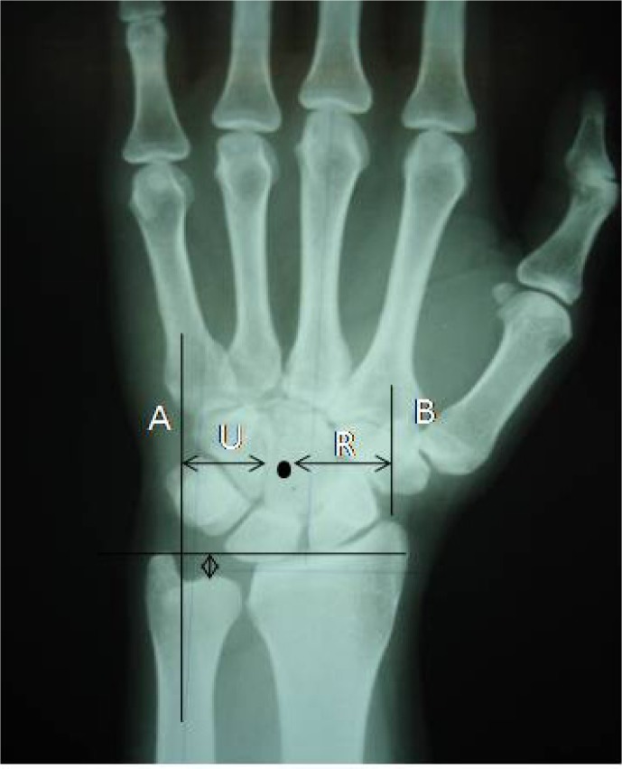
Fig. 2.
Determination of ulnar-carpal and radialcarpal distance, ulnar variance.
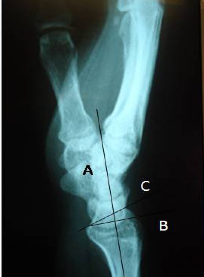
Fig. 3.
Line A, longitudinal axis of the distal end of the radius. Line B, horizontal line perpendicular to line A. Line C alignment of articular surface of the distal radius.The angle between lines B and C is the radial tilt.
Measurements
Both methods of Youm and Nattrass were used to measure carpal height and carpal height ratio. The third metacarpal axis continued to the distal radial articular surface. Carpal height was considered the distance between the base of the third metacarpal and the point of intersection of the metacarpal axis with the radiocarpal joint (Fig. 1).
Ulnar carpal distance (Fig. 2) was measured as a perpendicular distance between the distal extension of ulnar axis and the theoretical center of rotation of, the wrist for radial – ulnar deviation.
The center of rotation of the wrist was located in the head of the capitate, one millimeter to the ulnar side of the principal axis of the bone. We used the method of Chamay et all (2) to measure radial carpal distance (Fig 2), i.e. perpendicular distance between the radial styloid and the center of rotation of wrist joint.
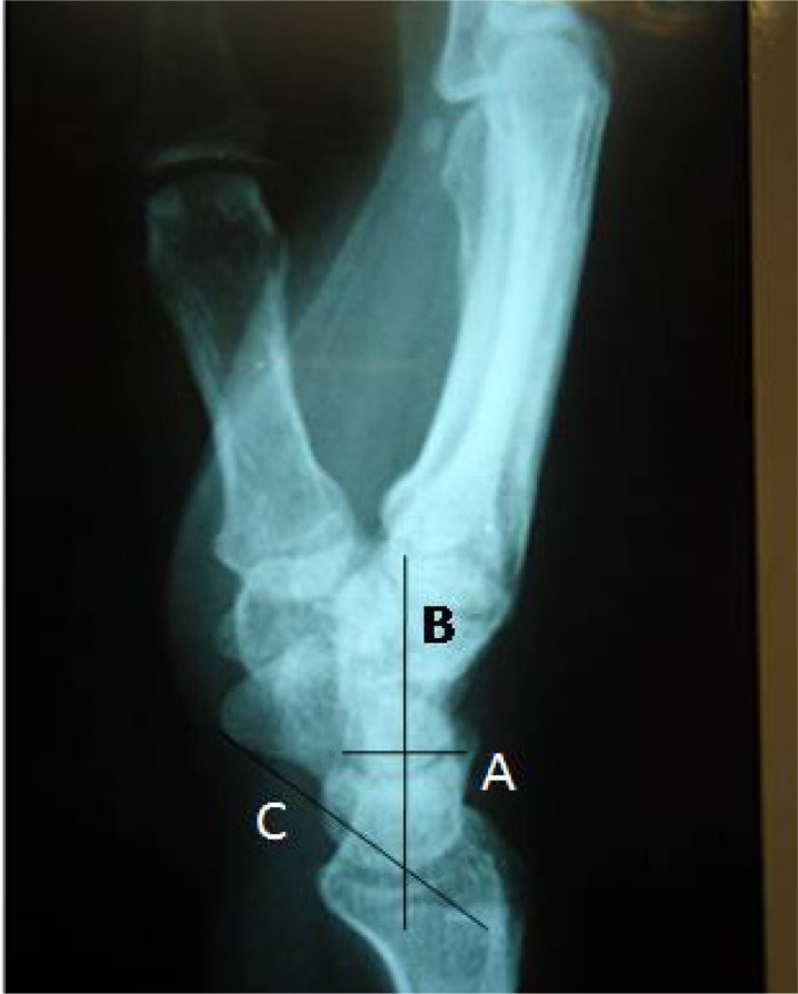
In lateral view, we drew the axis of radius, lunate and scaphoid, radiocarpal joint line, and used these lines to calculate radial tilt and scapholunate angles (Figs. 3 and 4).
Fig. 4.
Line A, drawn from the anterior to the posterior pole of the lunate bone. Line B, perpendicular to line A, is the axis of the lunate bone. Line C, drawn from the anterior surface of the scaphoid bone and parallel to the scaphoid axis. The angle between lines B and C is the scapholunate angle.
Data analysis
We used mean, median and standard deviation (SD), in order to describe continuous variables. The data were analyzed with linear regression and Student t-test. We compared men and women, and two age groups (20 to 40 and 40 to 60 years). The level of significance was p< 0.05.
Results
Measurements from one hundred fifty X-rays of normal wrist (men and women), were used to determine normal dimensions and ratios. Ulnar variance ranged from -3 mm (negative ulnar variance) to +5 mm, with a mean of + .99±1.6 mm. The mean radial inclination was 25 degrees (±4 degrees), and median radial length 12 mm (±1.7). Capitate length ranged from 15 to 28 mm (median 22 mm), and radial tilt from 0 to 24 degrees. Carpal height ratio was 52 ± 3 with the Youm method and 1.5± .09 with the Nattrass method (Table 1).
Table 1.
Radiographic measurements in one hundred fifty normal wrists.
| Mean | Median * | Range | Normal limits | |
|---|---|---|---|---|
| Ulnar variance | .99 | 1.0 ± 1.6 | -3.0 -5.0 | -2-3 |
| Radial length | 12.1 | 12.0 ± 1.7 | 8.0 -18.0 | 10-15 |
| Radial inclination | 25.3 | 25.0 ± 4.0 | 12.0 – 34.0 | 18-32 |
| Carpal height | 32.2 | 32.0 ± 2.8 | 26.0 -40.0 | 28-37 |
| Third metacarpal length | 61.4 | 60.0 ± 5.6 | 28-76 | 54-71 |
| Radial carpal distance | 17.5 | 17.0 ± 2.1 | 10.0 -25.0 | 14.5-21 |
| Ulnar carpal distance | 18.7 | 19.0 ± 2.4 | 13-26 | 15-23 |
| Distal radioulnar joint angle | 3.0 | .00 ± 10.3 | -20-30 | -13-21 |
| Lunate uncoverage length | 4.1 | 4.0 ± 1.3 | 1-9 | 2-7 |
| Lunate width | 14.2 | 14.0 ± 1.7 | 10-20 | 11.5-17.4 |
| Capitate length | 21.8 | 22.0 ± 2.3 | 15-28 | 19-27 |
| Radial tilt | 10.2 | 10.0 ± 5.1 | 0-24 | 0-20 |
| Scapholunate angle | 51.8 | 50.0 ± 8.4 | 30-80 | 40-67 |
| Carpal height ratio (Youm) | 52.3 | 52.0 ± 3.1 | 46- 60 | 47-50 |
| Carpal height ratio (Nattrass) | 1.4 | 1.5 ± .09 | 1.2 -2.0 | 1.2-1.6 |
|
| ||||
| Lunate uncoverage ratio | 28.7 | 28.0 ± 7.3 | 9-58 | 16-41 |
Median and standard deviation
Changes according to age
When the two age subgroups (20 to 40 years and 41 to 60 years) was compared we found significant difference in carpal height, third metacarpal length, radial carpal distance, capitate length, lunate uncoverage length and lunate width (Table 2).
Table 2.
Differences in the wrist Radiographic measurements between adults aged 20.
| To 40 and adults 41 to 60 years. | 20-40 years (n = 75) Mean±SD* | 41-60 years (n = 75) Mean±SD | p value |
|---|---|---|---|
| Ulnar variance | 0.8 ± 1.6 | 1.1 ± 1.6 | NS |
| Radial length | 12.2 ± 1.9 | 12.1 ± 1.5 | NS |
| Radial inclination | 25.1± 4.2 | 25.5 ± 3.8 | NS |
| Carpal Height | 33.4± 2.7 | 31.0 ± 2.3 | .0001 |
| Third metacarpal length | 63.5 ± 5.1 | 59.3 ± 5.3 | .0001 |
| Radial carpal distance | 18 ± 2.2 | 17 ± 2.0 | .03 |
| Ulnar carpal distance | 19.0 ± 2.6 | 18.4 ± 2.9 | NS |
| Distal Radioulnar joint Angle | 3.1 ± 10.9 | 3.0 ± 9.7 | NS |
| Lunate uncoverage length | 4.5 ±1.4 | 3.6 ± 1.2 | .0001 |
| Lunate width | 14.8 ± 2.4 | 13.6 ± 1.3 | .0001 |
| Capitate length | 22.8 ± 1.8 | 20 ± 1.9 | .0001 |
| Radial tilt | 10.0 ± 5.1 | 10.4 ± 5.1 | NS |
| Scapholunate Angle | 51.1 ± 8.8 | 52.4 ± 8.0 | NS |
| Carpal Height ratio (youm) | 52.6 ± 3.1 | 52.0 ± 3.1 | NS |
| Carpal Height ratio (nattrass) | 1.4 ± 0.1 | 1.4 ± .08 | NS |
| Lunate uncoverage ratio | 30.3 ± 8.2 | 26.3 ± 8.6 | NS |
| Right , Hand grip | 22.7 ± 15.9 | 14.3 ± 5.0 | NS |
| Left – Hand grip | 25.9 ± 11.9 | 14.6 ± 5.2 | .02 |
| Radial – carpal ratio | 28.3 ± 2.8 | 28.4 ± 2.3 | NS |
| Ulnar – carpal ratio | 29.7 ± 3.6 | 30.8 ± 3.3 | NS |
Relationships between variables
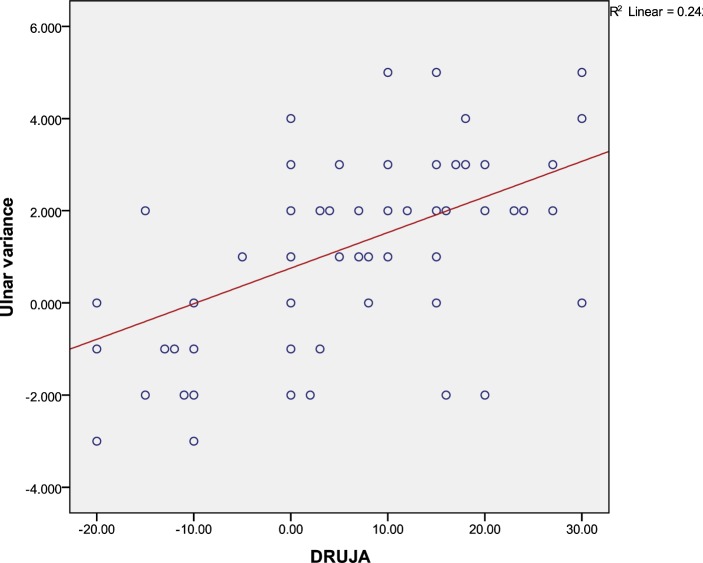
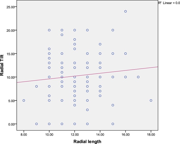
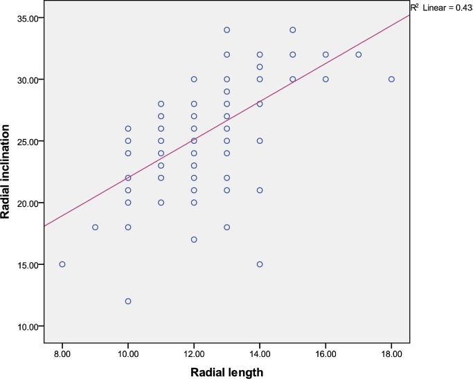
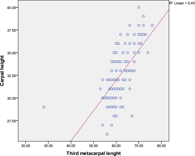
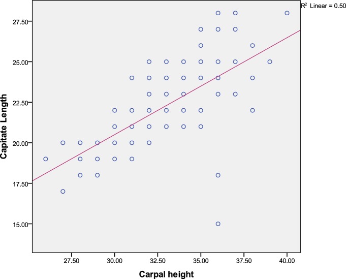
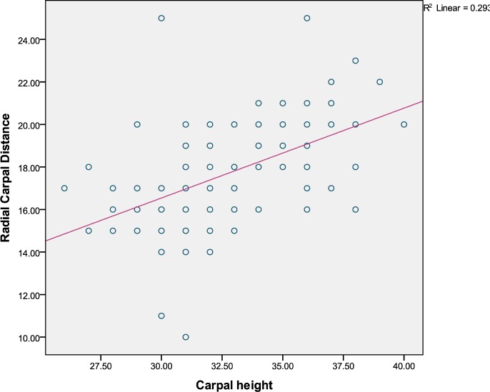
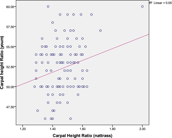
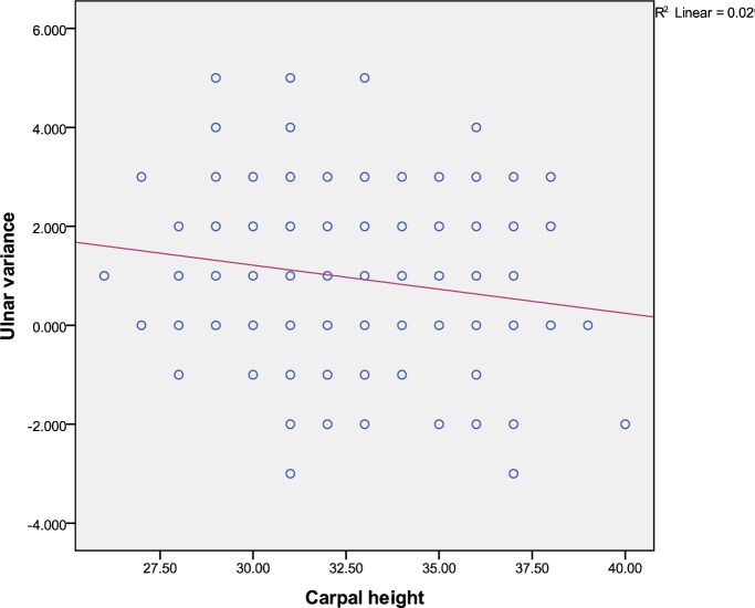
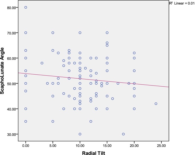
We assessed the relationships between all measurements and indices, and found a significant relationship (p< .05) between ulnar variance and distal radioulnar joint angle (Figure 5). There was no significant relationship between radial tilt and radial inclination (Fig 6). A positive linear relationship was found between radial inclination and radial length (Fig. 7), carpal height and third metacarpal length (Fig. 8), capitate length and carpal height (Fig. 9), radial carpal distance and carpal height (Fig. 10), Youm and Nattrass carpal height ratio (Fig. 11), and ulnar variance and carpal height (Fig. 12). There was no linear relationship between scapholunate angle and radial tilt (Fig. 13).
Fig. 5.
Linear relationship between ulnar variance and Distal Radioulnar Joint Angle (DRUJA). The equation of the regression line is: Y = 0.75 + (.07(x)) p = 0001
Fig. 6.
Linear relationship between radial tilt and radial length. The equation of the regression line is Y = 6.58 + (.30 (x)) p = .507
Fig. 7.
Linear relationship between radial inclination and radial length. The equation of the regression line is Y = 6.59 + (1.54 (x)) p = .001
Fig. 8.
Linear relationship between carpal height and third metacarpal length. The equation of the regression line is Y =11.8 + (.3(x)) p = .001.
Fig. 9.
Linear relationship between capitate length and carpal height. The equation of the regression line is Y= 2.5 + (.5(x)) p = .001
Fig. 10.
Linear relationship between radial carpal distance and carpal height. The equation of the regression line is Y = 14.3 + (.06 (x)) p = .001
Fig. 11.
Linear relationship between youm and nattrass carpal height ratio. The equation of the regression line is Y = 41.4 + (7.3 (x)) p = .005.
Fig. 12.
Linear relationship between ulnar variance and carpal height. The equation of the regression line is Y = 1.9 - (.1 (x)) p = .001.
Fig. 13.
Linear relationship between scapholunate angle and radial tilt. The equation of the regression line is Y = 53.8 - (.2 (x)) p = .388.
Discussion
Carpal instability, osteonecrosis (Kienbock's disease), osteoarthritis and rheumatoid arthritis can cause carpal collapse or a decreased carpal height ratio. These conditions are usually associated with ulnar translocation of the wrist which can be measured with ulnar- carpal and radial- carpal ratios. Ulnar displacement of the carpal bones and their center of rotation can alter the biomechanics and range of motion for the wrist joint.
Ulnar variance
Two methods were used to determine ulnar variance. Palmer's method uses concentric circles whereas the alternative method involves drawing two horizontal lines, from the dome of the ulnar head and ulnar-most part of the radiocarpal joint. The relationship between ulnar variance and keinbock's disease has been supported by Many studies (3–8). Our study confirms observations of Fredric and Ronald (9) and Forstner(10) regarding the relationship between ulnar variance and distal radioulnar joint angle.
Ricardo and Biemer (11) studied peak mineralization patterns in the distal radius and showed that ulnar variance can alter load transmission on lunate or scaphoid bones.
We found a relationship between ulnar variance and length of the lunate covered by the radius. When ulna is shorter, the distal radius covers a smaller part of the lunate. The resulting larger lunate uncoverage ratio can increase force transmission to the covered lunate area.
Horii (12) et al showed that differential loading is increased as the contact surface between the radius and lunate decreases. These studies can help shed light on biomechanical factors that may lead to Kienbock's disease. Razemon (13) noted that as for the epiphyseal extrusion quotient used in the hip, the degree of uncoverge of the lunate by the radius can be measured. Fredric and Ronald (9) found an average lunate uncoverage ratio of 33% with a wide range of variability among normal individuals. In our sample lunate uncoverage ratio was 28%.
Radial inclination and radial tilt
To evaluate the reduction of distal radius fractures, the angles of radial inclination and radial tilt were commonly used. Radial length and ulnar variance are important, both at the time of reduction and during follow-up. The normal angle of radial inclination is 25 degrees According to Freiberg and Lundstorm (14). Frederic and Ronald (9) found a mean value of 23.8 degrees with no difference between men and women. We found a mean angle of radial inclination of 25.3 degrees and a mean radial tilt of 10.2 degrees.
Comparisons of radial inclination between men and women and between the two age groups revealed no significant difference, but radial tilt in men (8.8 degrees) was smaller than in women (10.8 degrees).
Carpal height ratio
The carpal height ratio was first described by Youm and Flatt (15), who reported a normal value of 54%, with a mean and standard deviation of 0.54 ± 0.03. Nattrass et al (16) calculated carpal height ratio by dividing the carpal height by capitate length and reported a normal values of 1.45.
In the present study mean carpal height ratio was 52% with the Youm and 1.4 with the Nattrass method. Carpal height ratio an important and sensitive index of carpal anatomy, was decreased in many wrist derangements such as advanced stage of Kienbock's disease, scaphoid nonunion, osteoarthritis and wrist instabilities.
The positioning of the wrist for radiography affects these measurements. For example, Palmarflexion or dorsiflexion leads to shortening of the third metacarpal in the posteroanterior view. In fifty x-rays studied by Youm and Flatt (15) the ulnar – carpal ratio was 30%. Fredric and Ronald (9) found an ulnar-carpal ratio of 27% in their study population with a lower ratio in women. In our study mean ulnar-carpal ratio was 30 ±3.5%.The radial-carpal ratio was 28 ± 2.6%, with a low coefficient of variation, making this index a more sensitive quantitative indicator of ulnar translocation of the wrist.
Scapholunate Angle
Abnormal scapholunate angles can be seen in carpal instabilities, malunion of distal radius fractures and carpal collapse. In present study population scapholunate angle was 50 degrees with a range of normality of 40 to 60 degrees.
Age and sex differences
All length measurements were shorter in women in other and present study. In our population women's hands were proportionately smaller in all dimensions. However the angles, ratios and indices were the same in men and women (Table 3). Carpal height, third metacarpal length, lunate width and uncover age length and capitate length were smaller in the older age groups a finding that contrasted with previous studies such as that of Frederic (9). These differences in length between two age subgroups probably were related to sex differences in these groups.
Table 3.
Differences in wrist Radiographic measurements between men and women.
| Men (Mean±SD) | women (Mean±SD) | P value | |
|---|---|---|---|
| Ulnar variance | .6 ± 1.6 | 1.1 ± 1.5 | NS |
| Radial length | 12.6 ± 2.0 | 11.9 ± 1.5 | NS |
| Radial inclination | 24.7 ± 4.9 | 25.6 ± 3.5 | NS |
| Carpal Height | 34.6 ± 2.8 | 31.2 ± 2.0 | .0001 |
| Third metacarpal length | 66.0 ±5.2 | 59.3 ± 4.5 | .0001 |
| Radial carpal distance | 19.2 ± 2.0 | 16.7 ± 1.8 | .0001 |
| Ulnar carpal distance | 20.3 ± 2.6 | 18.1 ± 2.5 | .0001 |
| Distal radioulnar joint angle | .5 ±10.7 | 4.1 ± 10.0 | NS |
| Lunate uncoverage length | 5 ± 1.5 | 3.7 ± 1.1 | .0001 |
| Lunate width | 15.4 ± 2.0 | 13.7 ± 1.3 | .0001 |
| Capitate length | 23.6 ± 2.8 | 21.1 ± 1.6 | .0001 |
| Radial tilt | 8.8 ± 5.1 | 10.8 ± 5.0 | .02 |
| Scapholunate angle | 52.4 ± 8.5 | 51.5 ± 8.4 | NS |
| Carpal Height ratio (Youm) | 52.3 ± 2.8 | 52.3 ± 3.3 | NS |
| Carpal Height ratio (Nattrass) | 1.4 ± .1 | 1.4 ± .09 | NS |
| Lunate uncoverage ratio | 32.0 ± 8.2 | 26.5 ± 8.2 | .0001 |
| Radial – carpal ratio | 28.7± 2.8 | 28.2 ± 2.5 | NS |
| Ulnar – carpal ratio | 30.4 ± 3.8 | 30.16 ± 3.4 | NS |
Each age subgroups includes75 individuals but most participants of the older subgroup (41-60) were women.
Proper positioning of the hand, forearm, elbow and shoulder for X-ray is mandatory to obtain films that yield accurate measurements and assessments of the relations between the bones of the wrist.
Conclusion
Carpal height, third metacarpal and capitate length were smaller in women than in men. There was a significant positive relationship between all dimensions. Our data base could use to follow-up for conditions such as carpal instability, scaphoid nonunion, Kienbock's disease, osteoarthritis and osteonecrosis, as well as clinical research.
Acknowledgments
We thank Saeid Hoseini (Hormozgan University of Medical Sciences, Department of Epidemiology and Statistics) for the statistical analysis and K. Shashok (Author AID in the Eastern Mediterranean) for improving the use of English in the manuscript.
This research was supported by funds from Tehran University of Medical Sciences (1012).
Conflict of interest
The authors declare no conflicts of interest.
References
- 1.Schuind F, Alemzadeh S, Stallenberg B. Dose the normal contralateral wrist provide the best reference for X-Ray film measurements of the pathologic wrist? J Hand Surg (Am) 1996;21:24–30. doi: 10.1016/s0363-5023(96)80150-x. [DOI] [PubMed] [Google Scholar]
- 2.Chamay A, Della Santa D, Vilaseca A. Radiolunate arthrodesis factor of stability for the rheumatoid wrist. Ann Chir Main. 1983;2:5–17. doi: 10.1016/s0753-9053(83)80073-8. [DOI] [PubMed] [Google Scholar]
- 3.Youm Y, Flatt AE. Kinematics of the wrist. Clin Orthop. 1980;149:21–32. [PubMed] [Google Scholar]
- 4.Gelber Man RH, Salamon PB, Jurist JM, Posch JL. Ulnar variance in kienbock's disease. J Bone and Joint Surg Am. 1975;57:674–676. [PubMed] [Google Scholar]
- 5.Osamu Soejima F. Lateral closing wedge osteotomy of the distal radius for advanced stages of keinbock's disease. J Hand Surg (Am) 2002;27:31–36. doi: 10.1053/jhsu.2002.30906. [DOI] [PubMed] [Google Scholar]
- 6.Watanab K, Nakamura R. Biomechanical analysis of radial wedge osteotomy for the treatment of kienbock's disease. J Hand Surg (Am) 1993;18:686–690. doi: 10.1016/0363-5023(93)90319-X. [DOI] [PubMed] [Google Scholar]
- 7.Lamas C, Mir X, Liusa M. Dorsalateral Biplane closing radial osteotomy in zero variant cases of kienbock's disease. J Hand surg (Am) 2000;25:700–709. doi: 10.1053/jhsu.2000.6929. [DOI] [PubMed] [Google Scholar]
- 8.Tsuge S, Nakamura R. Anatomical risk factors for keinbock's disease. J Hand Surge (Br) 1993;18:70–75. doi: 10.1016/0266-7681(93)90201-p. [DOI] [PubMed] [Google Scholar]
- 9.Frederic A, Ronald L. A normal data base of posteroanterior roentgenoghraphic measurements of the wrist. J Bone and joint surg Am. 1992;74:1418–1429. [PubMed] [Google Scholar]
- 10.Forstner H. Zur morphologie desdistalen radioulnar gelenkes chirurgisch- orthopeadische kon sequenzen Handchir Mikrochir. Plast Chir. 1990;22:296–303. [PubMed] [Google Scholar]
- 11.Riccardo E, Edgar Biemer. Ulnar variance and subchondral bone mineralization patterns in the distal articular surface of the radius. J Hand Surg. 2004;29:835–840. doi: 10.1016/j.jhsa.2004.05.015. [DOI] [PubMed] [Google Scholar]
- 12.Horii E, Garcia E, Bishop K.N, Cooney AT, Linscheid WP. Effect on force transmission across the carpus in procedures used to treat kienbock's disease. J hand surg (Am) 1990;15:393–400. doi: 10.1016/0363-5023(90)90049-w. [DOI] [PubMed] [Google Scholar]
- 13.Razemon J.P. Kienbock's disease radiographic and therapeutic study: A review of 22 cases of shortening of the radius in the wrist. In: Razemon J.P, Fisk GR, editors. Edinburg: Churchill Livingstone; 1998. pp. 188–193. [Google Scholar]
- 14.Friberg S, Lundstorm Bo. Radiographic measurements of the radiocarpal joint in normal adults. Acta Radiol Diagn. 1976;17:249–256. doi: 10.1177/028418517601700212. [DOI] [PubMed] [Google Scholar]
- 15.Youm Y, MCMurtry R. Kienmatics of the wrist: An experimental study of radial – ulnar deviation and flexion – extension. J Bone Joint Surg (Am) 1978;60:423–431. [PubMed] [Google Scholar]
- 16.Nattrass GR, King GJ, Mc Murtry RY, Brant RF. An alternative method for determination of the carpal height ratio. J Bone Joint Surg (Am) 1994;76:88–94. doi: 10.2106/00004623-199401000-00011. [DOI] [PubMed] [Google Scholar]


