

Abstract
In the title compound, C11H10INO3, an addition product of itaconic acid anhydride and 4-iodoaniline, the least-squares planes defined by the atoms of the aromatic moiety and the non-H atoms of the carboxylic acid group enclose an angle of 74.82 (11)°. In the crystal, classical O—H⋯O hydrogen bonds formed by carboxylic groups, as well as N—H⋯O hydrogen bonds formed by amide groups, are present along with C—H⋯O contacts. Together, these connect the molecules into dimeric chains along the b-axis direction.
Related literature
For applications of itaconic acid anhydride, see: Oishi (1980 ▶); Urzua et al. (1998 ▶); Shetgiri & Nayak (2005 ▶); Katla et al. (2011 ▶); Hanoon (2011 ▶). For graph-set analysis of hydrogen bonds, see: Etter et al. (1990 ▶); Bernstein et al. (1995 ▶).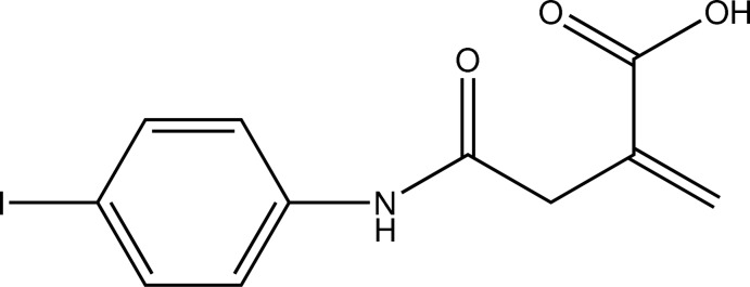
Experimental
Crystal data
C11H10INO3
M r = 331.10
Monoclinic,

a = 23.1111 (6) Å
b = 4.7863 (1) Å
c = 10.4262 (2) Å
β = 97.071 (1)°
V = 1144.54 (4) Å3
Z = 4
Mo Kα radiation
μ = 2.79 mm−1
T = 200 K
0.29 × 0.16 × 0.10 mm
Data collection
Bruker APEXII CCD diffractometer
Absorption correction: multi-scan (SADABS; Bruker, 2008 ▶) T min = 0.503, T max = 0.776
12435 measured reflections
2839 independent reflections
2612 reflections with I > 2σ(I)
R int = 0.015
Refinement
R[F 2 > 2σ(F 2)] = 0.018
wR(F 2) = 0.047
S = 1.06
2839 reflections
150 parameters
H atoms treated by a mixture of independent and constrained refinement
Δρmax = 0.73 e Å−3
Δρmin = −0.61 e Å−3
Data collection: APEX2 (Bruker, 2010 ▶); cell refinement: SAINT (Bruker, 2010 ▶); data reduction: SAINT; program(s) used to solve structure: SHELXS97 (Sheldrick, 2008 ▶); program(s) used to refine structure: SHELXL97 (Sheldrick, 2008 ▶); molecular graphics: ORTEP-3 (Farrugia, 2012 ▶) and Mercury (Macrae et al., 2008 ▶); software used to prepare material for publication: SHELXL97 (Sheldrick, 2008 ▶) and PLATON (Spek, 2009 ▶).
Supplementary Material
Crystal structure: contains datablock(s) I, global. DOI: 10.1107/S160053681205012X/gk2533sup1.cif
Supplementary material file. DOI: 10.1107/S160053681205012X/gk2533Isup2.cdx
Structure factors: contains datablock(s) I. DOI: 10.1107/S160053681205012X/gk2533Isup3.hkl
Supplementary material file. DOI: 10.1107/S160053681205012X/gk2533Isup4.cml
Additional supplementary materials: crystallographic information; 3D view; checkCIF report
Table 1. Hydrogen-bond geometry (Å, °).
| D—H⋯A | D—H | H⋯A | D⋯A | D—H⋯A |
|---|---|---|---|---|
| O1—H1⋯O2i | 0.84 | 1.83 | 2.6597 (19) | 170 |
| N1—H71⋯O3ii | 0.83 (2) | 2.10 (2) | 2.8963 (18) | 161 (2) |
| C3—H3B⋯O3ii | 0.99 | 2.51 | 3.266 (2) | 134 |
Symmetry codes: (i) 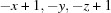 ; (ii)
  .
.
Acknowledgments
BN thanks the UGC for financial assistance through a BSR one-time grant for the purchase of chemicals. PSN thanks Mangalore University for research facilities and the DST–PURSE for financial assistance.
supplementary crystallographic information
Comment
Itaconic acid anhydride is a monomeric building block obtained from renewable sources. Copolymers containing both hydrophilic and hydrophobic segments are drawing considerable attention because of their possible use in biological systems. In this aspect, N-substituted itaconamic acid derivatives have attracted attention due to their amphiphilic properties. Being more reactive than maleic anhydride, itaconic anhydride has already been applied for introducing polar functional groups into polymers (Oishi, 1980; Urzua et al.,1998). In addition, the basic skeleton of itaconic anhydride is useful for the synthesis of cyclic derivatives of imides (Shetgiri & Nayak, 2005), pyridazines (Katla et al., 2011), oxazepines and diazepines (Hanoon, 2011) which show pharmacological activity. In continuation of our studies of pharmacologically active compounds, the crystal structure of the title compound was determined.
The C=C bond is present as its anti-Saytzeff tautomer. The N–C(=O) bond length of 1.351 (2) Å is indicative of amide-type resonance. The least-squares plane defined by the atoms of the aromatic moiety on the one hand and the non-hydrogen atoms of the carboxylic acid group on the other hand enclose an angle of 74.82 (11) ° (Fig. 1).
In the crystal, C–H···O contatcs whose range falls by more than 0.1 Å below the sum of van-der-Waals radii are observed next to classical hydrogen bonds of the N–H···O and C–H···O type. The N–H···O hydrogen bonds are supported by the carbonyl oxygen atom of the amide functionality as acceptor. Simultaneously, one of the hydrogen atoms of the methylene group forms a C–H···O contact to the same oxygen atom which, therefore, acts as twofold acceptor. The carboxylic acid groups engage in the common dimeric hydrogen bonding pattern frequently encountered for many carboxylic acids. In total, the molecules are connected to dimeric chains along the crystallographic b axis. Metrical parameters as well as information about the symmetry of these contacts are summarized in Table 1. In terms of graph-set analysis (Etter et al., 1990; Bernstein et al., 1995), the descriptor for these contacts is C11(4)C11(4)R22(8) on the unary level. The shortest intercentroid distance between two aromatic systems was found at 4.7863 (11) Å which is about the length of axis b (Fig. 2).
The packing of the title compound in the crystal structure is shown in Figure 3.
Experimental
Itaconic anhydride (0.112 g, 1 mmol) was dissolved in acetone (30 ml) and 4-iodoaniline (0.219 g, 1 mmol) was added in small portions under stirring at room temperature over a timespan of 30 minutes. The mixture turned into a yellow slurry. Stirring was continued for 1.5 h after which the slurry was filtered and the solid obtained was washed with acetone and dried to yield the title compound. Single crystals suitable for the X-ray diffraction study were grown from methanol by slow evaporation at room temperature.
Refinement
Carbon-bound H atoms were placed in calculated positions (C–H 0.95 Å for aromatic and vinylic carbon atoms, C–H 0.99 Å for methylene groups) and were included in the refinement in the riding model approximation, with U(H) set to 1.2Ueq(C). The H atom of the hydroxyl group was allowed to rotate with a fixed angle around the C–O bond to best fit the experimental electron density (HFIX 147 in the SHELX program suite (Sheldrick, 2008)), with U(H) set to 1.5Ueq(O). The nitrogen-bound H atom was located on a difference Fourier map and refined freely.
Figures
Fig. 1.
The molecular structure of the title compound, with atom labels and anisotropic displacement ellipsoids drawn at 50% probability level.
Fig. 2.

Intermolecular contacts, viewed along [0 0 - 1]. Symmetry operators: ix, y - 1, z; iix, y + 1, z; iii -x + 1, -y, -z + 1.
Fig. 3.

Molecular packing of the title compound, viewed along [0 1 0] (anisotropic displacement ellipsoids drawn at 50% probability level). Blue dashed lines indicate hydrogen bonds between the carboxylic acid groups.
Crystal data
| C11H10INO3 | F(000) = 640 |
| Mr = 331.10 | Dx = 1.921 Mg m−3 |
| Monoclinic, P21/c | Melting point = 447–445 K |
| Hall symbol: -P 2ybc | Mo Kα radiation, λ = 0.71073 Å |
| a = 23.1111 (6) Å | Cell parameters from 9343 reflections |
| b = 4.7863 (1) Å | θ = 2.7–28.3° |
| c = 10.4262 (2) Å | µ = 2.79 mm−1 |
| β = 97.071 (1)° | T = 200 K |
| V = 1144.54 (4) Å3 | Rectangular, colourless |
| Z = 4 | 0.29 × 0.16 × 0.10 mm |
Data collection
| Bruker APEXII CCD diffractometer | 2839 independent reflections |
| Radiation source: fine-focus sealed tube | 2612 reflections with I > 2σ(I) |
| Graphite monochromator | Rint = 0.015 |
| φ and ω scans | θmax = 28.3°, θmin = 2.7° |
| Absorption correction: multi-scan (SADABS; Bruker, 2008) | h = −30→30 |
| Tmin = 0.503, Tmax = 0.776 | k = −6→6 |
| 12435 measured reflections | l = −8→13 |
Refinement
| Refinement on F2 | Primary atom site location: structure-invariant direct methods |
| Least-squares matrix: full | Secondary atom site location: difference Fourier map |
| R[F2 > 2σ(F2)] = 0.018 | Hydrogen site location: inferred from neighbouring sites |
| wR(F2) = 0.047 | H atoms treated by a mixture of independent and constrained refinement |
| S = 1.06 | w = 1/[σ2(Fo2) + (0.0202P)2 + 0.979P] where P = (Fo2 + 2Fc2)/3 |
| 2839 reflections | (Δ/σ)max = 0.002 |
| 150 parameters | Δρmax = 0.73 e Å−3 |
| 0 restraints | Δρmin = −0.61 e Å−3 |
Fractional atomic coordinates and isotropic or equivalent isotropic displacement parameters (Å2)
| x | y | z | Uiso*/Ueq | ||
| I1 | 0.056006 (5) | −0.19408 (3) | 0.680940 (13) | 0.03784 (5) | |
| O1 | 0.48205 (6) | −0.1867 (3) | 0.34828 (13) | 0.0334 (3) | |
| H1 | 0.5045 | −0.1852 | 0.4180 | 0.050* | |
| O2 | 0.43726 (6) | 0.1500 (3) | 0.44728 (13) | 0.0335 (3) | |
| O3 | 0.30129 (6) | −0.2166 (2) | 0.35760 (14) | 0.0301 (3) | |
| N1 | 0.26626 (6) | 0.2123 (3) | 0.39810 (15) | 0.0238 (3) | |
| H71 | 0.2679 (10) | 0.382 (5) | 0.380 (2) | 0.031 (6)* | |
| C1 | 0.43994 (7) | −0.0058 (4) | 0.35485 (16) | 0.0232 (3) | |
| C2 | 0.39444 (7) | 0.0026 (3) | 0.24115 (16) | 0.0225 (3) | |
| C3 | 0.34191 (7) | 0.1767 (3) | 0.25648 (16) | 0.0232 (3) | |
| H3A | 0.3198 | 0.2104 | 0.1703 | 0.028* | |
| H3B | 0.3547 | 0.3601 | 0.2937 | 0.028* | |
| C4 | 0.30202 (7) | 0.0375 (3) | 0.34331 (15) | 0.0202 (3) | |
| C5 | 0.40223 (8) | −0.1299 (4) | 0.13366 (18) | 0.0330 (4) | |
| H5A | 0.4371 | −0.2318 | 0.1286 | 0.040* | |
| H5B | 0.3729 | −0.1235 | 0.0614 | 0.040* | |
| C11 | 0.21964 (7) | 0.1204 (3) | 0.46527 (16) | 0.0218 (3) | |
| C12 | 0.22773 (7) | −0.0842 (4) | 0.55994 (17) | 0.0263 (3) | |
| H12 | 0.2652 | −0.1644 | 0.5823 | 0.032* | |
| C13 | 0.18104 (8) | −0.1721 (4) | 0.62227 (18) | 0.0284 (4) | |
| H13 | 0.1864 | −0.3137 | 0.6865 | 0.034* | |
| C14 | 0.12659 (7) | −0.0515 (4) | 0.58994 (16) | 0.0265 (3) | |
| C15 | 0.11850 (8) | 0.1576 (4) | 0.49805 (19) | 0.0308 (4) | |
| H15 | 0.0813 | 0.2421 | 0.4777 | 0.037* | |
| C16 | 0.16521 (8) | 0.2432 (4) | 0.43571 (18) | 0.0290 (4) | |
| H16 | 0.1599 | 0.3867 | 0.3724 | 0.035* |
Atomic displacement parameters (Å2)
| U11 | U22 | U33 | U12 | U13 | U23 | |
| I1 | 0.02465 (7) | 0.04793 (9) | 0.04370 (9) | −0.00628 (5) | 0.01530 (5) | −0.00051 (6) |
| O1 | 0.0276 (6) | 0.0373 (7) | 0.0342 (7) | 0.0105 (5) | −0.0003 (5) | −0.0044 (6) |
| O2 | 0.0336 (7) | 0.0386 (8) | 0.0276 (6) | 0.0087 (6) | 0.0003 (5) | −0.0064 (5) |
| O3 | 0.0333 (7) | 0.0172 (5) | 0.0428 (7) | 0.0000 (5) | 0.0167 (6) | 0.0006 (5) |
| N1 | 0.0239 (7) | 0.0160 (6) | 0.0331 (7) | 0.0002 (5) | 0.0096 (6) | 0.0033 (6) |
| C1 | 0.0217 (7) | 0.0230 (7) | 0.0258 (7) | 0.0002 (6) | 0.0066 (6) | 0.0026 (6) |
| C2 | 0.0204 (7) | 0.0216 (7) | 0.0262 (7) | −0.0017 (6) | 0.0060 (6) | 0.0036 (6) |
| C3 | 0.0205 (7) | 0.0223 (8) | 0.0274 (8) | 0.0007 (6) | 0.0050 (6) | 0.0052 (6) |
| C4 | 0.0179 (7) | 0.0194 (7) | 0.0230 (7) | −0.0010 (5) | 0.0011 (5) | 0.0009 (6) |
| C5 | 0.0271 (9) | 0.0400 (10) | 0.0320 (9) | 0.0016 (7) | 0.0040 (7) | −0.0066 (8) |
| C11 | 0.0201 (7) | 0.0201 (7) | 0.0261 (7) | −0.0020 (6) | 0.0064 (6) | −0.0024 (6) |
| C12 | 0.0194 (7) | 0.0279 (8) | 0.0318 (8) | 0.0017 (6) | 0.0046 (6) | 0.0035 (7) |
| C13 | 0.0251 (8) | 0.0316 (9) | 0.0294 (8) | −0.0007 (7) | 0.0072 (7) | 0.0045 (7) |
| C14 | 0.0194 (7) | 0.0323 (9) | 0.0291 (8) | −0.0046 (6) | 0.0083 (6) | −0.0042 (7) |
| C15 | 0.0182 (7) | 0.0390 (10) | 0.0356 (9) | 0.0038 (7) | 0.0055 (7) | 0.0012 (8) |
| C16 | 0.0246 (8) | 0.0306 (9) | 0.0324 (9) | 0.0044 (7) | 0.0060 (7) | 0.0070 (7) |
Geometric parameters (Å, º)
| I1—C14 | 2.0994 (16) | C3—H3B | 0.9900 |
| O1—C1 | 1.311 (2) | C5—H5A | 0.9500 |
| O1—H1 | 0.8400 | C5—H5B | 0.9500 |
| O2—C1 | 1.226 (2) | C11—C12 | 1.387 (2) |
| O3—C4 | 1.2258 (19) | C11—C16 | 1.388 (2) |
| N1—C4 | 1.351 (2) | C12—C13 | 1.392 (2) |
| N1—C11 | 1.425 (2) | C12—H12 | 0.9500 |
| N1—H71 | 0.83 (2) | C13—C14 | 1.388 (2) |
| C1—C2 | 1.485 (2) | C13—H13 | 0.9500 |
| C2—C5 | 1.319 (2) | C14—C15 | 1.382 (3) |
| C2—C3 | 1.497 (2) | C15—C16 | 1.389 (3) |
| C3—C4 | 1.523 (2) | C15—H15 | 0.9500 |
| C3—H3A | 0.9900 | C16—H16 | 0.9500 |
| C1—O1—H1 | 109.5 | C2—C5—H5B | 120.0 |
| C4—N1—C11 | 123.75 (14) | H5A—C5—H5B | 120.0 |
| C4—N1—H71 | 117.5 (16) | C12—C11—C16 | 119.74 (15) |
| C11—N1—H71 | 117.8 (16) | C12—C11—N1 | 121.60 (15) |
| O2—C1—O1 | 123.55 (16) | C16—C11—N1 | 118.65 (15) |
| O2—C1—C2 | 120.76 (15) | C11—C12—C13 | 120.12 (16) |
| O1—C1—C2 | 115.69 (15) | C11—C12—H12 | 119.9 |
| C5—C2—C1 | 120.52 (16) | C13—C12—H12 | 119.9 |
| C5—C2—C3 | 123.74 (16) | C14—C13—C12 | 119.52 (16) |
| C1—C2—C3 | 115.70 (14) | C14—C13—H13 | 120.2 |
| C2—C3—C4 | 112.22 (13) | C12—C13—H13 | 120.2 |
| C2—C3—H3A | 109.2 | C15—C14—C13 | 120.72 (16) |
| C4—C3—H3A | 109.2 | C15—C14—I1 | 120.15 (13) |
| C2—C3—H3B | 109.2 | C13—C14—I1 | 119.13 (13) |
| C4—C3—H3B | 109.2 | C14—C15—C16 | 119.43 (16) |
| H3A—C3—H3B | 107.9 | C14—C15—H15 | 120.3 |
| O3—C4—N1 | 123.00 (15) | C16—C15—H15 | 120.3 |
| O3—C4—C3 | 121.66 (15) | C11—C16—C15 | 120.43 (17) |
| N1—C4—C3 | 115.28 (14) | C11—C16—H16 | 119.8 |
| C2—C5—H5A | 120.0 | C15—C16—H16 | 119.8 |
| O2—C1—C2—C5 | 168.65 (18) | C4—N1—C11—C16 | −131.82 (18) |
| O1—C1—C2—C5 | −11.0 (2) | C16—C11—C12—C13 | 2.0 (3) |
| O2—C1—C2—C3 | −9.2 (2) | N1—C11—C12—C13 | −179.01 (16) |
| O1—C1—C2—C3 | 171.13 (14) | C11—C12—C13—C14 | −0.7 (3) |
| C5—C2—C3—C4 | 108.2 (2) | C12—C13—C14—C15 | −1.0 (3) |
| C1—C2—C3—C4 | −74.03 (18) | C12—C13—C14—I1 | 178.29 (14) |
| C11—N1—C4—O3 | −7.6 (3) | C13—C14—C15—C16 | 1.4 (3) |
| C11—N1—C4—C3 | 169.68 (15) | I1—C14—C15—C16 | −177.91 (14) |
| C2—C3—C4—O3 | −25.0 (2) | C12—C11—C16—C15 | −1.6 (3) |
| C2—C3—C4—N1 | 157.71 (14) | N1—C11—C16—C15 | 179.35 (17) |
| C4—N1—C11—C12 | 49.1 (2) | C14—C15—C16—C11 | −0.1 (3) |
Hydrogen-bond geometry (Å, º)
| D—H···A | D—H | H···A | D···A | D—H···A |
| O1—H1···O2i | 0.84 | 1.83 | 2.6597 (19) | 170 |
| N1—H71···O3ii | 0.83 (2) | 2.10 (2) | 2.8963 (18) | 161 (2) |
| C3—H3B···O3ii | 0.99 | 2.51 | 3.266 (2) | 134 |
Symmetry codes: (i) −x+1, −y, −z+1; (ii) x, y+1, z.
Footnotes
Supplementary data and figures for this paper are available from the IUCr electronic archives (Reference: GK2533).
References
- Bernstein, J., Davis, R. E., Shimoni, L. & Chang, N.-L. (1995). Angew. Chem. Int. Ed. Engl. 34, 1555–1573.
- Bruker (2008). SADABS. Bruker Inc., Madison, Wisconsin, USA.
- Bruker (2010). APEX2 and SAINT Bruker AXS Inc., Madison, USA.
- Etter, M. C., MacDonald, J. C. & Bernstein, J. (1990). Acta Cryst. B46, 256–262. [DOI] [PubMed]
- Farrugia, L. J. (2012). J. Appl. Cryst. 45, 849–854.
- Hanoon, H. D. (2011). Nat. J. Chem. 41, 77–89.
- Katla, S., Pothana, P., Gubba, B. & Manda, S. (2011). Der Chem. Sin. 2, 47–53.
- Macrae, C. F., Bruno, I. J., Chisholm, J. A., Edgington, P. R., McCabe, P., Pidcock, E., Rodriguez-Monge, L., Taylor, R., van de Streek, J. & Wood, P. A. (2008). J. Appl. Cryst. 41, 466–470.
- Oishi, T. (1980). Polym. J. 12, 719–727.
- Sheldrick, G. M. (2008). Acta Cryst. A64, 112–122. [DOI] [PubMed]
- Shetgiri, N. P. & Nayak, B. K. (2005). Indian J. Chem. Sect. B, 44, 1933–1936.
- Spek, A. L. (2009). Acta Cryst. D65, 148–155. [DOI] [PMC free article] [PubMed]
- Urzua, M., Opazo, A., Gargallo, L. & Radic, D. (1998). Polym. Bull. 40, 63–67.
Associated Data
This section collects any data citations, data availability statements, or supplementary materials included in this article.
Supplementary Materials
Crystal structure: contains datablock(s) I, global. DOI: 10.1107/S160053681205012X/gk2533sup1.cif
Supplementary material file. DOI: 10.1107/S160053681205012X/gk2533Isup2.cdx
Structure factors: contains datablock(s) I. DOI: 10.1107/S160053681205012X/gk2533Isup3.hkl
Supplementary material file. DOI: 10.1107/S160053681205012X/gk2533Isup4.cml
Additional supplementary materials: crystallographic information; 3D view; checkCIF report