Abstract
Purpose
To compare Orbscan II and ultrasonic pachymetry for measurement of central corneal thickness (CCT) in eyes scheduled for keratorefractive surgery.
Methods
CCT was measured using Orbscan II (Bausch & Lomb, USA) and then by ultrasonic pachymetry (Tomey SP-3000, Tomey Ltd, Japan) in 100 eyes of 100 patients with no history of ocular surgery scheduled for excimer laser refractive surgery.
Results
Mean CCT was 544.7±35.5 (range 453–637) μm by ultrasonic pachymetry versus 546.9±41.6 (range 435–648) μm measured by Orbscan II applying an acoustic factor of 0.92 (P=0.14). The standard deviation of measurements was greater with Orbscan pachymetry but the difference was not statistically significant.
Conclusion
CCT measurements by Orbscan II (applying an acoustic factor) and by ultrasonic pachymetry are not significantly different; however, when CCT readings by Orbscan II are in the lower range, it is advisable to recheck the measurements using ultrasonic pachymetry.
INTRODUCTION
Corneal thickness measurement is indispensable in the diagnosis and management of corneal disorders and is an important parameter in predicting the long term complications of keratorefractive procedures. The increase in the number of refractive procedures such as photorefractive keratectomy and laser in situ keratomileusis(LASIK) and the concomitant increase in the rate of post-surgical keratectasia, underscore the importance of accurate corneal thickness measurements. 1–3. Recently, the importance of corneal pachymetry has been highlighted in other conditions such as side effects of contact lenses,4 glaucoma,5 dry-eye6 and diabetes mellitus.7
Despite the introduction of several new imaging devices, ultrasonic pachymetry remains the standard method for corneal thickness measurement. This method requires corneal anesthesia and instrument contact with the globe therefore entails disadvantages such as corneal injury and transmission of microorganisms. Furthermore, the results obtained by this technique are technician-dependent and care must be taken to apply the probe perpendicularly in the center of the cornea with minimal pressure. 8 In contrast, non-contact methods including Orbscan II scanning slit corneal topography, 9 specular microscopy10 and confocal laser scanning microscopy11 avoid these limitations and drawbacks. Orbscan II is widely used in evaluating the refractive properties of the anterior and posterior corneal surfaces before and after keratorefractive procedures. This instrument determines corneal thickness in the center and periphery and is the most commonly employed device in Iran for the preoperative evaluation of refractive surgery patients. Several studies have compared Orbscan II and ultrasonic pachymetry for measuring corneal thickness.12–15 Most of them have reported that Orbscan II yields higher measurements with a mean overestimation of 30 μm. To compensate for this difference, the manufacturer suggests applying a correction (acoustic) factor of 0.92.16 The current study was undertaken to compare central corneal thickness measurement by Orbscan II and ultrasonic pachymetry in an Iranian patient population scheduled for refractive surgery and to determine the accuracy of the acoustic factor (AF) for Orbscan II.
METHODS
One-hundred eyes of 100 patients referred for refractive surgery to Negah Eye Clinic, Tehran, Iran were enrolled in this study. None of them had history of ocular surgery and most subjects had myopia with or without astigmatism. Only data from the right eye of each patient was considered for the study.
First, corneal imaging was performed using Orbscan II (Bausch & Lomb, USA) and the measured corneal thickness was corrected applying an acoustic factor of 0.92. In the next step, ultrasonic pachymetry (Tomey SP-3000, Tomey Ltd, Japan) was performed by one experienced operator under topical anesthesia with tetracaine 1%. Measurements were obtained 5 times in the center of the cornea and the average reading was recorded. The results were compared using paired t-test with significance set at 0.05.
RESULTS
Overall, 100 right eyes of 100 patients including 39 male and 61 female subjects with mean age of 28.6±8.0 (range 19–46) years were studied.
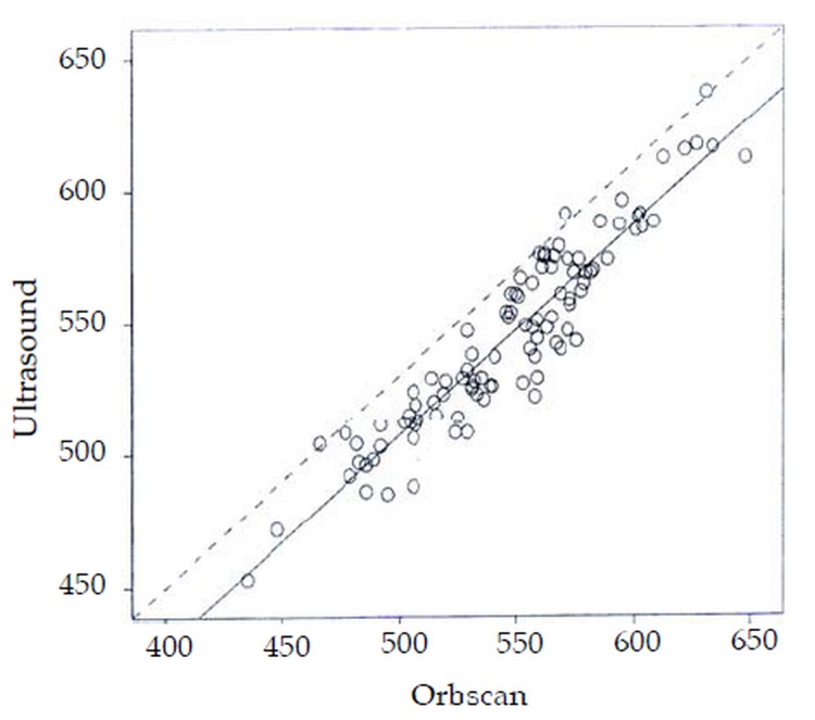
Mean central corneal thickness was 544.7±35.5(range 453–63) μm by ultrasonic pachymetry and 546.9±41.6 (range 435–648) μm by Orbscan II (P=0.14). The standard deviation of measurements was higher with Orbscan II as compared to ultrasonic pachymetry, but the difference was not statistically significant (Fig. 1).
Figure 1.
Scatter diagram showing the regression line of central corneal thickness (μm) measurements by Orbscan II and ultrasonic pachymetry.
DISCUSSION
Ultrasonic pachymetry has been the standard method for measurement of corneal thickness for several years. More recently, other devices have been used for this purpose, one of which is the Orbscan II by Bausch and Lomb. Advantages of this device include simultaneous topographical analysis of the anterior and posterior corneal surfaces and corneal pachymetry, use of a non-contact method, measurement of several parts of the cornea, being less techniciandependent, and not being affected by errors inherent to contact methods such as non-perpendicularity or eccentricity of the probe. Furthermore, repeatability of the measurements in normal eyes has been reported to be superior with Orbscan II as compared to ultrasonic pachymetry.13,17
Several studies have compared central corneal thickness measurements by Orbscan II and Ultrasound Yaylali et al 12 studied 51 normal eyes and reported that corneal thickness readings were 23–28 μm higher with Orbscan II without applying an AF. In a similar study on 101 normal and 30 post-LASIK eyes, corneal thickness measurements by Orbscan II without applying AF were 28 μm greater in normal eyes but 13 μm smaller in post-LASIK eyes as compared to measurements obtained by ultrasonic pachymetry.14 In another comparison on 99 normal eyes, measurements were 90 μm higher with Orbscan II without applying a correction factor.15
The reason for higher measurements by Orbscan II pachymetry as compared to ultrasound is the difference in the manner the instruments operate.18 Orbscan II optically measures the distance between the tear film including the hydrated epithelial layer of the cornea and the posterior corneal surface while ultrasonic pachymetry utilizes ultrasound echoes from the posterior cornea to calculate corneal thickness.8 The origin of this posterior echo is not clear and might be at level of Descemet's membrane or the anterior chamber. The ultrasound probe may displace the tear film 7 to 40 microns and therefore underestimate corneal epithelial thickness.19 Because of these discrepancies, an AF should be applied for Orbscan readings to bring them closer to conventional ultrasonic measurements. The amount of the AF as suggested by the manufacturer is 0.92.16
The current study demonstrated that mean corneal thickness readings using ultrasonic pachymetry and Orbscan II (applying an AF of 0.92) were comparable. Although not statistically significant, the standard deviation of measurements was higher with Orbscan II indicating the possibility of significant over- and underestimations with this device. In keratorefractive surgery using data from Orbscan II, it is prudent to recheck corneal thickness by ultrasonic pachymetry when the estimated residual corneal thickness is borderline in order to minimize the risk of excessive ablation and keratectasia. The present study was conducted on eyes with virgin corneas. However, in postsurgical cases and keratoconic corneas, the mentioned AF may not be appropriate and corneal thickness readings obtained by Orbscan topography, even without applying any correction factor, have been reported to be less than ultrasonic pachymetry.20,21
REFERENCES
- 1.Pallikaris IG, Kymionis GD, Astyrakakis NI. Corneal ectasia induced by LASIK. J Cataract Refract Surg. 2001;27:1796–1802. doi: 10.1016/s0886-3350(01)01090-2. [DOI] [PubMed] [Google Scholar]
- 2.Malecaze F, Coullet J, Calvas P, Fournie P, Ame JL, Brodaty C. Corneal ectasia after PRK for low myopia. Ophthalmology. 2003;110:267–275. doi: 10.1016/j.ophtha.2005.11.023. [DOI] [PubMed] [Google Scholar]
- 3.Auffarth GU, Wang L, Volcker HE. Keratoconus evaluation using the Orbscan topography system. J Cataract Refract Surg. 2000;26:222–228. doi: 10.1016/s0886-3350(99)00355-7. [DOI] [PubMed] [Google Scholar]
- 4.Liu Z, Pflugfelder SC. The effects of long-term contact lens wear on corneal thickness, curvature, and surface irregularity. Ophthalmology. 2000;107:105–111. doi: 10.1016/s0161-6420(99)00027-5. [DOI] [PubMed] [Google Scholar]
- 5.Shah S, Chatterjee A, Mathai M. Relationship between corneal thickness and measured IOP in a general ophthalmology clinic. Ophthalmology. 1999;106:2154–2160. doi: 10.1016/S0161-6420(99)90498-0. [DOI] [PubMed] [Google Scholar]
- 6.Liu S, Pflugfelder SC. Corneal thickness is reduced in dry eye. Cornea. 1999;18:403–407. doi: 10.1097/00003226-199907000-00002. [DOI] [PubMed] [Google Scholar]
- 7.Larsson LI, Bourne WM, Pach J M. Structure and function of the corneal endothelium in diabetes mellitus type I & type II. Arch Ophthalmol. 1996;114:9–14. doi: 10.1001/archopht.1996.01100130007001. [DOI] [PubMed] [Google Scholar]
- 8.Suzuki S, Oshika T, Oki K, Sakabe I, Iwase A, Amano S, et al. Corneal thickness measurements: Scanning-Slit corneal topography and non-contact specular microscopy versus ultrasonic pachymetry. J Cataract Refract Surg. 2003;29:1313–1318. doi: 10.1016/s0886-3350(03)00123-8. [DOI] [PubMed] [Google Scholar]
- 9.Liu Z, Huang AJ, Pflugfelder SC. Evaluation of corneal thickness & topography in normal eyes using the orb-scan corneal topography system. Br J Ophthlamol. 1999;83:774–778. doi: 10.1136/bjo.83.7.774. [DOI] [PMC free article] [PubMed] [Google Scholar]
- 10.Bovelle R, Kaufman SC, Thompson HW, Hamano H. Corneal thickness measurements with the Topcon Sp-2000P specular microscope and an ultrasound pachymeter. Arch Ophthalmol. 1999;117:868–870. doi: 10.1001/archopht.117.7.868. [DOI] [PubMed] [Google Scholar]
- 11.Cavanagh HD, El-Agha MS, Petroll WM, Jester JV. Specular microscopy, confocal microscopy, and ultrasound biomicroscopy: diagnostic tools of the past quarter century. Cornea. 2000;19:712–722. doi: 10.1097/00003226-200009000-00016. [DOI] [PubMed] [Google Scholar]
- 12.Yaylali V, Kaufman SC, Thompson HW. Corneal thickness measurements with the Orbscan topography system and ultrasonic pachmetry. J Cataract Refract Surg. 1997;23:1345–1350. doi: 10.1016/s0886-3350(97)80113-7. [DOI] [PubMed] [Google Scholar]
- 13.Marsich MW, Bullimore MA. The repeatability of Orbscan II vs Ultrasound for CCT Measurement. Cornea. 2000;19:792–795. doi: 10.1097/00003226-200011000-00007. [DOI] [PubMed] [Google Scholar]
- 14.Chakrabarti HS, Criag JP, Brahma A, Malik TY, McGhee CN. Comparison of corneal thickness measurements using ultrasound and orbscan slitscanning topography in normal and post-LASIK eyes. J Cataract Refract Surg. 2001;27:1823–1828. doi: 10.1016/s0886-3350(01)01089-6. [DOI] [PubMed] [Google Scholar]
- 15.Giraldez-Fernandez MJ, Diaz Rey A, Cervino A, Yebra-Pimental E. A comparison of two pachymetric systems: Slit-Scanning and ultrasound. CLAOJ. 2002;28:221–223. doi: 10.1097/01.ICL.0000034556.15901.CC. [Abstract] [DOI] [PubMed] [Google Scholar]
- 16.Gonzalez-Meijome JM, Cervino A, Yebra-Pimentel E, Parafita MA. Central and peripheral corneal thickness measurement with orbscan II and topographical ultrasound pachymetry. J Cataract Refract Surg. 2003;29:125–132. doi: 10.1016/s0886-3350(02)01815-1. [DOI] [PubMed] [Google Scholar]
- 17.Lattimore MR Jr, Kaupp S, Schallhorn S, Lewis R. Orbscan pachymetry; implications of a repeated measure and diurnal variation analysis. Ophthalmology. 1999;106:977–981. doi: 10.1016/S0161-6420(99)00519-9. [DOI] [PubMed] [Google Scholar]
- 18.Modis L Jr, Langenbucher A, Seitz B. Scanning-slit and specular microscopic pachymetry in comparison with ultrasonic determination of corneal thickness. Cornea. 2001;20:711–714. doi: 10.1097/00003226-200110000-00008. [DOI] [PubMed] [Google Scholar]
- 19.Nissen J, Hjortdal JQ, Ehlers N. A clinical of optical and ultrasonic pachymetry. Acta Ophthalmol (Copenh) 1991;69:659–663. doi: 10.1111/j.1755-3768.1991.tb04857.x. [DOI] [PubMed] [Google Scholar]
- 20.Fakhry AM, Artola A, Belda JI, Ayala MJ, Alio JL. Comparison of corneal pachymetry using ultrasound and orbscan II. J Cataract Refract Surg. 2002;28:248–253. doi: 10.1016/s0886-3350(01)01277-9. [DOI] [PubMed] [Google Scholar]
- 21.Prisant O, Calderon N, Chastang P, Gatinel D, Hoang-Xuan T. Reliability of pachymetric measurements using orbscan after excimer refractive surgery. Ophthalmology. 2003;110:511–515. doi: 10.1016/S0161-6420(02)01298-8. [DOI] [PubMed] [Google Scholar]


