Abstract
The value of coronal incisions in maxillofacial surgery has been well documented. The incision provides excellent access to the upper facial skeleton aiding in adequate access, good anatomic reduction of fractures and hidden scar. The associated bleeding with raising a bicoronal flap is a matter of concern to beginner surgeons. This often prevents the regular use of this approach. Textbooks have recommended the use of Raney clips but these are not available routinely in India and are expensive. We have utilized simple stationary paper clips, autoclaved and used for surgery. This not only provide haemostasis but also aids in holding the flap during dissection. This technique would be of great help to young surgeons and in developing countries where economics plays a major role in surgery.
Keywords: Bicoronal flap, Haemostasis, Stationary paper clips
Various authors have reported the technique, advantages, morbidity and modifications of bicoronal flaps for surgeries ranging from temporomandibular joints to zygomatic complex fractures [1–4].
The associated bleeding from the scalp during surgery is a major concern for young and beginner surgeons. We present a simple method to control bleeding during coronal incisions.
Technique
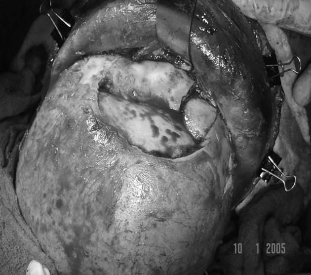
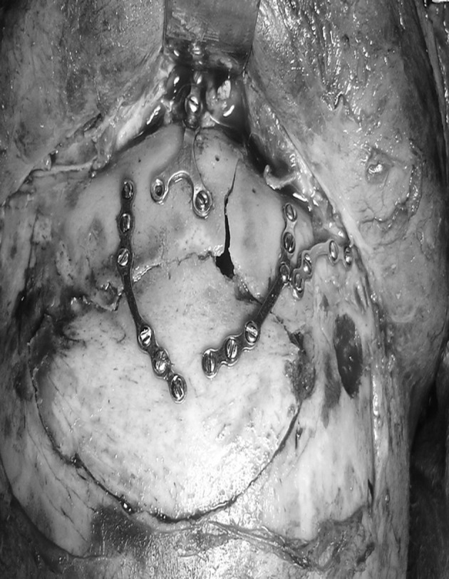
Prophylactic routine antibiotics were administered with pre-operative head shave for all patients. The incision was marked at the upper attachment of the helix extending transversely over the vault of the skull, curved slightly forward at the vertex following the hairline but staying posterior to it keeping in mind future baldness pattern in males. 10 ml of 2 percent Lignocaine hydrochloride containing 1:200,000 was taken, diluted with equal amount of normal saline and infiltrated along the incision. This helps in defining planes during dissection for young surgeons. The incision is then begun at the vertex carrying through it the skin, subcutaneous tissue and underlying layer to the loose aponeurotic layer. Electrocautery was avoided for the initial incision to avoid damage to hair follicles [5]. Textbooks [5] have recommended the use of Raney clips to control bleeding from the scalp. At this point the autoclaved stationary paper clip was applied to the flap and the flap was undermined along the plane in an anterior direction. A second clip was then applied at a distance and these clips helped to hold the flap during dissection. 4–5 clips suffice for the entire flap. The flap was then reflected in a standard fashion. The clips were intermittently released to prevent pinching or pressure on the skin (Figs. 1, 2).
Fig. 1.

Flap with clips in position
Fig. 2.

Fixation of fractures
After completion of the surgery the clips were released and bleeders were treated by electrocautery. A large vacuum suction drain was placed and the wound closed in two layers. The pericranium and muscle are closed with 2-0 vicryl and staples used for the skin. An external pressure dressing was given which helps to prevent haematoma and staples removed after 2 weeks. All patients had an uneventful healing. The clips were discarded after use.
Discussion
The idea of replacement of the Raney clips germinated with the thought that the Raney clip is essentially a clip which holds the coronal flap tightly in position, just like a paper clip holds a wad of papers. The paper clips were then taken from reputed stationary stores and sealed in pouches. They were then autoclaved and checked for any damage, bending, running of colour, rusting or any other problem. Once this was confirmed they were then discarded and fresh batch selected for each patient. An evaluation of personal records from 1st Jan 2000 to 31st Dec 2009 showed 12 patients (10 male, 2 female) operated with coronal incisions taken in this fashion. No case had complications of bleeding or blood transfusion post-operatively. There was no instance of rusting of the clips or chipping off of the paint of the clips after autoclaving since they were brand new, autoclaved only once prior to surgery and immediately discarded. All patients were followed up for a minimum of 6 months and showed no problems which could be associated with the use of the clips. There would be a logical argument that these are not originally meant for surgery and do not warrant use. We feel that since there is no non medical material which we have implanted inside the body and it is just used to hold the flap, this is acceptable. Moreover all patients were informed that we would be using these clips just to hold the flap and nowhere near or inside the vital structures like eyes, brain, ears and had taken a consent for the same.
Conclusion
Haemostasis during coronal incisions can be done by a continuous running suture also. But this takes time as compared to our method of using clips which not only prevents bleeding but also aids to hold the flap during dissection. This method will reduce the learning curve for young surgeons and help them to attempt the coronal incision with confidence. Once expertise is achieved then the surgeon can decide whether he wants to continue their regular use.
Acknowledgments
Conflict of interest
None.
References
- 1.Abubaker AO, Sotereanos G, Patteson GT. Use of the coronal incision for reconstruction of severe craniofacial injuries. J Oral Maxillofac Surg. 1990;48(6):579–586. doi: 10.1016/S0278-2391(10)80470-7. [DOI] [PubMed] [Google Scholar]
- 2.Pogrel MA, Perott DH, Kaban LB. Bicoronal flap approach to the temporomandibular joints. Int J Oral Maxillofac Surg. 1991;20(4):219–222. doi: 10.1016/S0901-5027(05)80179-1. [DOI] [PubMed] [Google Scholar]
- 3.Kerawala CJ, Grime RJ, Stassen LF, Perry M. The bicoronal flap (craniofacial access): an audit of morbidity and a proposed surgical modification in male pattern baldness. Br J Oral Maxillofac Surg. 2000;38(5):441–444. doi: 10.1054/bjom.2000.0315. [DOI] [PubMed] [Google Scholar]
- 4.Zhang QB, Dong YJ, Li ZB, Zhao JH. Coronal incision for treating zygomatic complex fractures. J Craniomaxillofac Surg. 2006;34(3):182–185. doi: 10.1016/j.jcms.2005.09.004. [DOI] [PubMed] [Google Scholar]
- 5.Fonseca RJ, Walker RV, Betts NJ, Barber HD, Powers MP, editors. Oral & Maxillofacial Trauma. 3. St. Louis: Elsevier Saunders; 2005. pp. 726–727. [Google Scholar]


