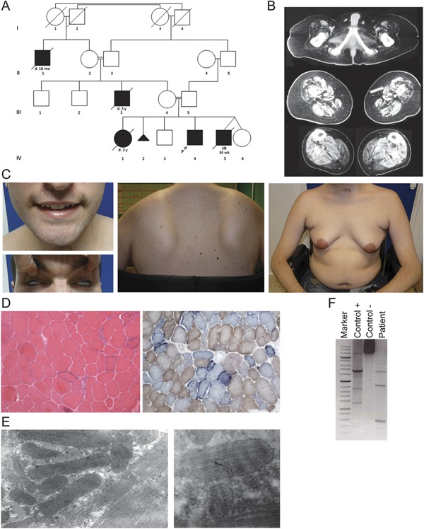
Figure 1. Clinical, pathologic, and molecular features of the patient.

(A) Pedigree (IV-4 is the index case). SB 34 wk under patient IV-5 indicates stillbirth at 34 weeks' gestation. (B) Muscle tomography of lower limbs. (C) Facial involvement with weakness of the orbicularis oculi (Bell phenomenon) and oris, pectoral atrophy, gynecomastia, and scapular winging. (D) The muscle biopsy shows a dystrophic pattern and 50% cytochrome c oxidase–negative fibers, most of which are “ragged-blue” with the succinate dehydrogenase stain (×10). (E) Electron microscopy shows enlarged bizarre mitochondria and paracrystalline inclusions (×39,000; ×46,000). (F) The long-range PCR reveals multiple deletions in mitochondrial DNA from muscle.
