Abstract
Autophagy is a catabolic mechanism that is important for many biological processes such as cell homeostasis, development and immunity. Though many molecular components of the autophagy pathway have been identified, the signaling pathways regulating the activity of essential autophagy mediators are still poorly defined. We recently demonstrated that the mitogen-activated protein kinase MAPK14 (p38α), when activated by the GADD45B (Gadd45β)-MAP3K4 (MEKK4) signaling complex (but not other MAPK14 activators), is directed to autophagosomes. Therefore, we demonstrated for the first time that MAPK14 operates at this subcellular compartment. Importantly, activation of MAPK14 impairs autophagosome-lysosome fusion and, thus, autophagy. This was demonstrated by increased autophagic flux in MAPK14-deficient as well as in GADD45B-deficient cells. Moreover, we identified a novel post-translational modification of the crucial autophagy mediator ATG5, since MAPK14 directly phosphorylates ATG5 at threonine 75, which is evolutionarily conserved from yeast to human. Using ATG5-deficient cells, which we reconstituted with either a phosphorylation-defective or a phospho-mimetic mutant of ATG5, we demonstrated that phosphorylation of ATG5 results in impaired autophagy.
Keywords: ATG5, autophagy, Gadd45, p38 MAPK, phosphorylation, post-translational modification
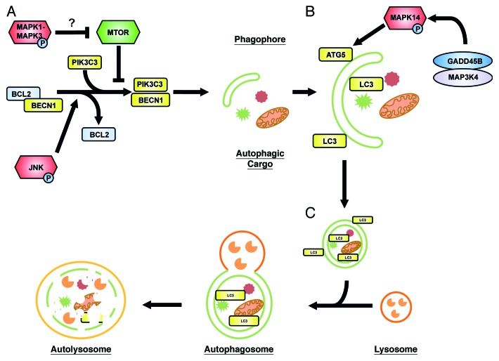
The human kinome consists of about 500 kinases that are involved in the regulation of proliferation, differentiation, cell death, immunity and other biological processes. Consistently, kinases such as the Atg1 homologs ULK1/2 and the class III phosphatidylinositol 3-kinase (PtdIns3KC3) Vps34 play an important role in autophagy induction. MAPKs constitute an evolutionarily conserved three-tier signaling module comprised of a MAPK kinase kinase (MAPKKK), a MAPK kinase (MEK or MKK) and a MAPK. Well-known MAPKs include MAPK1-MAPK3 (ERK2-ERK1), JNK and p38 MAPKs that all have been implicated in autophagy regulation. At the molecular level, the best studied example is MAPK8/JNK1, which phosphorylates BCL2 upon starvation or ceramide treatment, thereby releasing BECN1 from BCL2. Subsequently, BECN1 initiates autophagy as part of the class III PtdIns3K complex. Similarly, MAPK1-MAPK3 appears to promote autophagy. For instance, Corcelle et al. showed that the carcinogen lindane induces prolonged MAPK1-MAPK3 activation and formation of large autophagosomes. Interestingly, a constitutively active mutant of MAP2K1/MEK1, the MAPK1-MAPK3-activating MAPK kinase, has the same effect. Furthermore, Codognos’s group demonstrated that MAPK1-MAPK3 stimulates autophagy via the G-protein regulator RGS19/GAIP and its activity is impaired by amino acids. Finally, Wang and colleagues showed that activation of MAP2K1 and MAPK1-MAPK3 by AMPK inactivates MTOR and, thus, induces autophagy. In summary, MAPK1-MAPK3 and MAPK8 affect an early step of autophagy while the GADD45B-MAP3K4-MAPK14 pathway described by us acts further downstream (Fig. 1).
Figure 1. A model of regulatory mechanisms of MAPK signaling in autophagy. In the absence of amino acids or in response to certain stimuli, the cell mounts an autophagic response. This can be influenced by a number of different intracellular mediators, one of these being the mitogen activated protein kinases (MAPKs). (A) The initiation phase of autophagy. MAPK1-MAPK3 was reported to inhibit MTOR activity and thus contribute to the initiation of autophagy. However, the exact mechanism is not fully understood. JNK phosphorylates BCL2 thereby disrupting the BECN1-BCL2 complex and allowing for the activation of autophagy through BECN1. A phagophore is formed at the phagophore assembly site. (B) The elongation phase of autophagy. The autophagosomal membrane is elongated in a LC3-II- and ATG5-dependent manner. Here, we could show that GADD45B and MAP3K4 together direct MAPK14 to the autophagosomal membrane, where it phosphorylates ATG5. (C) The maturation phase of autophagy. The autophagosome fuses with a lysosome, leading to vesicle acidification and subsequent cargo degradation. MAPKs are shown in red, ATG proteins in yellow, MTOR in green and other, important regulators are depicted in gray/blue.
Regarding p38 MAPKs, autophagy promoting as well as inhibiting functions have been reported. For instance, Tang et al. suggested that the accumulation of glial fibrillary acidic protein (GFAP) in astrocytes activates p38 ΜΑΠΚσ, resulting in the direct inhibition of MTOR and the induction of autophagy. On the other hand, Häusinger and colleagues reported that exposure of hepatocytes or yeast cells to hypo-osmotic conditions activates p38 ΜΑΠΚσ and Hog1, the yeast p38 homolog, respectively, resulting in the suppression of autophagic proteolysis. Similarly, GABARAP, a mammalian Atg8 homolog, is upregulated in colon cancer cell lines by pharmacological inhibition of MAPK14, leading to autophagy and cell death. Although this dual role of MAPK14 seems puzzling, the biological outcome of MAPK signaling depends on strength, duration and localization. For instance, transient vs. sustained activation of MAPK1-MAPK3 downstream of different receptor tyrosine kinases such as EGFR and NGFR leads to proliferation vs. differentiation, respectively. Interestingly, GADD45B mediates the sustained activation of MAPK14. Furthermore, we observed phosphorylated MAPK14 at autophagosomes upon activation of the GADD45B-MAP3K4 pathway, in contrast to nuclear MAPK14 localization upon UV irradiation, a classical MAPK14 stimulus. How MAPKs are directed to specific locations within the cell is not well understood, but supposedly it involves scaffold proteins. In that respect, Webber and Tooze showed that the scaffold FAM48A/p38IP interacts with ATG9 at membranous vesicles, probably endosomes. However, the localization of MAPK14 in this context was not investigated. As the role of active MAPK14 is rather to sequester FAM48A away from ATG9, one might speculate that a different scaffold protein is involved in the GADD45B-MAP3K4 pathway.
In which physiological setting could the GADD45B-MAP3K4-MAPK14-pathway play a role in vivo? Autophagy is important in liver physiology. Using hepatectomy as a model, Guido Franzoso and colleagues reported that GADD45B increases hepatocyte survival and, thus, liver regeneration. They showed that GADD45B is induced upon partial hepatectomy and inhibits pro-apoptotic JNK signaling. Interestingly, Ulrich Pfeifer demonstrated already in 1979 that autophagy is inhibited during partial hepatectomy. It is, thus, conceivable that GADD45B, induced by partial hepatectomy, inhibits autophagy to allow for liver regeneration. Another setting, in which the MAP3K4-MAPK14-ATG5 pathway could play a role, is Crohn disease. This autoimmune disease is characterized by chronic inflammation of the gut and has been linked to autophagy. Crohn disease is associated with mutations in ATG16L1, a component of the ATG12–ATG5 complex, and NOD2, an intracellular pattern recognition receptor activated by muramyl dipeptide. NOD2 interacts with the kinase RIPK2 to activate the NFKB transcription factor, and NOD2 recruits ATG16L1 to the plasma membrane to induce autophagy. Of note, Derek Abbott’s group demonstrated that MAP3K4 sequesters RIPK2 from NOD2 to switch downstream signaling from NFKB to p38 MAPK activation. Therefore, MAP3K4 might be involved in the development of Crohn disease by influencing autophagy through activation of MAPK14 and inhibition of ATG5. As a third scenario, the GADD45B-MAP3K4-MAPK14-ATG5 pathway might act during infection as GADD45B is induced by lipopolysaccharide (LPS). Since LPS transfers signals via TLR4, which can induce autophagy, we addressed the question whether GADD45B affects LPS-induced autophagy. Indeed, we could show that autophagy is increased in GADD45B-deficient fibroblasts and macrophages suggesting that the induction of GADD45B by the LPS-TLR4-NFKB pathway acts as a negative feedback loop to dampen TLR-induced autophagy.
In summary, we identified a novel signaling pathway mediating autophagy regulation, which provides a potential new target for pharmacological intervention in diseases associated with the deregulation of autophagy, such as Crohn disease. Future studies will unravel additional physiological roles, in which the GADD45B-MAP3K4-MAPK14-ATG5 pathway functions as a molecular brake on autophagy.
Acknowledgments
This work is supported by grants from Deutsche Forschungsgemeinschaft (SCHM1586/3-1 to I.S.) and by the President’s Initiative and Networking Fund of the Helmholtz Association of German Research Centers (HGF) under contract number VH-GS-202.
Disclosure of Potential Conflicts of Interest
No potential conflicts of interest were disclosed.
Footnotes
Previously published online: www.landesbioscience.com/journals/autophagy/article/22924


