Abstract
Hydatid cysts rarely appear isolated in the orbital cavity without involvement of other organs. Most of these are situated in the superolateral and superomedial angles of the orbit. Inferiorly located cysts are very uncommon. The authors report a case of a primary hydatid cyst of the orbit with inferiolateral localization. The cyst was enucleated surgically via a rhinotomy approach. This case was considered as a primary infection, because there was no previous history of hydatid disease and no findings of liver and lung cysts on radiological examination. Physicians should include orbital hydatid cyst in the differential diagnosis of unilateral proptosis. To avoid complications that might occur during surgery, the cyst can be easily removed using a gentile enucleation technique.
Keywords: Exopthalmia investigation, follow-up, intraorbital hydatid cyst, proptosis, surgical treatment
INTRODUCTION
Hydatid disease is a parasitic infestation by a tapeworm of the genus Echinococcus. Hydatid disease (Echinococcus granulosus) is endemic in the Middle East as well as other parts of the world, including India, Africa, South America, New Zealand, Australia, Turkey, and Southern Europe. The incidence of intraorbital hydatid disease is extremely low in all hydatid cysts.[1,2,3,4] The symptoms include progressive exophthalmos with proptosis with or without pain, disturbance in ocular motility, visual deterioration, and chemosis.[3–5] Surgery is the primary treatment in these cases,[2,6] but chemotherapy should be used if a cyst ruptures or for prevention.[2,4,6] We present a new case of a hydatid cyst of the orbit located inferiorly that we removed via a right lateral rhinotomy approach. The patient had uneventful recovery.
CASE REPORT
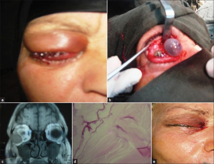
A 42-year-old female patient presented with headache gradually worsening, right eye swelling increased and visual deterioration. The swelling was not responded to systemic antibiotic. There was no history of trauma or systemic disease [Figures 1a and b].
Figure 1.

(a) Right eye swelling with lid edema, (b) Intraoperative view showing the cystic lesion, (c) Orbital MRI showing the cystic lesion, (d) Hisopathologic slide (H&E, 10x), (e) Postoperative view
On examination there was proptosis of the right eye, the globe was displaced medially and inferiorly, and there was significant lid edema. Ocular movements were restricted in all direction especially in abductions. Visual acuity was reduced to finger counting at 1 feet with accurate projection of light.
The mass was nontender, irreducible, nonpulsative, no bruits audible, and was felt in lateral aspect of the orbit; posterior limit of the mass could not be reached. There was chemosis of both the conjunctives; on the upper eyelid eversion the anterior lob of the lacrimal gland was prominently seen. The fundus revealed papilledema.
A magnetic resonance imaging (MRI) revealed a well-defined cyst lesion sized 25 × 25 × 30 mm located lateral to the right orbit [Figure 1c]; the cyst appeared as a low-intensity signal on T1-weighted images and as a high-intensity signal on T2-weighted images, after an IV injection of gadolinium ethoxybenzyl diethylenetriamine pentaacetic. Acid-enhanced magnetic resonance imaging findings of borderline lesions exhibited capsular contrast enhancement on T1-weighted images.
A chest radiography and abdominal ultrasonography showed no hydatid cyst. The diagnosis was intraorbital hydatid cyst. The patient informed in detail about all the possible intraoperative or postoperative complications.
The patient underwent surgery via a right lateral Rhinotomy approach. The patient was anesthetized via an orotracheal route and was positioned supine with the neck hyperextended and turned 30° toward the surgeon. The cyst was removed completely without rupture, and the wound was washed with normal saline. The wound was reapproximated with sutures. A systemic oral albendazole 200 mg was administered for 2 months after surgery for prevention. The patient recovered without complications.
The histopathologic report HE confirmed the hydatid cyst [Figure 1d].
The patient's postoperative course was uneventful, and she was discharged on the third postoperative day [Figure 1e]. Eight months after surgery the patient was doing well and her ocular motility and vision had improved. On physical examination, no enophthalmos or proptosis was detected. Follow-up MRI revealed no evidence of the cyst.
DISCUSSION
Hydatid cysts of the orbit are rare and account for 1% of all hydatid cysts.[2,3] Altýnörs et al.[1] surveyed the literature on central nervous system hydatidosis in Turkey and found that 22 of 336 cases were located in the intraorbital space. Typically, an orbital hydatid cyst is unilateral and occurs with or without hydatid cysts located elsewhere in the body.[4,5] The most frequent clinical findings are exophthalmos, chemosis, lid edema, visual impairment, and restriction of extraocular motility.[3–5] An orbital hydatid cyst tends to involve the retrobulbar tissues either within the muscle cone or outside in the superolateral or superomedial angle.[3,7,8,14,15]
Definitive treatment is total surgical excision.[1,2,4] Various surgical approaches have been used to expose the orbital mass.[7,9–13] An understanding of the microanatomy of the orbit and a proper surgical approach are very important in preventing surgical complications during intraorbital hydatid cyst treatment.
Because of the possibility of the cyst rupturing, we dissected the mass and orbital fat over the cyst wall with hypertonic saline-soaked cotton. The cyst ruptured during removal, but the cystic contents floated into the maxillary sinus due to the effect of gravity. Thus, we believe that the risks of spreading the infection and recurrences associated with our technique are minimal.
Albendazole treatment is useful, especially if begun 14–28 days before surgery and is used as an adjunctive therapy to surgery.[1,4,6] We administered albendazole to decrease the risk of a relapse. The cosmetic outcome is excellent. Finally, the early diagnosis of orbital hydatid cyst and its prompt surgical management have excellent results in the vast majority of these patients. This makes the importance of its appropriate diagnosis even more crucial. The clinician should always include the diagnosis of hydatid cyst in the differential diagnosis list of orbital mass, especially in children. The most important complication in surgical treatment is rupture of the cyst during excision, which can cause a relapse. However, complete extirpation of the cyst without rupture is almost impossible. Regarding our case complete extirpation was completed without rupturing the cyst; histological examination is the only confirmatory feature.
Footnotes
Source of Support: Nil
Conflict of Interest: None declared.
REFERENCES
- 1.Altınörs N, Bavbek M, Caner HH, Erdoðan B. Central nervous system hydatidosis in Turkey: A cooperative study and literature survey analysis of 458 cases. J Neurosurg. 2000;93:1–8. doi: 10.3171/jns.2000.93.1.0001. [DOI] [PubMed] [Google Scholar]
- 2.Ergün R, Ökten A, Yüksel M, Gül B, Evliyaoðlu C, Ergüngör F, et al. Orbital hydatid cysts: Report of four cases. Neurosurg Rev. 1997;20:33–7. doi: 10.1007/BF01390523. [DOI] [PubMed] [Google Scholar]
- 3.Benazzou S, Arkha Y, Derraz S, Elouahabi A, Elkhamlichi A. Orbital hydatid cyst: Review of 10 cases. J Craniomaxillofac Surg. 2010;38:274–8. doi: 10.1016/j.jcms.2009.10.001. [DOI] [PubMed] [Google Scholar]
- 4.Cooney RM, Flanagan KP, Zehyle E. Review of surgical management of cystic hydatid disease in a resourse limited setting: Turkana, Kenya. Eur J Gastroenterol Hepatol. 2004;16:1233–6. doi: 10.1097/00042737-200411000-00024. [DOI] [PubMed] [Google Scholar]
- 5.Gomez Morales A, Craxatto JO, Cravetto L, Ebner R. Hydatid cyst of the orbit. A review of 35 cases. Ophthalmology. 1998;95:1027–32. doi: 10.1016/s0161-6420(88)33067-8. [DOI] [PubMed] [Google Scholar]
- 6.Jimenez-Mejios ME, Alarcon-Cruz JC, Marquez-Rivas FJ, Palamino-Nicas J, Montero JM, Pachon J. Orbital hydatid cyst: Treatment and prevention of recurrences with albendazole plus praziquntel. J Infect. 2000;41:105–7. doi: 10.1053/jinf.2000.0687. [DOI] [PubMed] [Google Scholar]
- 7.Farioli B, Esposito V, Santoro A, Iannetti G, Giuffre R, Cantare G. Transmaxillosphenoidal approach to tumors invading the medial compartments of the cavernous sinus. J Neurosurg. 1995;82:63–9. doi: 10.3171/jns.1995.82.1.0063. [DOI] [PubMed] [Google Scholar]
- 8.Rabadan A, Conesa H. Transmaxillary-transnasal approach to the anterior clivus: A microsurgical anatomical model. Neurosurgery. 1992;30:473–82. doi: 10.1227/00006123-199204000-00001. [DOI] [PubMed] [Google Scholar]
- 9.Handousa AB. Proptosis caused by hydatid disease. Br J Ophthalmol. 1951;35:607–13. doi: 10.1136/bjo.35.10.607. [DOI] [PMC free article] [PubMed] [Google Scholar]
- 10.Akhan O, Bilgiç S, Akata D, Kýratlý H, Özmen MN. Percutaneous treatment of an orbital hydatid cyst: A new therapeutic approach. Am J Ophthalmol. 1998;125:877–9. doi: 10.1016/s0002-9394(98)00055-5. [DOI] [PubMed] [Google Scholar]
- 11.Baghdassarian SA, Zakharia H. Report of three cases of hydatid cyst of orbit. Am J Ophthalmol. 1971;71:1081–4. doi: 10.1016/0002-9394(71)90579-4. [DOI] [PubMed] [Google Scholar]
- 12.Talib H. Orbital hydatid disease in Iraq. Br J Surg. 1972;59:391–4. doi: 10.1002/bjs.1800590517. [DOI] [PubMed] [Google Scholar]
- 13.Aksoy F, Tanrikulu S, Kosar U. Inferiorly located retrobulbar Hydatid cyst: CT and MRI features. Comput Med Imaging Graph. 2001;25:535–40. doi: 10.1016/s0895-6111(00)00077-x. [DOI] [PubMed] [Google Scholar]
- 14.Mahesh L, Biswas J, Subramanian N. Role of ultra sound and CT scan in diagnosis of hydatid cyst of the orbit. Orbit. 2000;19:179–88. doi: 10.1076/orbi.19.3.179.2657. [DOI] [PubMed] [Google Scholar]
- 15.Zaidi M. An unusual case of orbital hydatid cyst: A surgical emergency. Ann Plast Surg. 1999;42:327–9. doi: 10.1097/00000637-199903000-00016. [DOI] [PubMed] [Google Scholar]


