Abstract
Human genetic analysis has recently identified Gpr177 as a susceptibility locus for bone-mineral-density and osteoporosis. Determining the unknown function of this gene is therefore extremely important to further our knowledge base of skeletal development and disease. The protein encoded by Gpr177 exhibits an ability to modulate the trafficking of Wnt similar to the Drosophila Wls/Evi/Srt. Because of a critical role in Wnt regulation, Gpr177 might be required for several key steps of skeletogenesis. To overcome the early lethality associated with the inactivation of Gpr177 in mice, conditional gene deletion is utilized to assess its functionality. Here we report the generation of four different mouse models with Gpr177 deficiency in various skeletogenic cell types. The loss of Gpr177 severely impairs development of the craniofacial and body skeletons, demonstrating its requirement for intramembranous and endochondral ossifications, respectively. Defects in the expansion of skeletal precursors and their differentiation into osteoblasts and chondrocytes suggest that Wnt production and signaling mediated by Gpr177 cannot be substituted. Because the Gpr177 ablation impairs the secretion of Wnt proteins, we therefore identify their sources essential for osteogenesis and chondrogenesis. The intercross of Wnt signaling between distinct cell types is carefully orchestrated and necessary for skeletogenesis. Our findings lead to a proposed mechanism by which Gpr177 controls skeletal development through modulation of autocrine and paracrine Wnt signals in a lineage-specific fashion.
Keywords: Gpr177, Wntless, mesenchyme, bone, cartilage, craniofacial development
Introduction
Osteoporosis is characterized by reduced bone mass along with micro-architectural deterioration of the skeleton increasing the risk of fragility fractures (1). In osteoporosis, bone mineral density (BMD) is reduced due to an imbalance in bone formation and resorption. Because of the strongly heritable nature of BMD and bone geometry, genes involved in the regulation of BMD as well as BMD associated loci have been described (2–6). Three of them, CTNNB1, LRP5 and Gpr177are intimately involved in the Wnt signal transduction pathway (3–5,7).
β-catenin, encoded by CTNNB1, is a master regulator for transducing the canonical Wnt pathway (8–9). It has been well established that Wnt/β-catenin regulates bone formation and remodeling through modulation of osteoblasts and osteoclasts (10–13). In development of skeletogenic mesenchyme, the upstream as well as downstream effectors of β-catenin, including Axin2 and cyclin D1, are tightly associated with calvarial morphogenesis in health and disease (14–17). Genetic studies have shown that not only osteoblastogenesis is causally affected by alteration of Wnt/β-catenin signaling (14–15,17), but also its interplay with FGF and BMP determines the stem cell fate (16). LRP5, encoding a Wnt receptor, has been strongly implicated in the regulation of bone mass (8–9,18). In humans, loss-of-function mutations in LRP5 are genetically linked to osteoporosis-pseudoglioma syndrome, characterized by low bone density and skeletal fragility (19). In contrast, gain-of-function mutations result in high bone mass (20). Successful development of mouse models, mimicking the observed phenotypes, further demonstrates that LRP5 controls bone formation through modulation of osteoblast proliferation (21).
We have recently shown that Gpr177 is the mouse orthologue of Drosophila Wls (also known as Evi and Srt) whose gene product is essential for Wnt sorting and secretion (22–24). Disruption of Gpr177 in mice causes defects in patterning of the embryonic anterior-posterior axis, a phenotype highly reminiscent to the loss of Wnt3 (22). The Wnt3 null phenotype is the earliest abnormality found in all Wnt knockouts, suggesting that the Gpr177-mediated regulation of Wnt cannot be substituted. As a transcriptional target of Wnt, Gpr177 is widely expressed during embryogenesis, leading to a hypothesis that reciprocal regulation of Wnt and Gpr177 is required for development of various organs in health and disease (22,25). However, the actual involvement of Grp177 in these processes, including skeletogenesis, remains unclear. The implication of Gpr177 in human BMD and osteoporosis-related traits prompts us to investigate the importance of Gpr177 in skeletal development. Because of the early lethality associated with the inactivation of Gpr177a mouse strain permitting its conditional deletion has been created (26). A number of mouse models were generated to determine its role in various skeletogenic cell types. This study not only reveals the requirement of Gpr177 in osteogenesis and chondrogenesis, but also identifies its essential function in modulating the interplay of Wnt signals across distinct cell types in skeletal development.
Materials and Methods
Mouse Strains
The Gpr177Fx, Dermo1-Cre, Osx-Cre, Cola1-Cre and Col2a1-Cre mouse strains and genotyping methods were reported previously (26–30). In brief, Dermo1-Cre and Osx-Cre are the Cre knock-in alleles of Dermo1 and Osx. Col1a1-Cre and Col2-Cre are transgenic lines expressing Cre under control of the murine 2.3 Kb Col1a1 and murine 6 kb Col2a1 promoters, respectively. Care and use of experimental animals described in this work comply with guidelines and policies of the University Committee on Animal Resources at the University of Rochester.
Histology, β-gal Staining, Immunostaining, In Situ Hybridization and Skeletal Analysis
Embryos were fixed, paraffin embedded and sectioned for histological evaluation as described (31–32). Details for β-gal staining in whole mounts and sections, and for skeletal preparation and staining were described previously (16,33). In situ hybridization and immunostaining analyses were performed as described (22,31,34–35). In brief, DNA plasmids, containing Col2a1, Ihh, Col10a1, Col1a1, MMP9, MMP13, Runx2, Osterix and Osteocalcin cDNAs, were linearized for in vitro transcription using T3 or T7 RNA polymerase (Promega, Wisconsin, WI, USA) to generate digoxigenin-labeled RNA probes for in situ hybridization (36–38). Embryos were then induced with the RNA probes, followed by recognition with an alkaline phosphatase conjugated anti-digoxigenin antibody (Roche, Indianapolis, IN, USA). To visualize the bound signals, samples were incubated with BM-purple (Roche) for 4–5 hours. For immunostaining, mouse monoclonal antibodies Runx2 (MBL International, Woburn, MA, USA) and ABC (Millipore, Billerica, MA, USA), rabbit polyclonal antibodies Gpr177 (22,25), Osterix (Abcam, Cambridge, MA, USA), PECAM-1 (Santa Cruz, Santa Cruz, CA, USA), and rabbit monoclonal antibody Ki67 (Thermo Fisher) were used. Proper controls for in situ hybridization with the sense RNA probes and immunostaining without the primary antibodies are shown in Figure S7. For statistical analysis, the number of proliferating skeletal precursors (Runx2-; Ki67/pHH3+; DAPI+) is divided by the number of skeletal precursors (Runx2−; Ki67/pHH3±; DAPI+) residing in the calvarial skeletogenic mesenchyme. To examine proliferation of the osteoprogenitors, the number of proliferating osteoprogenitors (Runx2+; Ki67/pHH3+; DAPI+) is divided by the total number of osteoprogenitors (Runx2+; Ki67/pHH3±; DAPI+).
Results
Gpr177 is essential for development of craniofacial and body skeletons
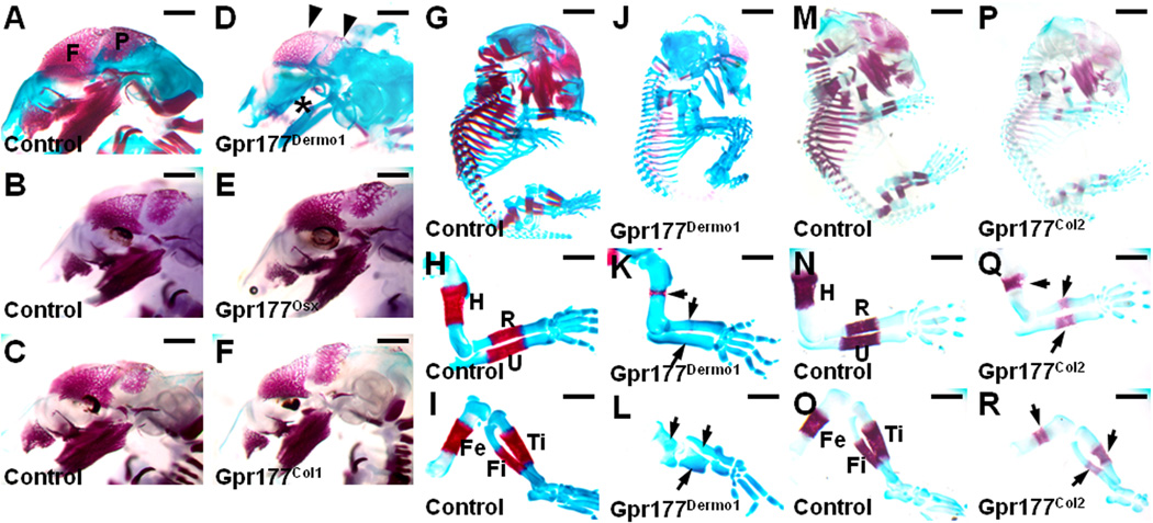
To determine the role of Gpr177 in skeletal development, conditional deletion was performed in various skeletogenic cell types. First, we generated Gpr177Dermo1 mutant mice in which Gpr177 was inactivated by the Dermo1-Cre transgene in the mesenchymal cells (30). The mesenchymal deletion of Gpr177 severely impaired development of the craniofacial skeleton at embryonic day 15.5 (E15.5). Alizarin red and alcian blue staining showed that formation of the calvarial, maxillary and mandibular bones mediated by intramembranous ossification is defective or completely missing in the Gpr177Dermo1 embryos (Figure 1A, D). Mineralization of the frontal bone was not detected in the mutants (Figure S1A, B). Because Dermo1-Cre induces recombination in the mesenchymal cells, Gpr177 is ablated not only in the precursors but also in their osteoblast derivatives (39–40). To further our assessment on the requirement of Gpr177 in the osteoprogenitors and osteoblasts, we generated Gpr177Osx and Gpr177Col1 mutants, in which Gpr177 was inactivated by Osx-Cre (29) and Col1a1-Cre (27), respectively. Compared to the control littermates, no obvious defect in development and mineralization of the craniofacial bones was detected in both mutants at E15.5 (Figure 1B, C, E, F and S1C–F). At newborn, the Gpr177Osx calvaria seemed to display delayed mineralization (Figure S2A–C). However, this is most likely caused by the Cre transgene but not the Gpr177 deletion as similar effects were also shown in the Osx-Cre; Gpr177+/+ calvaria (Figure S2A–C). Moreover, the Gpr177Col1 calvaria did not show any deformities, confirming that Gpr177 is dispensable in the osteoblasts (Figure S2D–F). The use of the R26R allele ensured the effectiveness of Cre lines (Figure S3A–D). Immunostaining of Gpr177 also revealed its loss of expression in the expected regions, including skeletogenic mesenchyme and osteogenic front (Figure S3E–L). As the deletion of β-catenin by Osx-Cre severely impairs calvarial development (29), the analysis of Gpr177 may provide new insight into the cell type responsible for Wnt production and signaling during intramembranous ossification. Although dispensable in the Osx-expressing osteoprogenitors and Col1-expressing osteoblasts cells, Gpr177 plays an important role in the mesenchymal cells essential for intramembranous ossification during calvarial development.
Figure 1.
Gpr177 is essential for development of the skeleton. (A–F) Skeletal staining of the E15.5 Gpr177Dermo1 (D), Gpr177Osx (E) and Gpr177Col1 (F) embryos, and their littermate controls (A–C) reveals the presence of Gpr177 in the mesenchymal but not the Osx-expressing osteoprogenitors and Col1-expressing osteoblasts is required for development of the craniofacial skeleton mediated by intramembranous ossification. Arrowheads and asterisk indicate impaired development of the calvarial bones (F, frontal; P, parietal) and the maxilla and mandible, respectively. Skeletal staining of the E15.5 Gpr177Dermo1 (J–L) and Gpr177Col2 (P–R) embryos, and their littermate controls (G–I, M–O) shows the requirement of Gpr177 in the mesenchymal and chondrocytes for development of the body skeleton mediated by endochondral ossification. Arrows indicate defective development of the forelimb and hindlimb. Fe, Femur; Fi, Fibula; H, Humerus; R, Radius; Ti, Tibia; U, Ulna. Scale bars, 1 mm (A–F); 2 mm (G, J, M, P); 500 µm (H, I, K, L, N, O, Q, R).
To determine the role of Gpr177 in endochondral ossification, we examined formation of the appendicular long bones in the Gpr177Dermo1 mutants. The mesenchymal deletion of Gpr177 causes severe defects in development of the body skeleton, including forelimbs and hindlimbs at E15.5 (Figure 1G–L). Mineralization occurred in the collar bones and primary spongiosa of control littermates, but missing in the Gpr177Dermo1 mutants (Figure S1G, H). This is also accompanied by the delay of chondrocyte maturation as hypertrophic chondrocytes were almost undetectable at this stage (Figure S1I, J). To examine whether the presence of Gpr177 in the mesenchymal cells is sufficient for chondrogenesis, we generated Gpr177Col2 mutants where Gpr177 was inactivated by the Col2a1-Cre transgene in chondrocytes (28). The removal of Gpr177 in the chondrocytes caused defects in the axial and appendicular bone formation (Figure 1M, P). In the E15.5 Gpr177Col2 mutants, the long bones were shortened and bone matrix formation was dramatically affected (Figure 1N, O, Q, R). The chondrogenic deletion of Gpr177 significantly reduced bone mineralization, and interfered with chondrocyte maturation (Figure S1K–N). Therefore, it is necessary to have Gpr177 present in the chondrocytes although we cannot rule out its requirement in the mesenchymal cells during endochondral ossification.
The role of Gpr177 in osteoblast development
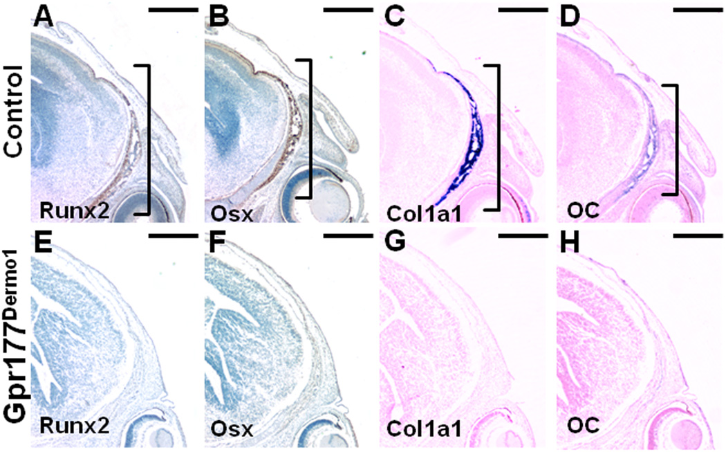
The development of calvarial bone plates, including the frontal and parietal bones, are mediated by intramembranous ossification (41–42). At about E12.5, the initial formation of a frontal bone primordium, which is sandwiched between the developing eye and brain, initiates osteogenesis (43). The osteogenic process then extends apically from the skull base to the midline within the skeletogenic mesenchyme, characterized by expression of the osteoprogenitor markers, Runx2 and Osterix (Osx), and the osteoblast marker, Col1a1 and Osteocalcin (OC) (Figure 2A–D). In contrast, the expression of these markers was strongly reduced or not detectable in the Gpr177Dermo1 mutants (Figure 2E–H), suggesting that the mesenchymal expression of Gpr177 is essential for osteoblast differentiation.
Figure 2.
The loss of Gpr177 impairs osteoblastogenesis. Coronal sections of the E15.5 control (A–D) and Gpr177Dermo1 (E–H) frontal bones were analyzed by immunostaining of Runx2 (A, E) and Osterix (Osx; B, F), and in situ hybridization of Col1a1 (C, G) and Osteocalcin (OC; D, H). Frontal bone formation occurs in the skeletogenic mesenchyme extending apically from the skull base to the midline in the controls (brackets), but absent in the mutants. Scale bars, 500 µm (A–H).
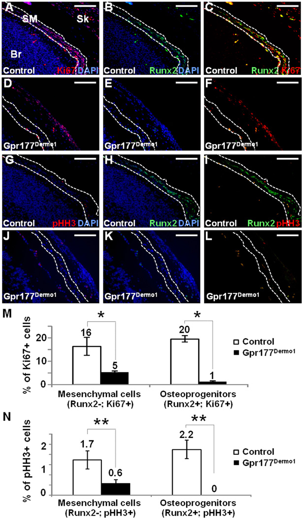
To determine if the expansion of osteoblast precursors was also affected by the Gpr177 ablation, cells undergoing mitotic division were detected by immunostaining of Ki67 (Figure 3A, D) and phosphorylated Histone H3 (Figure 3G, J). Consistent with our prior observation (17), there are two populations of precursors, Runx2-negative and Runx2-positive, actively expanding in the skeletogenic mesenchyme during intramembranous ossification (Figure 3A–C, G–I). However, proliferation of these two populations was severely affected in the Gpr177Dermo1 mutants (Figure 3D–F, J–L). Statistical analysis showed that the Gpr177 deletion significantly reduces the numbers of actively proliferating Runx2-negative and Runx2-positive cells (Figure 3M, N). The mesenchymal expression of Gpr177 is therefore essential for expansion of precursor cells and their differentiation into osteoblast cell types.
Figure 3.
Expansion of osteoblast precursors is affected by the Gpr177 ablation. Coronal sections of the E15.5 control (A–C, G–I) and Gpr177Dermo1 (D–F, J–L) frontal bones were double labeled with either Ki67 (A, C, D, F) or phosphorylated Histone H3 (pHH3; G, I, J, L), and Runx2 (B, C, E, F) to detect cells undergoing mitotic division and osteoprogenitors, respectively. Graphs illustrate the percentage of mitotic cells positive for Ki67 (M) or pHH3 (N) that are Runx2 negative (undifferentiated mesenchymal cells) or Runx2 positive (committed osteoprogenitors) affected by the deletion of Gpr177 (*, p value < 0.005; **, p value < 0.05, n=3 mice). Br, brain; SM, skeletogenic mesenchyme; Sk, skin. Scale bars, 100 µm (A–L).
Gpr177 in mesenchymal but not osteoblast cells is necessary for Wnt production in activation of β-catenin signaling during intramembranous ossification
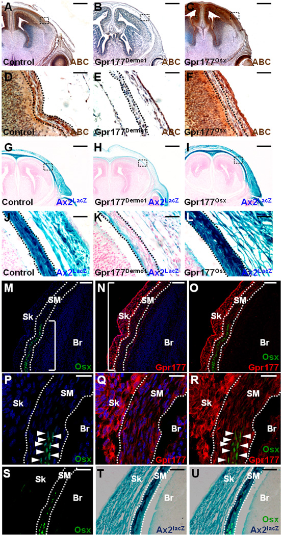
The loss of Gpr177 might affect Wnt signaling during skeletal development. To assess this question, the activation of β-catenin and its transcriptional target, Axin2, was examined by immunostaining with anti-activated form of β-catenin (ABC) and β-gal staining of the Axin2lacZ (Ax2lacZ) knock-in allele, respectively (14,16,22,34). Nuclear expression of β-catenin and uniform activation of Axin2 were evident in the E15.5 skeletogenic mesenchyme (Figure 4A, D, G, J). The expression of β-catenin and Axin2 was highly diminished in the Gpr177Dermo1 (Figure 4B, E, H, K), but not the Gpr177Osx (Figure 4C, F, I, L) mutants. In the Osx-expressing osteoprogenitors, although β-catenin is required for Wnt signal transduction (29), the Gpr177-mediated production of Wnt is apparently dispensable. The mesenchymal cells are the major and essential source of Wnt in osteoblastogenesis during calvarial morphogenesis.
Figure 4.
Gpr177 is required for Wnt production and signaling in osteoblastogenesis. Coronal sections of the E15.5 control (A, D, G, J), Gpr177Dermo1 (B, E, H, K) and Gpr177Osx (C, F, I, L) frontal bones were analyzed by immunostaining of activated β-catenin (ABC) (A–F) and β-gal staining (G–L). Immunostaining of ABC examines the signaling activity of the canonical Wnt pathway (A–F). Embryos heterozygous for the Axin2lacZ (Ax2lacZ) allele examine the expression of Axin2, a direct downstream target of Wnt/β-catenin signaling, in the control (G, J) and mutants (H, I, K, L). Enlargements of the insets in panels, A-C and G-I, are shown in D-F and J-L, respectively. Broken lines define the skeletogenic mesenchyme in the calvaria. (M–R) Coronal sections of the E15.5 frontal bone were analyzed by double labeling of Gpr177 and Osx. Osx-positive osteoprogenitors are localized to the skeletogenic mesenchyme extending apically from the skull base to the midline (M, O). Gpr177 is uniformly expressed in the skeletogenic mesenchyme (N, O). Brackets indicate the skeletogenic region positive for staining. Br, brain; Sk, skin; SM, skeletogenic mesenchyme. Higher power images (P–R) show the expression of Gpr177 in Osx-negative mesenchymal cells and Osx-positive osteoprogenitors (arrowheads). Coronal sections of the E15.5 frontal bone heterozygous for Axin2lacZ were used for expression analysis of Axin2 and Osx by β-gal staining (T, U) and fluorescent imaging (S, U), respectively. Broken lines define the skeletogenic mesenchyme in the calvaria. Scale bars, 500 µm (A–C, G–I); 50 µm (D–F, J–L); 100 µm (M–O, S–U); 10 µm (P-R).
To examine the signal producing and receiving cells of Wnt, we investigated the expression of Gpr177 and Axin2, respectively. While the expression of Osx was restricted to osteoprogenitors at the osteogenic front, Gpr177 showed a uniform expression pattern in the skeletogenic mesenchyme (Figure 4M–R). Therefore, Gpr177 is expressed in the osteoprogenitors even though their production of Wnt is not necessary for calvarial development. In agreement with β-catenin required for both mesenchymal and osteoprogenitor cells (29,38,40,44), we found that canonical Wnt signaling is uniformly activated in the skeletogenic mesenchyme using the Axin2lacZ allele (Figure 4S–U). Our findings suggest that mesenchymal production of Wnt activates β-catenin signaling in mesenchymal and osteoblast cell types in calvarial bone development.
Gpr177 is required for endochondral ossification
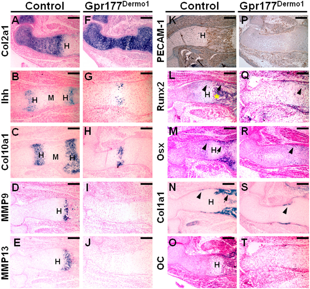
To determine the role of Gpr177 in development of the body skeleton, we performed a comprehensive molecular analysis examining the key steps of endochondral ossification, including chondrocyte proliferation, chondrogenesis, extracellular matrix (ECM) remodeling, vascular invasion and osteoblast differentiation. The number of cells undergoing mitotic division is significantly reduced in the columnar zone, but not epiphyses of the Gpr177Dermo1 and Gpr177Col2 humeruses, suggesting that expansion of the proliferating and prehypertrophic but not the resting chondrocytes was affected by the loss of Gpr177 (Figure S4). In the Gpr177Dermo1 mutants, the expression of Col2a1 in the chondrocytes was not affected at E15.5 (Figure 5A, F). Two expression zones of Ihh and Col10a1 separated by the marrow cavity were evident in the control littermates (Figure 5B, C). However, Ihh and Col10a1 just began to be expressed in center of the Gpr177Dermo1 humerus, suggesting severe delay of chondrocyte maturation (Figure 5G, H). The expression of MMP9 and MMP13 in the hypertrophic chondrocytes during ECM remodeling (45–46) was also absent in the mutants (Figure 5D, E, I, J). Vascular invasion, characterized by immunostaining of PECAM-1, did not occur as well (Figure 5K, P). Furthermore, strong expression of Runx2 in the perichondrium, collar bone and primary spongiosa, as well as hypertrophic chondrocytes of control was diminished significantly in the mutant (Figure 5L, Q). This was accompanied by decreased expression of Osx and Col1a1 in the collar bone and perichondrium of Gpr177Dermo1 (Figure 5M, N, R, S). The expression of Osteocalcin (OC) in the bone collar region was not detectable (Figure 5O, T). These results indicate that the disruption of endochondral ossification starts at chondrocyte maturation, and the subsequent events, including ECM remodeling, vascular invasion and osteoblastogenesis, are impaired in the Gpr177Dermo1 mutants.
Figure 5.
Deletion of Gpr177 in the skeletogenic mesenchyme disrupts endochondral ossification. Sections of the E15.5 control (A–E, K–O) and Gpr177Dermo1 (F–J, P–T) humeruses were analyzed by in situ hybridization of Col2a1 (A, F), IHH (B, G), Col10a1 (C, H), MMP9 (D, I), MMP13 (E, J), Runx2 (L, Q), Osx (M, R), Col1a1 (N, S) and OC (Osteocalcin; O, T), and immunostaining of PECAM-1 (K, P). Arrows, arrowheads and asterisk indicate collar bone, perichondrium and primary spongiosa, respectively. H, hypertrophic chondrocytes; M, marrow cavity. Scale bars, 200 µm (A–T).
Endochondral ossification requires the presence of Gpr177 in chondrocytes
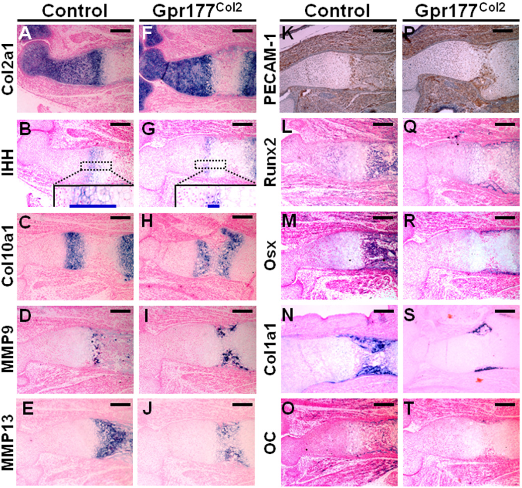
We then examined the role of Gpr177 in the chondrocytes during long bone development. A comprehensive molecular analysis was carried out to examine the key steps of endochondral ossification in the E15.5 Gpr177Col2 embryos. Similar to those of Gpr177Dermo1the expression of Col2a1 remained unchanged while the expression of Ihh was greatly reduced in the Gpr177Col2 mutants (Figure 6A, B, F, G). The marrow cavity and its surroundings, defined by two hypertrophic zones expressing Col10a1, were significantly smaller in the Gpr177Col2 humerus (Figure 6C, H). In addition, the expression of MMP9, MMP13 and PECAM-1 indicated that ECM remodeling and vascular invasion are defective in the Gpr177Col2 mutants (Figure 6D, E, I, J, K, P). The expression of Runx2, Osx and Col1a1 showed that osteoprogenitor cells are dramatically reduced and translocation of osteoblasts from the perichondrium to the nascent primary ossification center did not occur (Figure 6L–N, Q–S). Furthermore, the mature osteoblasts expressing OC were missing in the Gpr177Col2 mutants (Figure 6O, T).
Figure 6.
The presence of Gpr177 in the chondrocytes is necessary for endochondral ossification. Sections of the E15.5 control (A–E, K–O) and Gpr177Col2 (F–J, P–T) humeruses were analyzed by in situ hybridization of Col2a1 (A, F), IHH (B, G), Col10a1 (C, H), MMP9 (D, I), MMP13 (E, J), Runx2 (L, Q), Osx (M, R), Col1a1 (N, S) and OC (O, T), and immunostaining of PECAM-1 (K, P). Insets in panels B and G show prehypertrophic and hypertrophic chondrocytes, respectively. Blue bars denote the Ihh-expressing zone. Scale bars, 200 µm (A–T).
At E17.5, bone formation remained severely impaired in the humerus of Gpr177Col2compared to the control (Figure S5). Collar bones normally extended from diaphysis to the perichondrial region (Figure S5A, B). However, no bone collar was formed in the perichondrial region of Gpr177Col2 although mineralization of the hypertrophic chondrocytes was evident (Figure S5G, H). In addition, chondrogenesis, ECM remodeling and osteoblastogenesis were severely defective in the mutants (Figure S5C–F, I–L, M–X), suggesting the presence of Gpr177 in the chondrocytes is essential for endochondral ossification. To determine if the presence of Gpr177 in the osteoblasts is required for endochondral ossification, we examined the Gpr177Col1 limbs. Similar to that observed in calvarial development (Figure 1), the deletion of Gpr177 by Col1a1-Cre also did not cause any abnormality in limb development (Figure S6). Thus, Gpr177 is dispensable in the osteoblasts during intramembranous and endochondral ossifications. Our findings suggest that the impairment of osteoblast differentiation in the Gpr177Dermo1 and Gpr177Col2 limbs is attributed to delay in chondrocyte maturation but not intrinsic defects of the osteoblasts.
Gpr177 regulates Wnt signaling in long bone development
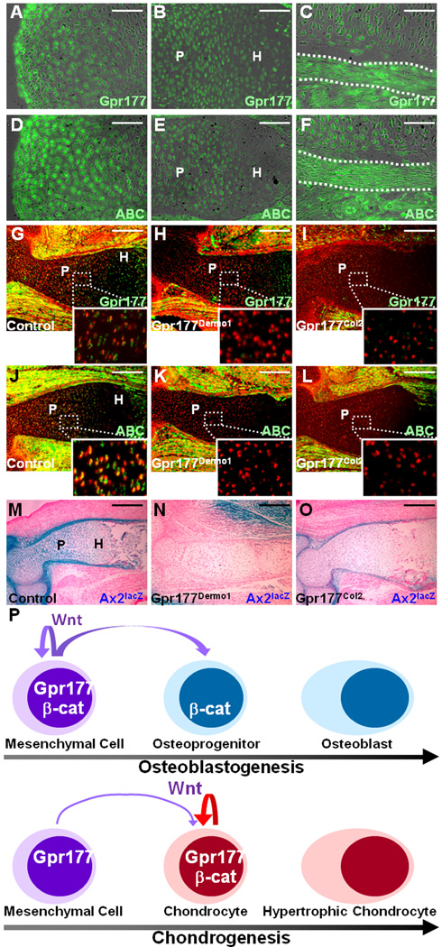
We next studied the effect of Gpr177 deficiency on the canonical Wnt pathway during limb development. The expression of Gpr177 was found in the resting, proliferating and hypertrophic chondrocytes, as well as the perichondrium in the developing limb (Figure 7A–C). Nuclear staining of the activated β-catenin was strong in the resting and proliferating chondrocytes, and in the perichondrium, but very weak in the hypertrophic chondrocytes (Figure 7D–F). Immunostaining of Gpr177 further showed the effective ablation of Gpr177 in the proliferating and hypertrophic chondrocytes in the Gpr177Dermo1 and Gpr177Col2 mutants (Figure 7G–I). The reduction of nuclear β-catenin staining further indicated that the Gpr177 deletion disrupts canonical Wnt signaling (Figure 7J-L). Crossing of the Ax2lacZ allele into the Gpr177Dermo1 and Gpr177Col2 backgrounds revealed that the expression of Axin2 is drastically reduced in the chondrocytes and perichondrium (Figure 7M–O). The data thus suggest that Gpr177 in the mesenchymal cells and chondrocytes is necessary for activation of canonical Wnt signaling during endochondral ossification.
Figure 7.
The canonical Wnt pathway is altered by the loss of Gpr177 during chondrogenesis. Sections of the E15.5 control (A–F, G, J, M), Gpr177Dermo1 (H, K, N) and Gpr177Col2 (I, L, O) humeruses were analyzed by immunostaining of Gpr177 (A–C, G–I) and ABC (D–F, J–L), and β-gal staining of the Axin2lacZ allele (Ax2lacZ; M–O). Insets in panels G–L show the significant reduction of Gpr177 and ABC staining in the proliferating chondrocytes. H, hypertrophic chondrocytes; P, proliferating chondrocytes. (P) Diagram illustrates the model for osteogenesis and chondrogenesis mediated by Wnt production and signaling. In osteoblast development, mesenchymal production of Wnt is essential for activation of β-catenin signaling in the mesenchymal cells and Osx-expressing osteoprogenitors through inter- and intra-cellular mechanisms, respectively. In contrast, Wnt produced in the mesenchymal cells may play an indispensable role but chondrocyte production of Wnt is mainly required for chondrogenesis. Scale bars, 50 µm (A, C–D, F); 100 µm (B, E); 200 µm (G–O).
Discussion
This study provides evidence that Gpr177, a gene closely linked to BMD and osteoporosis-related traits, is required for skeletogenesis. Genetic inactivation of Gpr177 in the mesenchymal cells severely impairs intramembranous and endochondral ossifications. Gpr177 plays an essential role in osteoblastogenesis and chondrogenesis through modulation of cell proliferation and differentiation. In the Gpr177 mutants, Wnt/β-catenin signaling is greatly reduced during development of the calvaria and limbs, suggesting that Gpr177-mediated regulation of Wnt production cannot be substituted. Genetic inactivation of Gpr177 in the osteogenic or chondrogenic progenitors further reveals its distinct role in osteogenesis and chondrogenesis. In osteoblast development, the presence of Gpr177 in the Osx-expressing osteoprogenitors and the Col1-expressing osteoblasts is dispensable as no skull defects associated with its ablation were detected in the mutants. In contrast, mesenchymal production of Wnt mediated by Gpr177 is necessary for intramembranous ossification. However, the deletion of β-catenin by Osx-Cre causes severe skull abnormalities, indicating that canonical Wnt signaling in the Osx-expressing osteoprogenitors is necessary for osteoblastogenesis (29). Our finding suggests that mesenchymal cells are the main cell type responsible for signal production. Their supply of Wnt activates β-catenin signaling in the signal-receiving mesenchymal and osteoblast cells. Both mesenchymal autocrine and paracrine signals of Wnt are essential for osteoblast development (Figure 7P). In development of the chondrocytes, however, chondrocyte production of Wnt is essential for chondrogenesis although the mesenchymal supply might also be necessary (Figure 7P). The chondrocyte-specific deletion of Gpr177 by Col2a1-Cre causes deficiencies in skeletal development although less severe than those of Gpr177Dermo1. Several key steps of endochondral ossification, including chondrocyte proliferation and maturation, ECM remodeling, vascular invasion, and osteoblast differentiation, are delayed in the developing humerus. These defects are reminiscent to those found in the mutants with ablation of β-catenin in the mesenchymal cells or chondrocytes (38,47), suggesting that Wnt proteins produced by chondrocytes are indispensable for endochondral ossification. Our findings lead us to propose a new mechanism underlying development of the skeleton mediated by the canonical Wnt pathway. The intercross of Wnt signaling between undifferentiated mesenchymal cells and skeletogenic progenitors is well orchestrated in a cell type-specific manner.
Canonical Wnt signaling has been demonstrated to play an important role in skeletogenesis, including the determination of mesenchymal cell fate (29,40,44) and chondrocyte maturation (38,47). Based on the molecular analysis of Gpr177Dermo1 and Gpr177Col2 mutants, specification of the Col2a1-expressing chondrocytes does not seem to be affected. However, chondrocyte proliferation and maturation are significantly reduced in both mutants. Our study of Gpr177 therefore supports an important function of β-catenin signaling in chondrogenesis at later stages. Because of ectopic chondrogenesis detected in the total β-catenin knockouts, it has been postulated that Wnt signaling possesses an inhibitory effect on chondrogenesis (29,40,44). We have never detected ectopic chondrogenesis in the Gpr177 mutants. This difference might be attributed to the dual role of β-catenin in cell adhesion and signaling (29,39–40,44,48) as the loss of Gpr177 would not interfere with cell-cell interaction. It is possible that β-catenin-mediated cell adhesion regulates the lineage commitment of mesenchymal cells to chondrocytes. Recent analysis of mice with deficiency in only the transcriptional activation of β-catenin has unexpectedly revealed its cell adhesion function, but not Wnt signaling activation, critical for neural development (49). Using the transcription-deficient mutants of β-catenin permits dissecting its signaling and structural functions, further analysis may uncover new mechanisms underlying lineage commitment of the skeletal progenitors.
The differential phenotypes caused by the deletion of Gpr177 and β-catenin might also be attributed to the involvement of noncanonical Wnt in lineage commitment of mesenchymal cells. Although our prior studies suggest that Gpr177 is a master regulator of Wnt production (22,26), there remains a lack of compelling evidence to support this theory. The noncanonical Wnt5a and Wnt5b are important regulators for chondrogenesis (50). Therefore, ectopic chondrogenesis detected in the β-catenin mutants might be caused by elevated signaling of noncanonical Wnt. Further investigation, focusing on the balance of canonical and noncanonical Wnt signaling, promises new insights into mesenchymal cell fate determination in skeletal development and disease.
Supplementary Material
Acknowledgements
This work is supported by National Institutes of Health grant DE015654 Supplemental data are included.
We thank Erestina Schipani and Matthew Hilton for reagents, Jiang Fu and Ansa Zahid for technical assistance, and Anthony Mirando for comments and suggestions. This work is supported by National Institutes of Health grant DE015654 to W.H. T.M. is a recipient of fellowship award from NYSTEM C026877.
Footnotes
Conflict of Interest
The authors declare no conflict of interest.
References
- 1.Manolagas SC. Birth and death of bone cells: basic regulatory mechanisms and implications for the pathogenesis and treatment of osteoporosis. Endocr Rev. 2000;21(2):115–137. doi: 10.1210/edrv.21.2.0395. [DOI] [PubMed] [Google Scholar]
- 2.Ralston SH, de Crombrugghe B. Genetic regulation of bone mass and susceptibility to osteoporosis. Genes Dev. 2006;20(18):2492–2506. doi: 10.1101/gad.1449506. [DOI] [PubMed] [Google Scholar]
- 3.Hsu YH, Zillikens MC, Wilson SG, Farber CR, Demissie S, Soranzo N, Bianchi EN, Grundberg E, Liang L, Richards JB, Estrada K, Zhou Y, van Nas A, Moffatt MF, Zhai G, Hofman A, van Meurs JB, Pols HA, Price RI, Nilsson O, Pastinen T, Cupples LA, Lusis AJ, Schadt EE, Ferrari S, Uitterlinden AG, Rivadeneira F, Spector TD, Karasik D, Kiel DP. An integration of genome-wide association study and gene expression profiling to prioritize the discovery of novel susceptibility Loci for osteoporosis-related traits. PLoS Genet. 2010;6(6):e1000977. doi: 10.1371/journal.pgen.1000977. [DOI] [PMC free article] [PubMed] [Google Scholar]
- 4.Richards JB, Rivadeneira F, Inouye M, Pastinen TM, Soranzo N, Wilson SG, Andrew T, Falchi M, Gwilliam R, Ahmadi KR, Valdes AM, Arp P, Whittaker P, Verlaan DJ, Jhamai M, Kumanduri V, Moorhouse M, van Meurs JB, Hofman A, Pols HA, Hart D, Zhai G, Kato BS, Mullin BH, Zhang F, Deloukas P, Uitterlinden AG, Spector TD. Bone mineral density, osteoporosis, and osteoporotic fractures: a genome-wide association study. Lancet. 2008;371(9623):1505–1512. doi: 10.1016/S0140-6736(08)60599-1. [DOI] [PMC free article] [PubMed] [Google Scholar]
- 5.Rivadeneira F, Styrkarsdottir U, Estrada K, Halldorsson BV, Hsu YH, Richards JB, Zillikens MC, Kavvoura FK, Amin N, Aulchenko YS, Cupples LA, Deloukas P, Demissie S, Grundberg E, Hofman A, Kong A, Karasik D, van Meurs JB, Oostra B, Pastinen T, Pols HA, Sigurdsson G, Soranzo N, Thorleifsson G, Thorsteinsdottir U, Williams FM, Wilson SG, Zhou Y, Ralston SH, van Duijn CM, Spector T, Kiel DP, Stefansson K, Ioannidis JP, Uitterlinden AG. Twenty bone-mineral-density loci identified by large-scale meta-analysis of genome-wide association studies. Nat Genet. 2009;41(11):1199–1206. doi: 10.1038/ng.446. [DOI] [PMC free article] [PubMed] [Google Scholar]
- 6.Styrkarsdottir U, Halldorsson BV, Gretarsdottir S, Gudbjartsson DF, Walters GB, Ingvarsson T, Jonsdottir T, Saemundsdottir J, Center JR, Nguyen TV, Bagger Y, Gulcher JR, Eisman JA, Christiansen C, Sigurdsson G, Kong A, Thorsteinsdottir U, Stefansson K. Multiple genetic loci for bone mineral density and fractures. N Engl J Med. 2008;358(22):2355–2365. doi: 10.1056/NEJMoa0801197. [DOI] [PubMed] [Google Scholar]
- 7.Ferrari SL, Karasik D, Liu J, Karamohamed S, Herbert AG, Cupples LA, Kiel DP. Interactions of interleukin-6 promoter polymorphisms with dietary and lifestyle factors and their association with bone mass in men and women from the Framingham Osteoporosis Study. J Bone Miner Res. 2004;19(4):552–559. doi: 10.1359/JBMR.040103. [DOI] [PubMed] [Google Scholar]
- 8.Cadigan KM, Peifer M. Wnt signaling from development to disease: insights from model systems. Cold Spring Harb Perspect Biol. 2009;1(2):a002881. doi: 10.1101/cshperspect.a002881. [DOI] [PMC free article] [PubMed] [Google Scholar]
- 9.MacDonald BT, Tamai K, He X. Wnt/beta-catenin signaling: components, mechanisms, and diseases. Dev Cell. 2009;17(1):9–26. doi: 10.1016/j.devcel.2009.06.016. [DOI] [PMC free article] [PubMed] [Google Scholar]
- 10.Krishnan V, Bryant HU, Macdougald OA. Regulation of bone mass by Wnt signaling. J Clin Invest. 2006;116(5):1202–1209. doi: 10.1172/JCI28551. [DOI] [PMC free article] [PubMed] [Google Scholar]
- 11.Hoeppner LH, Secreto FJ, Westendorf JJ. Wnt signaling as a therapeutic target for bone diseases. Expert Opin Ther Targets. 2009;13(4):485–496. doi: 10.1517/14728220902841961. [DOI] [PMC free article] [PubMed] [Google Scholar]
- 12.Hartmann C. A Wnt canon orchestrating osteoblastogenesis. Trends Cell Biol. 2006;16(3):151–158. doi: 10.1016/j.tcb.2006.01.001. [DOI] [PubMed] [Google Scholar]
- 13.Long F. Building strong bones: molecular regulation of the osteoblast lineage. Nat Rev Mol Cell Biol. 2012;13(1):27–38. doi: 10.1038/nrm3254. [DOI] [PubMed] [Google Scholar]
- 14.Yu HM, Jerchow B, Sheu TJ, Liu B, Costantini F, Puzas JE, Birchmeier W, Hsu W. The role of Axin2 in calvarial morphogenesis and craniosynostosis. Development. 2005;132(8):1995–2005. doi: 10.1242/dev.01786. [DOI] [PMC free article] [PubMed] [Google Scholar]
- 15.Liu B, Yu HM, Hsu W. Craniosynostosis caused by Axin2 deficiency is mediated through distinct functions of beta-catenin in proliferation and differentiation. Dev Biol. 2007;301(1):298–308. doi: 10.1016/j.physletb.2003.10.071. [DOI] [PMC free article] [PubMed] [Google Scholar]
- 16.Maruyama T, Mirando AJ, Deng CX, Hsu W. The balance of WNT and FGF signaling influences mesenchymal stem cell fate during skeletal development. Sci Signal. 2010;3(123):ra40. doi: 10.1126/scisignal.2000727. [DOI] [PMC free article] [PubMed] [Google Scholar]
- 17.Mirando AJ, Maruyama T, Fu J, Yu HM, Hsu W. Beta-catenin/cyclin D1 mediated development of suture mesenchyme in calvarial morphogenesis. BMC Dev Biol. 2010;10(1):116. doi: 10.1186/1471-213X-10-116. [DOI] [PMC free article] [PubMed] [Google Scholar]
- 18.Baldridge D, Shchelochkov O, Kelley B, Lee B. Signaling pathways in human skeletal dysplasias. Annu Rev Genomics Hum Genet. 2010;11:189–217. doi: 10.1146/annurev-genom-082908-150158. [DOI] [PubMed] [Google Scholar]
- 19.Gong Y, Slee RB, Fukai N, Rawadi G, Roman-Roman S, Reginato AM, Wang H, Cundy T, Glorieux FH, Lev D, Zacharin M, Oexle K, Marcelino J, Suwairi W, Heeger S, Sabatakos G, Apte S, Adkins WN, Allgrove J, Arslan-Kirchner M, Batch JA, Beighton P, Black GC, Boles RG, Boon LM, Borrone C, Brunner HG, Carle GF, Dallapiccola B, De Paepe A, Floege B, Halfhide ML, Hall B, Hennekam RC, Hirose T, Jans A, Juppner H, Kim CA, Keppler-Noreuil K, Kohlschuetter A, LaCombe D, Lambert M, Lemyre E, Letteboer T, Peltonen L, Ramesar RS, Romanengo M, Somer H, Steichen-Gersdorf E, Steinmann B, Sullivan B, Superti-Furga A, Swoboda W, van den Boogaard MJ, Van Hul W, Vikkula M, Votruba M, Zabel B, Garcia T, Baron R, Olsen BR, Warman ML. LDL receptor-related protein 5 (LRP5) affects bone accrual and eye development. Cell. 2001;107(4):513–523. doi: 10.1016/s0092-8674(01)00571-2. [DOI] [PubMed] [Google Scholar]
- 20.Boyden LM, Mao J, Belsky J, Mitzner L, Farhi A, Mitnick MA, Wu D, Insogna K, Lifton RP. High bone density due to a mutation in LDL-receptor-related protein 5. N Engl J Med. 2002;346(20):1513–1521. doi: 10.1056/NEJMoa013444. [DOI] [PubMed] [Google Scholar]
- 21.Kato M, Patel MS, Levasseur R, Lobov I, Chang BH, Glass DA, 2nd, Hartmann C, Li L, Hwang TH, Brayton CF, Lang RA, Karsenty G, Chan L. Cbfa1-independent decrease in osteoblast proliferation, osteopenia, and persistent embryonic eye vascularization in mice deficient in Lrp5, a Wnt coreceptor. J Cell Biol. 2002;157(2):303–314. doi: 10.1083/jcb.200201089. [DOI] [PMC free article] [PubMed] [Google Scholar]
- 22.Fu J, Jiang M, Mirando AJ, Yu HM, Hsu W. Reciprocal regulation of Wnt and Gpr177/mouse Wntless is required for embryonic axis formation. Proc Natl Acad Sci U S A. 2009;106(44):18598–18603. doi: 10.1073/pnas.0904894106. [DOI] [PMC free article] [PubMed] [Google Scholar]
- 23.Banziger C, Soldini D, Schutt C, Zipperlen P, Hausmann G, Basler K. Wntless, a conserved membrane protein dedicated to the secretion of Wnt proteins from signaling cells. Cell. 2006;125(3):509–522. doi: 10.1016/j.cell.2006.02.049. [DOI] [PubMed] [Google Scholar]
- 24.Bartscherer K, Pelte N, Ingelfinger D, Boutros M. Secretion of Wnt ligands requires Evi, a conserved transmembrane protein. Cell. 2006;125(3):523–533. doi: 10.1016/j.cell.2006.04.009. [DOI] [PubMed] [Google Scholar]
- 25.Yu HM, Jin Y, Fu J, Hsu W. Expression of Gpr177, a Wnt trafficking regulator, in mouse embryogenesis. Dev Dyn. 2010;239(7):2102–2109. doi: 10.1002/dvdy.22336. [DOI] [PMC free article] [PubMed] [Google Scholar]
- 26.Fu J, Ivy Yu HM, Maruyama T, Mirando AJ, Hsu W. Gpr177/mouse Wntless is essential for Wnt-mediated craniofacial and brain development. Dev Dyn. 2011;240(2):365–371. doi: 10.1002/dvdy.22541. [DOI] [PMC free article] [PubMed] [Google Scholar]
- 27.Liu F, Woitge HW, Braut A, Kronenberg MS, Lichtler AC, Mina M, Kream BE. Expression and activity of osteoblast-targeted Cre recombinase transgenes in murine skeletal tissues. Int J Dev Biol. 2004;48(7):645–653. doi: 10.1387/ijdb.041816fl. [DOI] [PubMed] [Google Scholar]
- 28.Ovchinnikov DA, Deng JM, Ogunrinu G, Behringer RR. Col2a1-directed expression of Cre recombinase in differentiating chondrocytes in transgenic mice. Genesis. 2000;26(2):145–146. [PubMed] [Google Scholar]
- 29.Rodda SJ, McMahon AP. Distinct roles for Hedgehog and canonical Wnt signaling in specification, differentiation and maintenance of osteoblast progenitors. Development. 2006;133(16):3231–3244. doi: 10.1242/dev.02480. [DOI] [PubMed] [Google Scholar]
- 30.Yu K, Xu J, Liu Z, Sosic D, Shao J, Olson EN, Towler DA, Ornitz DM. Conditional inactivation of FGF receptor 2 reveals an essential role for FGF signaling in the regulation of osteoblast function and bone growth. Development. 2003;130(13):3063–3074. doi: 10.1242/dev.00491. [DOI] [PubMed] [Google Scholar]
- 31.Chiu SY, Asai N, Costantini F, Hsu W. SUMO-Specific Protease 2 Is Essential for Modulating p53-Mdm2 in Development of Trophoblast Stem Cell Niches and Lineages. PLoS Biol. 2008;6(12):e310. doi: 10.1371/journal.pbio.0060310. [DOI] [PMC free article] [PubMed] [Google Scholar]
- 32.Hsu W, Mirando AJ, Yu HM. Manipulating gene activity in Wnt1-expressing precursors of neural epithelial and neural crest cells. Dev Dyn. 2010;239(1):338–345. doi: 10.1002/dvdy.22044. [DOI] [PMC free article] [PubMed] [Google Scholar]
- 33.Yu HM, Liu B, Chiu SY, Costantini F, Hsu W. Development of a unique system for spatiotemporal and lineage-specific gene expression in mice. Proc Natl Acad Sci U S A. 2005;102(24):8615–8620. doi: 10.1073/pnas.0500124102. [DOI] [PMC free article] [PubMed] [Google Scholar]
- 34.Liu B, Yu HM, Huang J, Hsu W. Co-opted JNK/SAPK signaling in Wnt/beta-catenin-induced tumorigenesis. Neoplasia. 2008;10(9):1004–1013. doi: 10.1593/neo.08548. [DOI] [PMC free article] [PubMed] [Google Scholar]
- 35.Jiang M, Chiu SY, Hsu W. SUMO-specific protease 2 in Mdm2-mediated regulation of p53. Cell Death Differ. 2011;18(6):1005–1015. doi: 10.1038/cdd.2010.168. [DOI] [PMC free article] [PubMed] [Google Scholar]
- 36.Provot S, Zinyk D, Gunes Y, Kathri R, Le Q, Kronenberg HM, Johnson RS, Longaker MT, Giaccia AJ, Schipani E. Hif-1alpha regulates differentiation of limb bud mesenchyme and joint development. J Cell Biol. 2007;177(3):451–464. doi: 10.1083/jcb.200612023. [DOI] [PMC free article] [PubMed] [Google Scholar]
- 37.Shimada M, Greer PA, McMahon AP, Bouxsein ML, Schipani E. In vivo targeted deletion of calpain small subunit, Capn4, in cells of the osteoblast lineage impairs cell proliferation, differentiation, and bone formation. J Biol Chem. 2008;283(30):21002–21010. doi: 10.1074/jbc.M710354200. [DOI] [PMC free article] [PubMed] [Google Scholar]
- 38.Hu H, Hilton MJ, Tu X, Yu K, Ornitz DM, Long F. Sequential roles of Hedgehog and Wnt signaling in osteoblast development. Development. 2005;132(1):49–60. doi: 10.1242/dev.01564. [DOI] [PubMed] [Google Scholar]
- 39.Tran TH, Jarrell A, Zentner GE, Welsh A, Brownell I, Scacheri PC, Atit R. Role of canonical Wnt signaling/ss-catenin via Dermo1 in cranial dermal cell development. Development. 2010;137(23):3973–3984. doi: 10.1242/dev.056473. [DOI] [PMC free article] [PubMed] [Google Scholar]
- 40.Day TF, Guo X, Garrett-Beal L, Yang Y. Wnt/beta-catenin signaling in mesenchymal progenitors controls osteoblast and chondrocyte differentiation during vertebrate skeletogenesis. Dev Cell. 2005;8(5):739–750. doi: 10.1016/j.devcel.2005.03.016. [DOI] [PubMed] [Google Scholar]
- 41.Rice DP. Developmental anatomy of craniofacial sutures. Front Oral Biol. 2008;12:1–21. doi: 10.1159/000115028. [DOI] [PubMed] [Google Scholar]
- 42.Opperman LA. Cranial sutures as intramembranous bone growth sites. Dev Dyn. 2000;219(4):472–485. doi: 10.1002/1097-0177(2000)9999:9999<::AID-DVDY1073>3.0.CO;2-F. [DOI] [PubMed] [Google Scholar]
- 43.Sasaki T, Ito Y, Bringas P, Jr, Chou S, Urata MM, Slavkin H, Chai Y. TGFbeta-mediated FGF signaling is crucial for regulating cranial neural crest cell proliferation during frontal bone development. Development. 2006;133(2):371–381. doi: 10.1242/dev.02200. [DOI] [PubMed] [Google Scholar]
- 44.Hill TP, Spater D, Taketo MM, Birchmeier W, Hartmann C. Canonical Wnt/beta-catenin signaling prevents osteoblasts from differentiating into chondrocytes. Dev Cell. 2005;8(5):727–738. doi: 10.1016/j.devcel.2005.02.013. [DOI] [PubMed] [Google Scholar]
- 45.Stickens D, Behonick DJ, Ortega N, Heyer B, Hartenstein B, Yu Y, Fosang AJ, Schorpp-Kistner M, Angel P, Werb Z. Altered endochondral bone development in matrix metalloproteinase 13-deficient mice. Development. 2004;131(23):5883–5895. doi: 10.1242/dev.01461. [DOI] [PMC free article] [PubMed] [Google Scholar]
- 46.Vu TH, Shipley JM, Bergers G, Berger JE, Helms JA, Hanahan D, Shapiro SD, Senior RM, Werb Z. MMP-9/gelatinase B is a key regulator of growth plate angiogenesis and apoptosis of hypertrophic chondrocytes. Cell. 1998;93(3):411–422. doi: 10.1016/s0092-8674(00)81169-1. [DOI] [PMC free article] [PubMed] [Google Scholar]
- 47.Akiyama H, Lyons JP, Mori-Akiyama Y, Yang X, Zhang R, Zhang Z, Deng JM, Taketo MM, Nakamura T, Behringer RR, McCrea PD, de Crombrugghe B. Interactions between Sox9 and beta-catenin control chondrocyte differentiation. Genes Dev. 2004;18(9):1072–1087. doi: 10.1101/gad.1171104. [DOI] [PMC free article] [PubMed] [Google Scholar]
- 48.Heuberger J, Birchmeier W. Interplay of cadherin-mediated cell adhesion and canonical Wnt signaling. Cold Spring Harb Perspect Biol. 2010;2(2):a002915. doi: 10.1101/cshperspect.a002915. [DOI] [PMC free article] [PubMed] [Google Scholar]
- 49.Valenta T, Gay M, Steiner S, Draganova K, Zemke M, Hoffmans R, Cinelli P, Aguet M, Sommer L, Basler K. Probing transcription-specific outputs of beta-catenin in vivo. Genes Dev. 2011;25(24):2631–2643. doi: 10.1101/gad.181289.111. [DOI] [PMC free article] [PubMed] [Google Scholar]
- 50.Yang Y, Topol L, Lee H, Wu J. Wnt5a and Wnt5b exhibit distinct activities in coordinating chondrocyte proliferation and differentiation. Development. 2003;130(5):1003–1015. doi: 10.1242/dev.00324. [DOI] [PubMed] [Google Scholar]
Associated Data
This section collects any data citations, data availability statements, or supplementary materials included in this article.


