Abstract
Change in carpal arch width (CAW) is associated with wrist movement, carpal tunnel release, or therapeutic tunnel manipulation. This study investigated the angular rotations of the distal carpal joints as the CAW was adjusted. The CAW was narrowed and widened by 2 and 4 mm in seven cadaveric specimens while the bone positions were tracked by a marker-based motion capture system. The joints mainly pronated during CAW narrowing and supinated during widening. Ranges of motion about the pronation axis for the hamate-capitate (H-C), capitate-trapezoid (C-Td), and trapezoid-trapezium (Td-Tm) joints were 8.1 ± 2.3°, 5.3 ± 1.3°, and 5.5 ± 3.5°, respectively. Differences between the angular rotations of the joints were found at ΔCAW = −4 mm about the pronation and ulnar-deviation axes. For the pronation axis, angular rotations of the H-C joint were larger than that of the C-Td and Td-Tm joints. Statistical interactions among the factors of joint, rotation axis, and ΔCAW indicated complex joint motion patterns. The complex three-dimensional motion of the bones can be attributed to several anatomical constraints such as bone arrangement, ligament attachments, and articular congruence. The results of this study provide insight into the mechanisms of carpal tunnel adaptations in response to biomechanical alterations of the structural components.
Keywords: carpal bones, kinematics, carpal arch width, distal carpal row, carpal tunnel
1. Introduction
The wrist complex has an intricate arrangement of eight irregularly shaped carpal bones connected by numerous interosseous ligaments. The carpal bones and ligaments form the carpal tunnel which encloses nine flexor tendons and the median nerve. The proximal row of the carpal tunnel consists of the pisiform, triquetrum, lunate, and scaphoid bones, while the distal row is comprised of the hamate, capitate, trapezoid, and trapezium bones. The distal level of the carpal tunnel tends to have an elevated pressure [1], and it is structurally stiffer than the proximal level [2]. The distal row is considered to be tightly bound with little intercarpal motion during wrist movement [3–5] especially at the trapezium-trapezoid and trapezoid-capitate joints [5]. However, evidence of intercarpal mobility within the distal row has been reported in several studies relating to changes in the carpal arch width [6–11].
Carpal arch width (CAW) is defined at the distal level of the carpal tunnel as the distance between the hook of hamate and the ridge of the trapezium. Narrowing and widening of CAW can occur during wrist motion [6], after carpal tunnel release [6–9], or during cadaveric experimentation [2,11–13]. For example, Garcia-Elias et al. demonstrated that CAW narrowing occurs during flexion and extension of the wrist [6]. Transection of the transverse carpal ligament, also known as carpal tunnel release, results in a widening of the CAW up to 3 mm [7] or 11% [6], and it resulted in angular rotations within the distal carpal row [8,10] such as slight external rotations of the trapezium and trapezoid [7]. Sucher et al. showed that the CAW can be altered by manipulation and sustained load bearing [12]. Xiu et al. further specified that the CAW can be modified with a force as small as 2 N in both a narrowing and widening direction [2]. In addition, Li et al. reported that the CAW narrowing is associated with an increase of carpal tunnel cross-sectional area [11,13]. The carpal tunnel expansion achieved through CAW narrowing could potentially reduce carpal tunnel pressure and alleviate the symptoms associated with carpal tunnel syndrome [2,11,13].
As previously stated, it has been shown that changes in the CAW occur under different wrist conditions [2, 6–8, 12]. However, the underlying kinematics of the joints within the distal carpal row during CAW widening and narrowing remained unclear. The aim of the study was to investigate the relative angular rotations of the distal carpal joints that contribute towards carpal arch widening and narrowing. We hypothesized that changes in the carpal arch width would result in larger angular rotations of the hamate-capitate and capitate-trapezoid joints than the trapezium-trapezoid joint. We further hypothesized that the intercarpal joints will exhibit three-dimensional angular rotations for a linear change of the carpal arch width.
2. Methods
Specimen Preparation.
Eight fresh frozen cadaver hands (51.3 ± 9.5 years) with no history of injury or degenerative disorders to the wrist were thawed overnight prior to experimentation. Each specimen was dissected volarly to remove the transverse carpal ligament. On the dorsal aspect, the carpal bones were exposed by removing the skin, fat, fascia, and extensor tendons. A hole was drilled into the volar aspect of hamate and trapezium, each, at the attachment sites of the transverse carpal ligament. Screws were inserted within the holes, fixed with glue, and wires were connected to them. The distal carpal bones were identified, dorsally under guidance of fluoroscopic imaging, and holes were drilled into the hamate, capitate, trapezoid, and trapezium. Care was taken to ensure that the intercarpal ligaments were left intact. Custom made marker clusters were then inserted into the hamate, capitate, trapezoid, and trapezium and fixed with the use of glue. An additional marker set was glued to the skin on the dorsal aspect of the radius.
Experimental Setup.
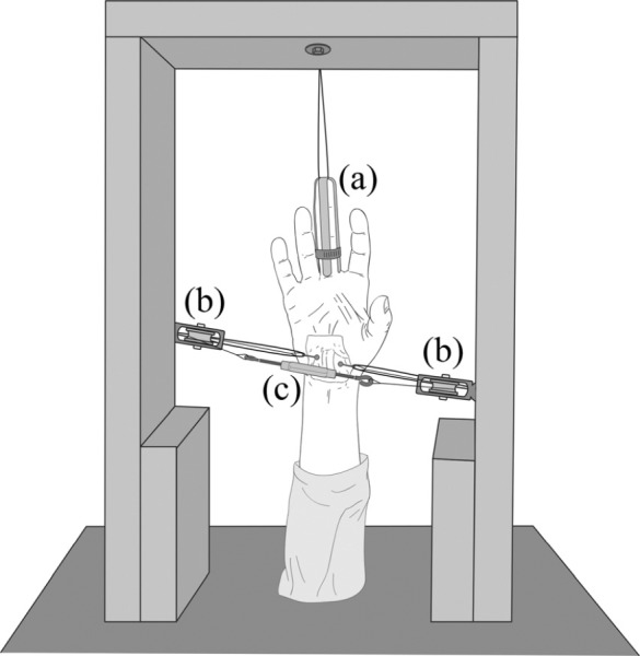
A custom apparatus was used to uniformly narrow and widen the CAW (Fig. 1). The apparatus consisted of a rigid frame, two height adjustable pulleys, a finger trap, and a turnbuckle. The specimens elbow was placed onto the base and an additional wire was attached to the third digit by means of the finger trap to hold the forearm in a vertical position. The CAW was then measured volarly, with a caliper at a resolution of 0.02 mm, as the distance between base of the screws inserted into the hamate and trapezium. The wires attached to the hamate and trapezium screws were looped around the two pulleys and attached to the turnbuckle. For CAW widening, the wires were looped around the nearest pulley as shown in Fig. 1. During CAW narrowing, the wires crossed each other to loop around the furthest pulley. The pulleys had the capability of rotating in the coronal plane of the specimen to ensure that narrowing and widening of the CAW was in an axis connecting the hook of hamate and the beak of the trapezium. The turnbuckle allowed for fine adjustment of the CAW during experimentation.
Fig. 1.

Experimental setup showing the (a) fingertrap, (b) pulleys, and (c) turnbuckle for widening of the carpal arch width (volar view)
Protocol, Kinematics, and Statistical Analysis.
Based on the initial CAW measurement, a total of five conditions were tested: −4, −2, 0, +2, and +4 mm changes in the CAW (ΔCAW); where negative changes reflected narrowing of the CAW and positive changes reflected widening of the CAW. The five conditions were repeated during three trials, and the order of the five conditions was randomized within each trial.
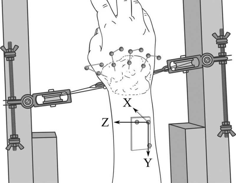
Kinematics of the carpal bones was investigated using a passive marker infrared motion capture system (VICON T40, Oxford Metrics Ltd., Oxford, England) to track the movement of the marker clusters. A coordinate system was created for each bone using the respective marker cluster. In the reference (ΔCAW = 0 mm) condition, each bone assumed an initial orientation identical to the wrist movement axes based on the radius marker set following the ISB recommendations [14] (Fig. 2). A relationship was established between this initial configuration and each marker cluster, and this relationship was used to back transform the orientation of the marker cluster into actual bone orientation during all conditions. Joint motion between adjacent bones was defined as the movement of the lateral bone relative to the medial bone, i.e., the capitate relative to the hamate (H-C), the trapezoid relative to the capitate (C-Td), and the trapezium relative to the trapezoid (Td-Tm). The Euler angles were calculated in the Y-Z-X sequence corresponding to rotations about the pronation(+)/supination(−), flexion(+)/extension(−), and ulnar(+)/radial(−) deviation axes, respectively. The pronation/supination (Y) axis was chosen as the first Euler angle because it was expected to be the primary rotation axis for manipulation of the CAW. Rotations about the flexion/extension and ulnar/radial deviation axes were secondary and therefore chosen as subsequent Euler angles.
Fig. 2.

Dorsal view of the specimen during experimentation showing the marker clusters and radial coordinate system
Initial joint orientation was calculated using the 0 mm ΔCAW condition of each trial. The relative angular rotations of each specimen were averaged among the three trials. Then a three-way repeated measures ANOVA (3 × 3 × 4) was performed with respect to independent variables of Joints (H-C, C-Td, Td-Tm), rotation axes (Y, Z′, X″) and ΔCAW (−4, −2, +2, +4). The significance level was set at α = 0.05.
3. Results
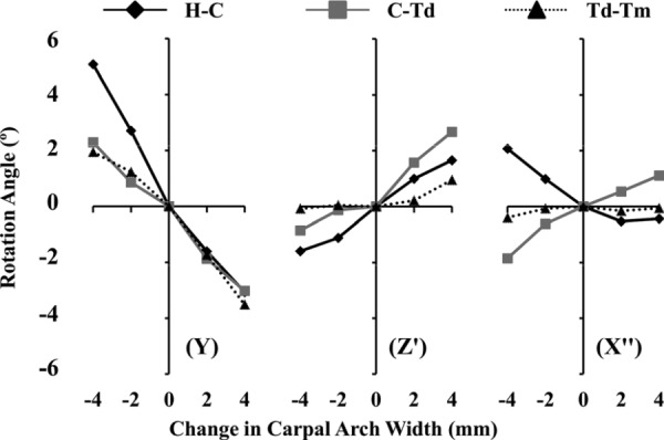
The hamate fractured in one specimen during experimentation; therefore the results are based on seven specimens (n = 7). The initial CAW, before dissection, was measured at 22.6 ± 2.7 mm. Angular rotations of the joints characteristically corresponded to the narrowing and widening of CAW with rotations about all three rotation axes (Fig. 3). The largest angular rotations occurred about the Y axis with ranges of motion of 8.1 ± 2.3 deg, 5.3 ± 1.3 deg, and 5.5 ± 3.5 deg for the H-C, C-Td, and Td-Tm joints, respectively, from a ΔCAW of −4 mm to +4 mm.
Fig. 3.

Mean rotation angles (deg) about the pronation/supination (Y), flexion/extension (Z′), and ulnar/radial deviation (X″) axes for each joint during changes in carpal arch width (mm). Pronation, flexion, and ulnar deviation are the positive directions for the respective axes.
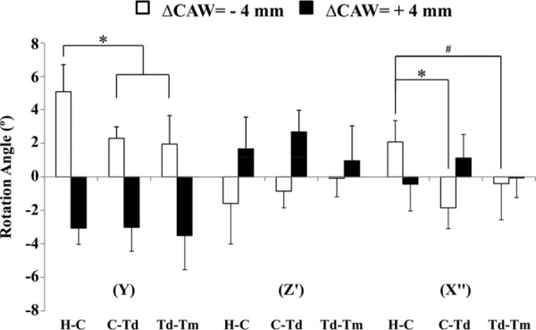
First, a significant difference in angular rotations occurred among the intercarpal joints (p < 0.05). Specifically, the H-C joint had significantly different angular rotations than that of the Td-Tm joint (p < 0.05). However, there was no significant difference in the angular rotations between the H-C and C-Td joints or between the C-Td and Td-Tm joints. Second, angular rotations were significantly affected by ΔCAW (p < 0.001) (Fig. 3). In particular, significant differences were found for angular rotations between ΔCAWs of −2 and +2 mm (p < 0.05), and between ΔCAWs of −2 and +4 mm (p < 0.001). Furthermore, the angular rotations at a ΔCAW of −4 mm were significantly different (p < 0.001) from that at ΔCAWs of +2 and +4 mm. In general, the angular rotations at a ΔCAW of −4 mm were asymmetrical to that at ΔCAW of +4 mm (Fig. 4). Third, rotation angles were not significantly dependent on the factor of rotation axis (p = 0.132).
Fig. 4.

Mean ± standard deviation of angular rotation for each joint at ΔCAW = −4 and +4 mm for the pronation/supination (Y), flexion/extension (Z′), and ulnar/radial deviation (X″) axes. (* denotes p < 0.001 and # denotes p < 0.05). Pronation, flexion, and ulnar deviation are the positive directions for the respective axes.
A significant three-way interaction was found among ΔCAW, joint, and axis of rotation (p < 0.05). There were significant two-way interactions between joint and ΔCAW (p < 0.001), as well as between ΔCAW and axis of rotation (p < 0.001). There was also a significant two-way interaction between axis of rotation and joint (p < 0.05) which was dependent on the level of ΔCAW, specifically at a level of ΔCAW = −4 mm (p < 0.001). These complex interactions were more explicit at specific levels of the factors. For example, at the level of ΔCAW = –4 mm the intercarpal joints had significant differences (p < 0.001) in the angular rotations about the Y or X″ axis (Fig. 4). For angular rotations about the Y axis, the H-C joint (5.1 ± 1.6 deg) had significantly larger (p < 0.001) angular rotation than the C-Td (2.3 ± 0.7 deg) or Td-Tm (1.9 ± 1.7 deg) joints. About the X″ axis, the rotations of the H-C joint (2.1 ± 1.3 deg) were significantly different than the C-Td joint (−1.9 ± 1.2 deg, p < 0.001) and the Td-Tm joint (−0.4 ± 2.2 deg, p < 0.05). However, about the Z′ axis there were no significant differences (p = 0.13) among the rotations of H-C (−1.6 ± 2.4 deg), C-Td (−0.9 ± 1.0 deg), and Td-Tm (−0.1 ± 1.1 deg) joints.
4. Discussion
This study investigated the relative angular rotations of the distal carpal joints in relation to changes of the CAW from −4 to +4 mm which primarily resulted in rotations about the pronation/supination (Y) axis for all joints. However, the joints also exhibited rotations about the flexion/extension (Z′) and ulnar/radial deviation (X″) axes. We found that the angular rotations were dependent on the joints and ΔCAW with complex statistical interactions among the factors of joint, rotation axis, and ΔCAW.
The rotational difference of individual joints may be attributable to anatomical constraints. The H-C joint was generally the most mobile in angular rotation while the Td-Tm joint rotated the least. The limited angular rotation of the Td-Tm joint may be attributable to the tight coupling in the scaphotrapezial-trapezoidal complex [5]. In particular, the trapezium interacts with the scaphoid through articular contact and the scaphotrapezium ligament. Anatomically, the orientation of the scaphoid within the wrist and the trapezium's interactions with the scaphoid would limit angular rotations of the Td-Tm joint about the ulnar/radial deviation and the flexion/extension axes [5]. Ulnar deviation of the Td-Tm joint may be restricted by scaphotrapezium ligament while radial deviation of the Td-Tm joint would be restricted by the scaphoid-trapezium articulation. Flexion/extension of the Td-Tm joint is dependent on an articulation chain among the trapezoid, trapezium and scaphoid [15]. Therefore, due to the ligamentous and articular restrictions the angular rotations of the Td-Tm joint may be limited in multiple rotation axes, specifically the ulnar/radial deviation and flexion/extension axes.
The result that angular rotation was not dependent on the factor of rotation axis indicates that the joints exhibit complex rotations about all three axes. Such three-dimensional rotations could be attributable to the anatomical arrangement of the carpal arch. The connection between the hook of hamate and the ridge of the trapezium, which define the CAW, is not aligned with the anatomically defined rotation axes. The hook of the hamate is generally more distal and dorsal than the ridge of the trapezium. As such, the orientation along the CAW is oblique to the flexion/extension axis and out of the frontal plane. Moreover, the intricate system of intercarpal ligaments [16,17] and articular contacts/surfaces [5,16] would constrain the joint rotations around particular axes that do not necessarily conform with the currently defined axes.
Biomechanically, the dorsal intercarpal ligaments would limit angular rotations, primarily about the pronation/supination axis, of the distal carpal joints during CAW narrowing. The dorsal hamate-capitate ligament is the stiffest of the distal row followed by the dorsal capitate-trapezoid and dorsal trapezoid-trapezium ligaments, respectively [17], implying that the angular rotations of H-C joint could be less than that of the Td-Tm joint. However, our results show that the H-C joint had the largest angular rotation about the pronation/supination and ulnar/radial deviation axes during narrowing of the CAW, specifically at ΔCAW = −4 mm, while the Td-Tm joint had the least angular rotation about the flexion/extension and ulnar/radial deviation axes. Furthermore, the H-C joint had a greater angular rotation than the Td-Tm joint about the pronation/supination axis. Therefore, it is likely that bone congruence plays a role in constraining the angular rotations of the distal carpal joints during CAW narrowing.
During CAW widening, the angular rotations would be limited by the palmar intercarpal ligaments. The palmar intercarpal ligaments, ordered by decreasing stiffness, are the palmar hamate-capitate, palmar capitate-trapezium, palmar capitate-trapezoid, and palmar trapezoid-trapezium ligaments [17]. The result that the Td-Tm joint had relatively small angular rotations about the flexion/extension and ulnar/radial deviation axes may be explained by the constraint of two palmar intercarpal ligaments acting on the trapezium, the palmar capitate-trapezium, and palmar trapezoid-trapezium ligaments. The combination of the two ligaments may have equivalent constraining forces to that of the palmar hamate-capitate ligament and thus explain why the Td-Tm and H-C joints have comparable angular rotations about the pronation/supination axis during CAW widening. The result of the small angular rotation about the ulnar/radial deviation axis for the H-C joint may be explained by the constraint of the palmar pisohamate ligament which would be in tension during CAW widening. Therefore, it is possible that during CAW widening the constrained angular rotations of the distal carpal joints may include contributions of the intercarpal ligaments.
Narrowing and widening of the CAW did not produce symmetric angular rotations for the joints, especially about the ulnar/radial deviation axis. For example, the H-C joint was more mobile about the ulnar/radial deviation axis during CAW narrowing than during CAW widening, which may be explained by different constraining factors (e.g., ligaments or bone congruence) that are associated with different directions of width change. During CAW widening all three joints of the distal carpal row exhibited nearly equivalent rotations about the pronation/supination axis while differences in rotations were observed during CAW narrowing. As in vivo studies have shown, CAW narrowing or widening occurs under different wrist conditions. For example, CAW narrowing occurred during flexion/extension of the wrist [6], but widening resulted from carpal tunnel release [6–8]. Hence, it is logical that CAW narrowing and widening would have different angular rotations for the distal carpal joints.
Carpal tunnel release leads to a widening of the CAW up to 3 mm [7] and also results in angular rotations of the distal carpal joints [8]. Our data agrees with the clinical observation of Flores et al. [8] who reported a 6.2 deg angular change, in the axial plane, between the hook of hamate and the ridge of the trapezium. This is comparable to the summation of the angular rotations about the pronation/supination axis for the H-C, C-Td, and Td-Tm joints. We found a total angular change about the pronation/supination axis of 5.2 deg for a ΔCAWs of +2 mm and 9.6 deg for ΔCAW of +4 mm (Fig. 3). Therefore, manually widening of the CAW, as in our study, results in similar carpal bone rotations as observed after carpal tunnel release.
This study has some limitations. First, the transverse carpal ligament was dissected prior to experimentation. The transverse carpal ligament would be in slack during CAW narrowing and have little influence on CAW manipulation. During CAW widening, the ligament would be in tension and would affect the amount of necessary force to move the joints but not the motion itself as its attachment sites were directly manipulated. Dissection of the transverse carpal ligament may have also introduced some CAW widening. However, we were investigating the relative angular rotations of the distal carpal joints as CAW was manipulated in different directions. If CAW widening occurred due to dissection of the transverse carpal ligament, the relative joint mobility during CAW narrowing and widening would still be expected. Second, dorsal dissection was needed for insertion of the marker clusters and there was a risk of cutting some interosseous ligaments. However, care was taken to do as minimal dissection as possible for bone identification and to ensure all interosseous ligaments were left intact. Third, the observed angular rotations apply only to a neutral wrist position. Future studies are needed to investigate the angular rotations of the distal carpal joints in other wrist positions. Fourth, the marker clusters could potentially interfere with natural bone movement. This effect would likely be negligible because the marker clusters were made of light weight materials with a weight about only 0.9 g.
In summary we found that the H-C joint showed the most angular rotations, specifically during narrowing of CAW. However, manipulation of the CAW resulted in a complex movement due to three anatomical factors such as the oblique orientation of the CAW, bone congruence, and ligamentous constraints. Overall, the results provide new insight of the carpal bones kinematics relative to changes in carpal arch width. The individual joint rotations are quantified during CAW widening, a result from carpal tunnel release, or during CAW narrowing, a result of wrist flexion/extension. Moreover this information may be useful for biomechanical manipulation of the carpal tunnel to enlarge the tunnel's cross sectional area and reduction of carpal tunnel pressure.
Acknowledgment
The project described was supported by Grant Number 1R21AR062753 from NIAMS/NIH.
Its contents are solely the responsibility of the authors and do not necessarily represent the official views of the NIAMS or NIH.
Contributor Information
Joseph N. Gabra, Departments of Biomedical Engineering, , Cleveland Clinic, , Cleveland, OH 44195; Department of Chemical and , Biomedical Engineering, , Cleveland State University, , Cleveland, OH 44115
Mathieu Domalain, Departments of Biomedical Engineering, , Cleveland Clinic, , Cleveland, OH 44195.
Zong-Ming Li, Departments of Biomedical Engineering, , Orthopaedic Surgery, and Physical , Medicine and Rehabilitation, , Cleveland Clinic, , Cleveland, OH 44195;; Department of Chemical and , Biomedical Engineering, , Cleveland State University, , Cleveland, OH 44115 , e-mail: liz4@ccf.org
References
- [1]. Goss, B. C. , and Agee, J. M. , 2010, “Dynamics of Intracarpal Tunnel Pressure in Patients With Carpal Tunnel Syndrome,” J. Hand Surg. Am., 35(2), pp. 197–206. [DOI] [PubMed] [Google Scholar]
- [2]. Xiu, K. H. , Kim, J. H. , and Li, Z. M. , 2010, “Biomechanics of the Transverse Carpal Arch Under Carpal Bone Loading,” Clin. Biomech. (Bristol, Avon), 25(8), pp. 776–780. [DOI] [PMC free article] [PubMed] [Google Scholar]
- [3]. de Lange, A. , Kauer, J. M. , and Huiskes, R. , 1985, “Kinematic Behavior of the Human Wrist Joint: A Roentgen-Stereophotogrammetric Analysis,” J. Orthop. Res., 3(1), pp. 56–64. [DOI] [PubMed] [Google Scholar]
- [4]. Ruby, L. K. , Cooney, W. P., 3rd , An, K. N. , Linscheid, R. L. , and Chao, E. Y. , 1988, “Relative Motion of Selected Carpal Bones: A Kinematic Analysis of the Normal Wrist,” J. Hand Surg., 13(1), pp. 1–10. [DOI] [PubMed] [Google Scholar]
- [5]. Moritomo, H. , Viegas, S. F. , Elder, K. , Nakamura, K. , Dasilva, M. F. , and Patterson, R. M. , 2000, “The Scaphotrapezio-Trapezoidal Joint. Part 2: A Kinematic Study,” J. Hand Surg., 25(5), pp. 911–920. [DOI] [PubMed] [Google Scholar]
- [6]. Garcia-Elias, M. , Sanchez-Freijo, J. M. , Salo, J. M. , and Lluch, A. L. , 1992, “Dynamic Changes of the Transverse Carpal Arch During Flexion-Extension of the Wrist: Effects of Sectioning the Transverse Carpal Ligament,” J. Hand Surg., 17(6), pp. 1017–1019. [DOI] [PubMed] [Google Scholar]
- [7]. Kuhlmann, N. , Tubiana, R. , and Lisfranc, R. , 1978, “Contribution of Anatomy to the Understanding of Carpal Tunnel Compression Syndromes and Sequelae of Decompression Operations,” Rev. Chir. Orthop. Reparatrice Appar. Mot., 64(1), pp. 59–70. [PubMed] [Google Scholar]
- [8]. Flores, L. P. , Cavalcante, T. F. , Neto, O. R. , and Alcantara, F. S. , 2009, “Quantitative Analysis of the Variation in Angles of the Carpal Arch After Open and Endoscopic Carpal Tunnel Release. Clinical Article,” J. Neurosurg., 111(2), pp. 311–316. [DOI] [PubMed] [Google Scholar]
- [9]. Gartsman, G. M. , Kovach, J. C. , Crouch, C. C. , Noble, P. C. , and Bennett, J. B. , 1986, “Carpal Arch Alteration After Carpal Tunnel Release,” J. Hand Surg., 11(3), pp. 372–374. [DOI] [PubMed] [Google Scholar]
- [10]. Richman, J. A. , Gelberman, R. H. , Rydevik, B. L. , Hajek, P. C. , Braun, R. M. , Gylys-Morin, V. M. , and Berthoty, D. , 1989, “Carpal Tunnel Syndrome: Morphologic Changes After Release of the Transverse Carpal Ligament,” J. Hand Surg., 14(5), pp. 852–857. [DOI] [PubMed] [Google Scholar]
- [11]. Li, Z. M. , Tang, J. , Chakan, M. , and Kaz, R. , 2009, “Carpal Tunnel Expansion by Palmarly Directed Forces to the Transverse Carpal Ligament,” J. Biomech. Eng., 131(8), p. 081011. [DOI] [PMC free article] [PubMed] [Google Scholar]
- [12]. Sucher, B. M. , and Hinrichs, R. N. , 1998, “Manipulative Treatment of Carpal Tunnel Syndrome: Biomechanical and Osteopathic Intervention to Increase the Length of the Transverse Carpal Ligament,” J. Am. Osteopath Assoc., 98(12), pp. 679–686. [PubMed] [Google Scholar]
- [13]. Li, Z. M. , Masters, T. L. , and Mondello, T. A. , 2011, “Area and Shape Changes of the Carpal Tunnel in Response to Tunnel Pressure,” J. Orthop. Res., 29(12), pp. 1951–1956. [DOI] [PMC free article] [PubMed] [Google Scholar]
- [14]. Wu, G. , van der Helm, F. C. , Veeger, H. E. , Makhsous, M. , Van Roy, P. , Anglin, C. , Nagels, J. , Karduna, A. R. , McQuade, K. , Wang, X. , Werner, F. W. , and Buchholz, B. , 2005, “ISB Recommendation on Definitions of Joint Coordinate Systems of Various Joints for the Reporting of Human Joint Motion—Part II: Shoulder, Elbow, Wrist and Hand,” J. Biomech., 38(5), pp. 981–992. [DOI] [PubMed] [Google Scholar]
- [15]. Kauer, J. M. , 1974, “The Interdependence of Carpal Articulation Chains,” Acta Anatom., 88(4), pp. 481–501. [DOI] [PubMed] [Google Scholar]
- [16]. Berger, R. A. , Crowninshield, R. D. , and Flatt, A. E. , 1982, “The Three-Dimensional Rotational Behaviors of the Carpal Bones,” Clin. Orthop., (167), pp. 303–310. [PubMed] [Google Scholar]
- [17]. Garcia-Elias, M. , An, K. N. , Cooney, W. P., 3rd , Linscheid, R. L. , and Chao, E. Y. , 1989, “Stability of the Transverse Carpal Arch: An Experimental Study,” J. Hand Surg., 14(2 Pt 1), pp. 277–282. [DOI] [PubMed] [Google Scholar]


