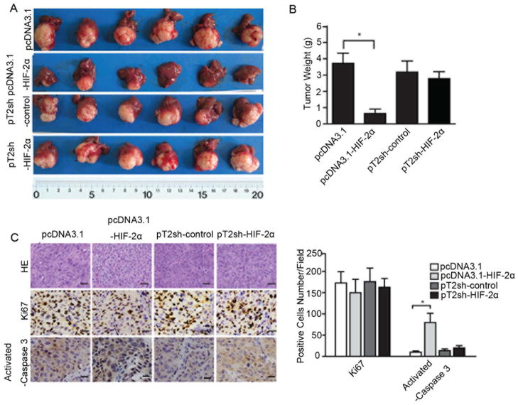
Figure 3.
HIF-2α-induced HCC growth arrest and high apoptosis rate in vivo. Monoclonal cells with either high or low HIF-2α expression were implanted into the livers of nude mice (4 weeks old). Tumor tissues were harvested 6 weeks after implantation. (A) Livers bearing tumors formed by implanted cells with different expression levels of HIF-2α. (B) Weights of the tumors in livers of nude mice. Error bars indicate standard deviation (n=6). *, p<0.01, two-tailed test. (C) Representative images of immunohistochemical staining of KI67 and activated caspase 3 in tumor xenografts, with counterstaining by hematoxylin and eosin. Bar = 2 mm.

