Abstract
Purpose
The purpose of this study was to compare kinetic and knee kinematic measurements from male and female ACL-intact (ACLINT) and ACL-reconstructed (ACLREC) subjects during a jump-cut maneuver using biplanar videoradiography.
Methods
Twenty subjects were recruited; 10 ACLINT (5 males, 5 females) and 10 ACLREC (4 males, 6 females; five years post surgery). Each subject performed a jump-cut maneuver by landing on a single leg and performing a 45° side-step cut. Ground reaction force was measured by a force plate and expressed relative to body weight. Six-degree-of-freedom knee kinematics were determined from a biplanar videoradiography system and an optical motion capture system.
Results
ACLINT female subjects landed with a larger peak vertical GRF (p<0.001) compared to ACLINT male subjects. ACLINT subjects landed with a larger peak vertical GRF (p≤0.036) compared to ACLREC subjects. Regardless of ACL reconstruction status, female subjects underwent less knee flexion angle excursion (p=0.002) and had an increased average rate of anterior tibial translation (0.05±0.01%/millisecond; p=0.037) after contact compared to male subjects. Furthermore, ACLREC subjects had a lower rate of anterior tibial translation compared to ACLINT subjects (0.05±0.01%/millisecond; p=0.035). Finally, no striking differences were observed in other knee motion parameters.
Conclusion
Women permit a smaller amount of knee flexion angle excursion during a jump-cut maneuver, resulting in a larger peak vertical GRF and increased rate of anterior tibial translation. Notably, ACLREC subjects also perform the jump cut maneuver with lower GRF than ACLINT subjects five years post surgery. This study proposes a causal sequence whereby increased landing stiffness (larger peak vertical GRF combined with less knee flexion angle excursion) leads to an increased rate of anterior tibial translation while performing a jump-cut maneuver.
Keywords: kinematics, kinetics, landing stiffness, ground reaction force, anterior tibial translation, biplanar videoradiography
INTRODUCTION
Injuries to the anterior cruciate ligament (ACL) are commonly associated with sport maneuvers involving jumping, landing, and cutting (16). These maneuvers result in a sudden loading of the ACL due to the deceleration of the tibia that occurs after landing but just prior to a rapid direction change (17). Approximately 70% of ACL injuries occur during deceleration maneuvers without contact from another athlete (23). Although males suffer non-contact deceleration injury, females are reported to be up to ten times more prone when participating in the same high-risk activities (19). Although many theories exist, the ACL failure mechanism and the associated gender bias remain unclear.
During normal function, the ACL restrains excessive anterior tibial translation and stabilizes secondary knee rotations (i.e., internal/external and abduction/adduction) (22). ACL reconstruction has become the gold standard of treatment for athletes with an ACL tear in an attempt to restore joint stability and to return patients to a high functional level (13). Unfortunately, of the 400,000 patients that undergo ACL reconstruction in the United States each year, up to 5% are at risk for re-injury (40), 45% fail to return to their pre-injury sport level (5), and 80% to 90% will develop radiographic evidence of osteoarthritis even as early as seven years post surgery (20).
Given the unexplained greater risk of non-contact deceleration ACL injury in female subjects, any differences between gender and ACL reconstruction status in the kinematic and kinetic factors during associated sport activities may point to root causes for injury, re-injury, and avenues for prevention and rehabilitation. Unfortunately, the biomechanics of male and female ACL-intact (ACLINT) and ACL-reconstructed (ACLREC) knees during high risk non-contact deceleration activities, such as a jump-cut maneuver, are not well understood. These data have previously been difficult to obtain, in part, because non-invasive measurement of kinematics has been limited to optical motion capture (OMC), which depend on surface markers that are prone to artifact from soft tissue oscillation immediately following landing (24).
Biplanar videoradiography, however, allows for direct measurement of in vivo bone motion, circumventing the effect of soft tissue artifact (14,28,29,33,34, 36, 37). Biplanar videoradiography has recently been used to study dynamic ACLINT and ACLREC knee motion during running (33,34), two-legged drop landings (28,29,36,37), and single-leg hopping (14). While these studies have made significant contributions to our understanding of both ACLINT and ACLREC knee function during running, drop landing, and hopping, the combined jump-cut maneuver, which is more commonly associated with non-contact deceleration ACL injury, has not been investigated (15,17). Additionally, the biomechanics of ACLREC subjects during these other dynamic tasks were investigated between 4 and 12 months after surgery (14,33,34). While these time points are crucial for quantifying the immediate effects of ACL reconstruction, understanding the biomechanics of the knee more than five years after surgery may provide further insight into the long-term recovery process.
The purpose of this study was to compare force plate kinetic data and knee kinematic measurements from male and female ACLINT and ACLREC recreational athletes during a jump-cut maneuver in hopes differences would point to plausible risk factors for injury. Knee kinematic measurements were primarily obtained from biplanar videoradiography; however, knee flexion/extension outside the field of view of the biplanar videoradiography system was obtained from traditional optical motion capture. The specific aims were to determine differences due to both gender and ACL reconstruction status between ACLREC patients who were at least five years post-surgery, and ACLINT control subjects. More specifically, it was anticipated that ACLINT women would tend to perform the jump-cut maneuver more upright with more landing stiffness than ACLINT men. This would be evident as decreased knee flexion angle excursion and increased peak ground reaction force (GRF), relative to their body weight, resulting in greater tibial translation (particularly anterior). In contrast, it was not known whether or not ACLREC females and males five years post reconstruction would follow a similar pattern, or if their injury and subsequent repair and rehabilitation would have resulted in altered kinetic and kinematic parameters (tested as an interaction between gender and ACL reconstruction status).
METHODS
Subjects
All experimental procedures were approved by the Institutional Review Board. Twenty recreational athletes were enrolled in this study. Of these subjects, 10 were ACLINT (5 males, 5 females) and 10 were ACLREC (4 males, 6 females; 7 bone-patellar tendon-bone autografts, 3 hamstring tendon autografts). Age, weight, and height for all subjects are displayed in Table 1. The inclusion criteria for the ACLINT subjects were: 1, no history of lower extremity injury; 2, no neurological disease(s); 3, no pregnancy; and 4, a Tegner activity score of five of greater (35). It should be noted that the ACLINT subjects were part of a separate study investigating the effects of soft tissue artifact on kinematic outcomes during a combined jump-cut maneuver (24). The inclusion criteria for the ACLREC patients were: 1, unilateral ACL reconstruction using bone-patellar tendon-bone or four-stranded hamstring tendon autograft (looped semitendinosus and gracilis); 2, at least five years post ACL reconstruction; 3, no systemic infection; 4, no neurological disease(s); 5, no pregnancy; and 6, a Tegner activity score of five or greater. The ACL reconstruction surgery type was confirmed from patient records. After granting their informed consent, each subject was outfitted with 23 retro-reflective surface markers on a single leg to permit measurement of foot, shank, and thigh motion using OMC (10). The outfitted leg was chosen at random for the ACLINT subjects (6L and 4R). For the ACLREC subjects, the ACL reconstructed leg was outfitted (7L and 3R).
Table 1.
ACLINT and ACLREC male and female age, mass, and height demographics. Ages are in years, mass is in kilograms, and height is in centimeters. A two-way analysis of variance was performed on each set of demographics data, and no statistically significant differences (p≤0.05) were found between gender and condition. Statistically significant differences (p≤0.05) are highlighted using a dark gray background fill.
| PARAMETER | ACL STATUS | MEAN | SEM | GENDER | MEAN | SEM | INTERACTION | MEAN | SEM | |||
|---|---|---|---|---|---|---|---|---|---|---|---|---|
|
AGE (YEARS) |
ACLINT | 25.20 | 1.64 | p=0.464 | M | 27.53 | 1.74 | p=0.235 | INT × M | 25.80 | 2.32 | p=0.481 |
| INT × F | 24.60 | 2.32 | ||||||||||
| ACLREC | 26.96 | 1.67 | F | 24.63 | 1.57 | REC × M | 29.25 | 2.59 | ||||
| REC × F | 24.67 | 2.12 | ||||||||||
|
MASS (KG) |
ACLINT | 73.16 | 2.93 | p=0.242 | M | 84.88 | 3.11 | p<0.001 | INT × M | 81.53 | 4.15 | p=0.617 |
| INT × F | 65.07 | 4.15 | ||||||||||
| ACLREC | 78.26 | 2.99 | F | 66.55 | 2.81 | REC × M | 88.49 | 4.64 | ||||
| REC × F | 68.03 | 3.79 | ||||||||||
|
HEIGHT (CM) |
ACLINT | 172.85 | 2.09 | p=0.736 | M | 178.70 | 2.21 | p=0.003 | INT × M | 177.40 | 2.95 | p=0.605 |
| INT × F | 168.30 | 2.95 | ||||||||||
| ACLREC | 173.88 | 2.13 | F | 168.03 | 2.00 | REC × M | 180.00 | 3.30 | ||||
| REC × F | 167.75 | 2.70 | ||||||||||
Jump-Cut Maneuver
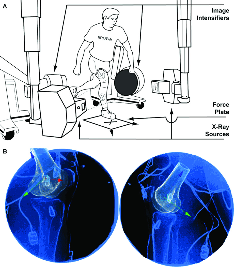
Each subject performed a jump-cut maneuver that was adapted from Ford et al (15), and previously described in detail (24). Briefly, three targets were placed on the floor within the testing environment (Figure 1A). The first target was located in the center of a force plate (Kistler model 9281B, Amherst, NY, USA). The other two targets were placed toward the left and right of the landing target at an angle of 45°. Before beginning the maneuver, the subject was asked to stand approximately one meter from the force plate with their knees bent approximately 45°. Upon hearing a verbal “GO” prompt, the subject jumped upward and forward toward the first landing target. At the same time as the verbal “GO” prompt, a visual directional prompt, left (L) or right (R), cued the subject as to which direction to cut after landing on the target with one leg. Upon landing, the subject performed a sidestep cut and then jogged past the respective angled targets. For example, if a subject was prompted to cut to the left they would land, cut, and push-off with their right leg. A trial was excluded if the subject incorrectly performed the jump-cut maneuver (landing outside the target area, incorrect cut direction, crossover cut, etc…). A total of ten correctly executed trials were performed, and the subject was unaware of the directional prompt prior to a given trial.
Figure 1.
A, illustration depicting the experimental set-up used to capture both biplanar videoradiography and OMC data during a jump-cut maneuver. A screen directly in front of the subject prompted them with the directional arrow. The subject would land and cut in the direction they were prompted using the opposite leg. For example, if prompted with the left arrow, the subject would land and cut to the left using their right leg. The four OMC cameras are not displayed in this figure; however, they were positioned to capture the retro-reflective markers shown on the subject’s right leg. B, example frame from the Autoscoper markerless tracking software. Each view represents one frame from each of the two videoradiographs generated from the two image intensifiers (Figure 1A). The blue and black portions of the images represent the actual radiographs. The orange femur represents the DRR. Both the DRR and videoradiographs have been enhanced with a sobel edge detection filter and a contact filter. This was done to create a strong visual match between the DRR and actual radiograph. The translational manipulator is shown. This manipulator allowed the user to translate the DRR within the 3-D environment. A rotational manipulator was also available to the user. The DRR is shown here after performing markerless registration. The knee shown in this image is from one of the ACLREC subjects. Both interference screws are visible in the femur and tibia.
Data Collection and Processing
The jump-cut maneuvers were carried out and kinetic and kinematic data were gathered in the W.M. Keck Foundation XROMM Facility at Brown University (Providence, RI, USA; http://www.xromm.org). A four camera OMC system (Qualisys Oqus 5, Gothenburg, Sweden) was used to track the retro-reflective surface markers (10 mm diameter) on each subject’s outfitted leg during the entire jump-cut maneuver at a capture rate of 250 Hz. A force plate (Kistler model 9281B, Amherst, NY, USA) was used to measure the GRF at 5,000 Hz. The biplanar videoradiography system was engaged for a maximum of six trials and measured motion at 250 Hz within a restricted field of view (FOV) above the force plate (26). This was done to reduce radiation exposure and maximize the likelihood that the jump-cut maneuver occurred within the FOV of the biplanar videoradiography system. All devices were time synchronized. Image de-distortion and 3-D space calibration followed established protocols using custom MATLAB software (XrayProject, Brown University, Providence, RI, USA; http://www.xromm.org) (9).
Additionally, a single static clinical computed tomography (CT) scan was collected for each subject’s outfitted knee. Image volumes were captured in the axial plane at 80 kVp while using GE’s SMART mA and Bone Plus reconstruction algorithms. The voxel resolution (slice thickness and in-plane resolution) for each scan was less than 0.625-0.465-0.465 mm3. The voxels corresponding to the femur and tibia were isolated from each CT volume using previously described methods (25) implemented in commercially available image segmentation software (Mimics v14, Materialise, Ann Arbor, MI, USA).
Custom markerless tracking software (Autoscoper, Brown University, Providence, RI, http://www.xromm.org) was used to process the biplanar videoradiography data (26). Briefly, isolated CT volumes for the femur and tibia were input into a virtual 3-D environment containing the biplanar videoradiography sequences and their calibration information. Digitally reconstructed radiographs (DRRs) were generated from the CT volumes, and the kinematic transforms from CT space to each radiograph frame were determined after optimally matching the DRRs with the two views from the biplanar videoradiography system (Figure 1B). It has previously been shown that in vivo bone motion can be determined within 0.25 mm and 0.25° using these methods (7,26). Furthermore, the rotational and translational tracking precision for this study was estimated at 0.08° and 0.45 mm, respectively.
The retroreflective marker data from the OMC system were filtered using a digital low-pass Butterworth filter with a cutoff frequency of 25 Hz. The kinematic transforms of the femur and tibia obtained from the biplanar videoradiography system were converted into quaternions. A quaternion is represented by four parameters that can be filtered (12). A digital Butterworth filter with a 25 Hz cutoff frequency was applied to the three kinematic translation parameters and the four quaternion parameters. The filtered quaternion parameters were converted back to rotation matrices and recombined with the filtered kinematic translations. The GRF data was filtered using a digital Butterworth filter with a 100 Hz cutoff frequency.
Data Analysis
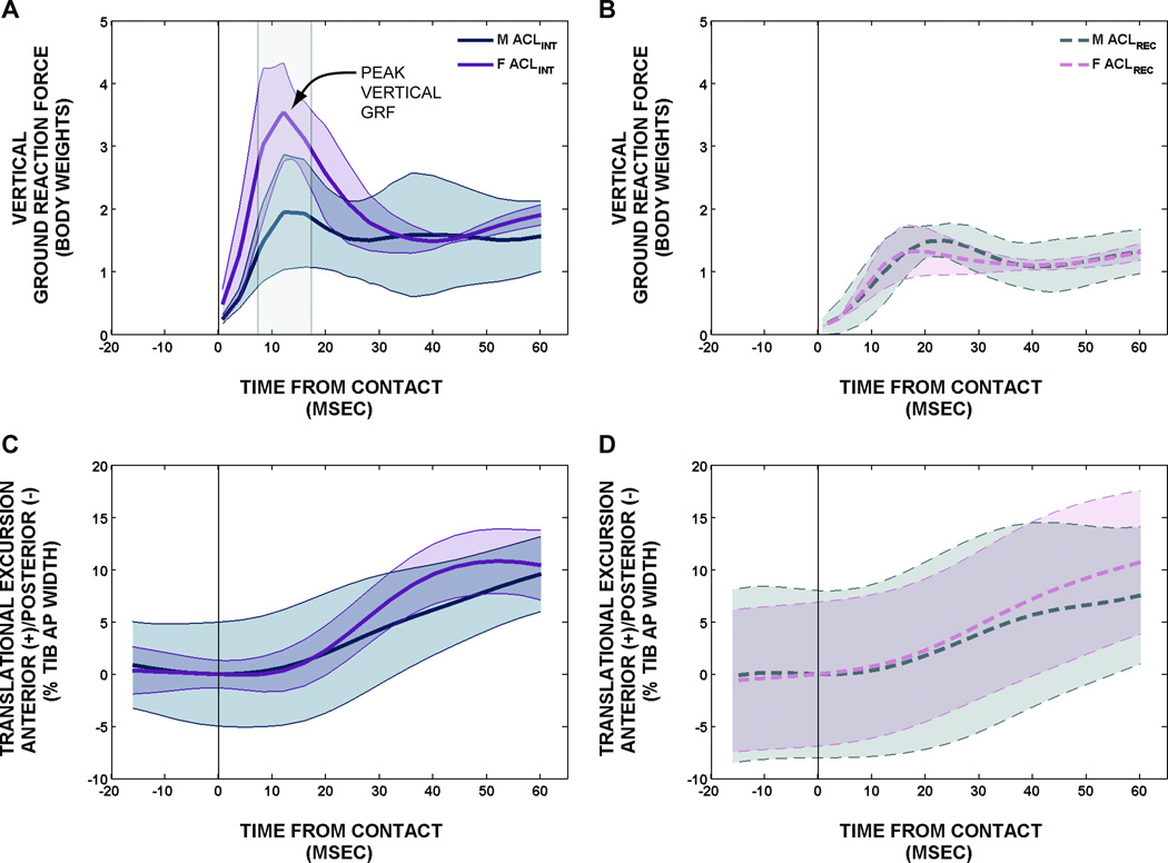
For comparison between subjects, the vertical GRF was normalized by body weight. A characteristic peak (Figure 2A) was observed in the vertical GRF within the first 25 milliseconds. This peak vertical GRF was quantified by its time after contact (peak vertical GRF time) and its magnitude (peak vertical GRF magnitude).
Figure 2.
A, ACLINT vertical GRF. B, ACLREC vertical GRF. Each subject’s GRF was normalized by their respective weight. Thus, vertical GRF units are in body weights. Notice the highlighted peak in the ACLINT vertical GRF graph. All curves are displayed as mean ± 1 SD. The vertical line on each graph represents the time at contact. C, ACLINT AN/PO translational excursion. D, ACLREC AN/PO translational excursion. Anterior is positive and posterior is negative. All AN/PO translations were normalized for each subject by their respective tibial plateau width. Thus, translational units are defined as a percent of the total tibial width in the anterior-posterior direction. It should be noted that the AN/PO translational excursion data were obtained from the biplanar videoradiography system.
The kinematics of the tibia with respect to the femur were described for both OMC and biplanar videoradiography data sets using two independent anatomical coordinate systems (ACSs). These ACSs were determined from the 3-D CT models of the femur and tibia using previously described methods (25). In order to use the same ACSs for both OMC and biplanar videoradiography, their global coordinate spaces were co-registered using a rigid lattice containing spherical markers that were radio-opaque and retro-reflective (24). The mean and standard deviation for the root mean square fit error of the co-registration transforms was 0.31±0.09 mm.
Knee joint rotations in flexion/extension (FL/EX), adduction/abduction (AD/AB), and internal/external (IN/EX) rotations of the tibia relative to the femur were interpreted using the method described by Grood and Suntay (18). Joint translations in medial/lateral (ME/LA) and anterior/posterior (AN/PO) displacements of the tibia relative to the femur were determined by a vector originating at the origin of femoral ACS and terminating at the origin of the tibial ACS (14). The ME/LA and AN/PO translations were normalized for each subject according to the ME/LA or AN/PO width of their tibial plateau, similar to the method reported by Tanifuji et al. (32). These translations are interpreted as percent ME/LA or AN/PO tibial plateau width. Normalization was performed in order to make kinematic evaluations on individuals of different sizes.
Due to the limited field of view (FOV) of the biplanar videoradiography system, the joint rotations and translations were time normalized from 16 milliseconds prior to contact to 60 milliseconds after contact. This window was selected since it was common to all subjects for at least one trial. For comparison, all joint rotations and translations were zeroed at contact and interpreted as excursion. The average rate and maximum rate of AN/PO excursion was determined for each subject. Average rate was calculated as the total range divided by the change in time, and the maximum rate was calculated as the maximum time derivative. Additionally, the area under the curve (AUC), which simplifies time-series curve comparisons, was calculated for each time-series kinematic excursion trace by integrating the signal with respect to time.
The FOV of the OMC system is significantly larger than that of the biplanar videoradiography system, permitting the measurement of knee FL/EX angle outside the time period containing the biplanar videoradiography data. Despite the soft tissue artifact observed in secondary rotations (AB/AD, IN/EX rotation) and translations (ME/LA, AN/PO) obtained from OMC, FL/EX remains relatively unaffected (24). Using the OMC data, the minimum flexion angle after contact was determined. Additionally, the change from minimum flexion angle to maximum flexion angle after contact was calculated and interpreted as excursion. The OMC data were presented for only knee joint FL/EX. The biplanar videoradiography data are presented for all other kinematic parameters (AD/AB and IN/EX rotations, ME/LA and AN/PO translations).
The described kinematic and GRF outcomes were determined for each applicable subject trial, and then all trials were ensemble averaged for each subject. Comparisons between gender (M and F) and ACL reconstruction status (ACLINT and ACLREC) were made for all kinematic and kinetic variables using two way analyses of variance. These tests were performed with a significance level (alpha) of 0.05. Pairwise multiple comparisons were made using the Holm-Sidak method when a significant gender and ACL reconstruction status interaction was determined. The Holm-Sidak method maintains alpha at 0.05 across a set of hypothesis tests and adjusts p-values differently depending on their values ranked against each other. This is effective at maintaining alpha and avoiding beta inflation.
RESULTS
A statistically significant interaction (p=0.003) between gender and ACL reconstruction status was observed for the peak vertical GRF (Figure 2). Within the ACLINT subjects, the females had a peak vertical GRF that was 1.45 body weights larger than the male subjects (p<0.001). In contrast, the ACLREC male and female subjects had nearly equal peak vertical GRFs. The ACLREC male subjects’ peak vertical GRF was 0.22 body weights larger than the female ACLREC subjects but was not statistically significant (p=0.522). When comparing within ACL reconstruction status, both the male and female ACLINT subjects had a larger peak vertical GRF than the male and female ACLREC subjects, respectively. The male ACLINT subjects’ peak vertical GRF were 0.80 body weights larger than the male ACLREC subjects (p=0.036), and the female ACLINT subjects’ peak vertical GRF were 2.46 body weights larger than the female ACLREC subjects (p<0.001).
The peak vertical GRF for the female subjects occurred 6.24 milliseconds earlier than the male subjects (p=0.021). The interaction between gender and ACL reconstruction status approached, but was not statistically significant (p=0.117); the peak vertical GRF for the female ACLREC subjects occurred only 2.21 milliseconds before the male ACLREC subjects. Conversely, the peak vertical GRF for the female ACLINT subjects occurred 10.27 milliseconds before the male ACLINT subjects. Moreover, the peak vertical GRF appears to occur earlier in ACLINT subjects than ACLREC subjects (4.80 milliseconds; p=0.066).
The average rate of AN/PO translational excursion (Figure 2) was 0.05 %/millisecond larger for ACLINT subjects compared to ACLREC subjects (p=0.035), and 0.05 %/millisecond larger for female subjects compared to male subjects (p=0.037). The maximum rate of AN/PO translational excursion was 0.13 %/millisecond larger in female ACLINT subjects as compared to male ACLINT subjects; however, the difference between genders in ACLREC subjects was only 0.01 %/millisecond. Pairwise multiple comparisons revealed that maximum AN/PO translational excursion rate differences were significant for males versus females within ACLINT subjects (p=0.027) and for ACLINT versus ACLREC within female (p=0.007). Additionally, the AUC of the AN/PO translational excursion was observed to be 55 %∙millisecond larger for ACLINT subjects than ACLREC subjects (p=0.180). The AUC for the female subjects was also larger than the AUC for the male subjects (64 %∙millisecond; p=0.122). No significant interaction between gender and ACL reconstruction status was observed (p=0.961). However, the AUC of the AN/PO translational excursion was observed to be 66 %∙millisecond larger for female ACLINT subjects than for male ACLINT subjects, and the AUC for the female ACLREC subjects was also larger than the AUC for the male ACLREC subjects (62.1 %∙millisecond). These AN/PO translational kinematic data were determined from the biplanar videoradiography system.
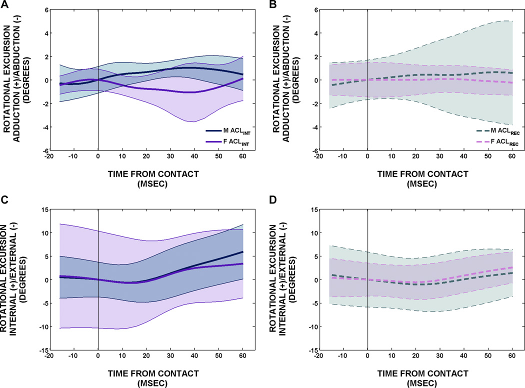
The AD/AB rotational excursion (Figure 3) was relatively constant after contact, changing less than 2 degrees for both male and female ACLREC and ACLINT subjects. Despite the minimal rotational change after contact, the female subjects were abducting (valgus) slightly after contact while the male subjects were adducting (varus) slightly after contact (average female abduction excursion equal to 1.02 degrees, average male adduction excursion equal to 1.07 degrees; p=0.033). The IN/EX rotational excursion (Figure 3) for all subjects followed a consistent pattern. Specifically, the male and female ACLINT and ACLREC subjects all began internally rotating after contact. While no statistically significant differences were observed in any group, the ACLINT male and female subjects had a 76 larger AUC than the ACLREC male and female subjects (p=0.171). These AD/AB and IN/EX rotational kinematic data were determined from the biplanar videoradiography system.
Figure 3.
A, ACLINT AD/AB rotational excursion. B, ACLREC AD/AB rotational excursion. Adduction is positive and abduction is negative. C, ACLINT IN/EX rotational excursion. D, ACLREC IN/EX rotational excursion. All rotational excursion units are in degrees. All curves are displayed as mean ± 1 SD. The vertical line on each graph represents the time at contact. It should be noted that these data were obtained from the biplanar videoradiography system.
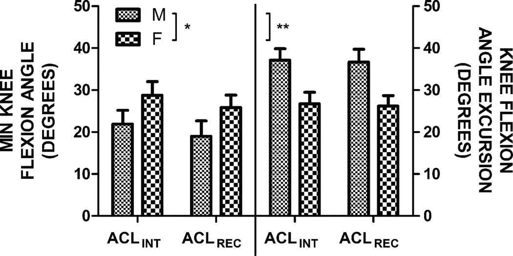
The minimum flexion angle occurred at or immediately following ground contact. Following this, all of the subjects absorbed the landing and continued the cut by flexing through stance phase to a maximum flexion angle. Using the OMC data to quantify the minimum flexion angle, maximum flexion angle, and the flexion angle excursion (change from minimum to maximum flexion angle), we observed that females tended to be more flexed at contact (p=0.054); but their total excursion was significantly less (p=0.002) (Figure 4). These FL/EX kinematic data were determined from the OMC system.
Figure 4.
Left y-axis, minimum knee flexion angle for ACLINT and ACLREC male and female subjects. Minimum flexion occurred at or immediately following ground contact. No statistically significant differences between gender and condition were observed for minimum knee flexion angle values; however, the * represents a p-value of 0.054 denoting an apparent gender difference. Right y-axis, knee flexion angle excursion for ACLINT and ACLREC male and female subjects. Knee flexion angle excursion was defined as the change in knee flexion angle from minimum flexion to maximum flexion. A statistically significant difference was observed between male and female subjects. This is highlighted by the **, which represents a p-value of 0.002. The minimum knee flexion angle and knee flexion angle excursion units are in degrees. It should be noted that these data were obtained from the OMC system.
We have included the following data as supplemental digital content in order to provide contextual reference for the above results: 1, the knee angles (in degrees) at contact and peak vertical GRF for knee FL/EX, AD/AB, and IN/EX obtained from OMC and biplanar videoradiography (see Table A, Supplemental Digital Content 1); 2, the peak AN/PO and ME/LA knee translations in millimeters and their respective time points in milliseconds (see Table B, Supplemental Digital Content 2); and 3, the AN/PO and ME/LA knee translation excursions in millimeters (see Table C, Supplemental Digital Content 3).
DISCUSSION
We have compared knee kinematic and kinetic measurements from male and female ACLINT and ACLREC recreational athletes during a jump-cut maneuver associated with non-contact deceleration ACL injury. Two major findings were observed in our study. First, female subjects who have never had an ACL reconstruction appeared to perform the jump-cut maneuver with greater landing stiffness (smaller amount of knee flexion angle excursion combined with larger peak vertical GRF (28)) than males with or without a history of ACL reconstruction and other females with a history of ACL reconstruction. This was evidenced by the differences observed in the knee flexion angle excursion, which translated to qualitatively comparable differences in peak vertical GRF. Second, the male and female ACLREC subjects appear to perform the jump-cut maneuver with less energy than the ACLINT subjects, resulting in a lower peak vertical GRF even five years or more after their reconstruction. This may be a result of differences in strength, confidence, habit, and/or training following their injury.
These kinetic differences likely influence the differences observed in the rate of anterior tibial translation after contact, which is a common instigator of ACL injury. Specifically, we observed that anterior tibial translation increased at a faster rate in female ACLINT subjects compared to their male ACLINT counterparts (Figure 2). Notably, peak anterior tibial translation for the ACLINT female subjects occurred within 60 milliseconds. A similar peak is not observed in the male ACLINT subjects or the male and female ACLREC subjects. Interestingly, the time to peak vertical GRF was significantly less in female subjects as compared to male subjects. Also, the time to peak vertical GRF appears to be smaller in ACLINT subjects as compared to ACLREC subjects. The increased rate of anterior tibial translation observed in female ACLINT subjects is likely a result of the larger and more rapid peak vertical GRF observed immediately after ground contact. This rapid and large peak vertical GRF appears to produce a ‘snapping’ motion that differs from the more gradual increase in peak vertical GRF and anterior tibial translation observed in male ACLINT subjects and male and female ACLREC subjects. It may be that there is a reliable tendency for females to absorb less energy upon landing, which, through greater peak vertical GRF, resultant forces, and/or abnormal kinematics may increase the risk for ACL injury.
Previous research has suggested that increased landing stiffness, as characterized by a smaller amount of knee flexion angle excursion combined with a high vertical GRF during landing and cutting activities, place individuals at increased risk of ACL injury (6,8,11). Attempts have been made to correlate increased landing stiffness with increased anterior tibial translation with the goal of developing knee injury prevention training and rehabilitation programs (28). During the jump-cut maneuver in our study, the female subjects landed and cut with less knee flexion angle excursion after contact. This result, when interpreted in the context of the faster and larger peak vertical GRF, confirms that the females are performing the jump-cut maneuver with more landing stiffness. This finding is in contrast to the observations made for the male subjects, who appear to be absorbing the energy they are applying at ground contact by flexing through the landing and subsequent cut. Moreover, the AN/PO translation never reached a maximum (within 60 milliseconds after contact) and increased at a lower rate after contact for the male ACLINT and male and female ACLREC subjects. This combination of increased knee flexion angle excursion and/or reduced peak vertical GRF (decreased landing stiffness) may contribute to the slower time to peak anterior tibial translation after contact for these subjects.
In a similar study investigating the knee kinematics of ACLINT females during four functional tasks, Myers et al observed that anterior tibial translation was increased in activities of increasing external loading (29). Specifically, they observed a 2.4 mm increase in anterior tibial translation during landing maneuvers as compared to walking. Moreover, their results show the same characteristic peak vertical GRF immediately following ground contact during the landing tasks. This peak is absent in the vertical GRF walking trace. Conversely, another study by Myers et al showed no differences in anterior tibial translation between soft and stiff drop landings (28). The authors attributed these findings to the ability of the ligaments and musculature about the knee to keep joint translations within a safe envelope of motion during controlled activities where external loading conditions are anticipated. While no excessive rotational or translation motion was observed in our study, the increased rate of anterior translation in female ACLINT subjects suggests that stiffer landings under more unanticipated cutting activities may affect kinematic translations more than controlled, anticipated activities. Furthermore, the lower rate of anterior tibial translation seen in the ACLREC subjects immediately following contact may be influenced by the lower peak vertical GRF.
This low peak vertical GRF observed in both male and female ACLREC subjects matches results from Paterno et al (30,31) and Vairo et al (38). In two separate studies, Paterno et al showed ACLREC male and female subjects decreased peak vertical GRF when performing landing activities two years after reconstruction. Vairo et al reported decreased vertical peak GRF upon landing from a vertical drop among ACLREC subjects approximately two years post surgery. These results are consistent with those reported in our study for ACLREC men and women who are at least five years post-reconstruction. Additionally, no gender differences were observed in the peak GRF within ACLREC subjects. Even after five years of strengthening activities, including formal rehabilitation, functional exercise and return to sports, the ACLREC subjects performed the jump-cut maneuver with less energy compared to the ACLINT subjects. While the exact mechanisms for this are unknown, it is possible that both behavioral and neuromechanical deficiencies are present in the ACLREC subjects when performing jump-cut maneuvers on their previously injured limb. Alternatively, it is possible that the altered mechanics are a result of protective habits obtained during the ACLREC subjects’ rehabilitation. Additional research investigating neuromuscular activity and contrallateral biomechanics may provide additional insight into the reduced vertical GRF observed in both male and female ACLREC subjects.
In addition to the larger peak vertical GRF and rate of anterior tibial translation, the female subjects were generally abducting after contact (Figure 3A–B). This is an interesting finding because videographic studies have suggested that a valgus (abduction) collapse is involved in the non-contact deceleration ACL injury mechanism (21). Furthermore, the results presented herein are consistent with previous reports suggesting that females land with more knee abduction compared to males (15). While the female subjects in our study were abducting after contact compared to the male subjects, the total amount of abduction (< 2 degrees) does not seem to correspond to a valgus collapse position. This may be a result of the subjects’ ability to safely perform the jump-cut maneuver, which was implemented in our study to challenge the ACL. A valgus collapse position was not observed, neither were any adverse events (injury).
No significant differences were observed between gender and ACL reconstruction status for both IN/EX rotation or ME/LA translation after contact (Table 2). In general, all subjects began internally rotating after contact and remained stable in the ME/LA direction. With exception to the gender difference observed in AD/AB angle, the similar kinematic outcomes observed between gender and ACL reconstruction status after contact does not support our hypothesis. Deneweth et al showed that, as compared to the ACLINT contralateral knees, ACLREC knees were more externally rotated and less laterally translated during a single-leg hopped landing (14). Unfortunately, obtaining contralateral limb kinematics was not feasible for our study. This makes direct comparisons difficult; however, Deneweth et al does report total IN/EX excursion to be approximately five degrees and total ME/LA translation to be less than 1 mm for both reconstructed and contralateral knees. These excursion values align with the results presented in our study.
Table 2.
ACLINT and ACLREC male and female kinematic AUC, rate, and peak vertical GRF results. A two-way analysis of variance was performed on each set of data. Statistically significant differences (p≤0.05) are highlighted using a dark gray background fill. Near statistical significance (p≤0.10) is highlighted using a light gray background fill. Pair-wise comparisons were made using the Holm-Sidak method when a significant (or near significant) gender and ACL reconstruction status interaction was determined. These results, denoted by the * or **, are shown in the bottom row of the table. The data presented in the parentheses (ME/LA and AN/PO AUC, AN/PO rates) are the raw translational values in millimeters. Finally, it should be noted that the kinematic data presented in Table 2 were obtained from the biplanar videoradiography system. Supplemental Digital Content 1 *.pdf
| OUTCOME | ACL STATUS | MEAN | SEM | GENDER | MEAN | SEM | INTERACTION | MEAN | SEM | |||
|---|---|---|---|---|---|---|---|---|---|---|---|---|
|
AD\AB EXCURSION AUC DEG·MSEC |
ACLINT | −5.18 | 13.74 | p=0.406 | M | 26.03 | 14.58 | p=0.033 | INT × M | 25.88 | 19.43 | p=0.415 |
| INT × F | −36.24 | 19.43 | ||||||||||
| ACLREC | 11.57 | 14.03 | F | −19.65 | 13.16 | REC × M | 26.19 | 21.73 | ||||
| REC × F | −3.05 | 17.74 | ||||||||||
|
IN\EX EXCURSION AUC DEG·MSEC |
ACLINT | 76.59 | 32.91 | p=0.171 | M | 38.06 | 34.21 | p=0.815 | INT × M | 86.81 | 45.61 | p=0.505 |
| INT × F | 66.37 | 45.61 | ||||||||||
| ACLREC | 10.59 | 32.25 | F | 49.04 | 30.88 | REC × M | −10.63 | 50.99 | ||||
| REC × F | 31.71 | 41.63 | ||||||||||
|
ME\LA EXCURSION AUC %·MSEC (MM·MSEC) |
ACLINT | 6.56 | 12.65 | p=0.153 | M | −17.35 | 13.41 | p=0.269 | INT × M | −6.85 (−5.72) | 17.89 (13.16) | p=0.739 |
| (4.06) | (9.31) | (−14.25) | (9.87) | INT × F | 19.97 (13.83) | 17.89 (13.16) | ||||||
| ACLREC | −20.57 | 12.91 | F | 3.34 | 12.11 | REC × M | −27.84 (22.79) | 20.00 (14.72) | ||||
| (−15.92) | (9.50) | (2.319) | (8.91) | REC × F | −13.292 (−9.20) | 16.33 (12.02) | ||||||
|
AN\PO EXCURSION AUC %·MSEC (MM·MSEC) |
ACLINT | 303.85 | 27.417 | p=0.180 | M | 244.38 | 29.08 | p=0.122 | INT × M | 270.85 (150.74) | 38.77 (21.33) | p=0.961 |
| (162.80) | (15.08) | (139.89) | (15.99) | INT × F | 336.84 (174.85) | 38.77 (21.33) | ||||||
| ACLREC | 248.96 | 27.98 | F | 308.42 | 26.25 | REC × M | 217.91 (129.04) | 43.35 (23.84) | ||||
| (138.56) | (15.39) | (161.47) | (14.44) | REC × F | 280.01 (148.08) | 35.40 (19.47) | ||||||
|
AN\PO AVERAGE RATE %/MSEC (MM/MSEC) |
ACLINT | 0.22 | 0.02 | p=0.035 | M | 0.17 | 0.02 | p=0.037 | INT × M | 0.18 (0.10) | 0.02 (0.01) | p=0.213 |
| (0.12) | (0.01) | (0.10) | (0.01) | INT × F | 0.26 (0.13) | 0.02 (0.01) | ||||||
| ACLREC | 0.17 | 0.02 | F | 0.22 | 0.01 | REC × M | 0.16 (0.10) | 0.02 (0.01) | ||||
| (0.10) | (0.01) | (0.12) | (0.01) | REC × F | 0.18 (0.10) | 0.02 (0.01) | ||||||
|
AN\PO MAXIMUM RATE %/MSEC* (MM/MSEC) |
ACLINT | 0.39 | 0.03 | p=0.035 | M | 0.31 | 0.03 | p=0.135 | INT × M | 0.32 (0.18) | 0.04 (0.02) | p=0.087 |
| (0.21) | (0.01) | (0.18) | (0.02) | INT × F | 0.45 (0.23) | 0.04 (0.02) | ||||||
| ACLREC | 0.30 | 0.03 | F | 0.37 | 0.03 | REC × M | 0.30 (0.18) | 0.04 (0.02) | ||||
| (0.17) | (0.02) | (0.19) | (0.01) | REC × F | 0.29 (0.16) | 0.03 (0.02) | ||||||
|
PEAK VIRT GRF MAG BW** |
ACLINT | 1.53 | 0.17 | NA | M | 2.04 | 0.17 | NA | INT × M | 2.44 | 0.23 | p=0.003 |
| INT × F | 3.88 | 0.23 | ||||||||||
| ACLREC | 3.16 | 0.16 | F | 2.65 | 0.16 | REC × M | 1.64 | 0.26 | ||||
| REC × F | 1.42 | 0.21 | ||||||||||
|
PEAK VIRT GRF TIME MSEC |
ACLINT | 16.73 | 1.70 | p=0.066 | M | 22.25 | 1.80 | p=0.021 | INT × M | 21.87 | 2.40 | p=0.117 |
| INT × F | 11.60 | 2.40 | ||||||||||
| ACLREC | 21.53 | 1.74 | F | 16.01 | 1.63 | REC × M | 22.63 | 2.69 | ||||
| REC × F | 20.42 | 2.19 | ||||||||||
MvF in I: p=0.027; MvF in R: p=0.863; IvR in M: p=0.751; IvR in F: p=0.007
MvF in I: p<0.001; MvF in R: p=0.522; IvR in M: p=0.036; IvR in F: p<0.001
Despite the kinematic similarities, additional research investigating surface interactions between the medial and lateral compartments of the knee may provide more specific insight into subtle kinematic differences between both gender and ACL reconstruction status. Specifically, methods have been developed to identify distance weighted proximity centroids, regions of closest proximity (1), and point based surface velocities (2,3). These techniques take advantage of the accuracy associated with biplanar videoradiography to make inferences about biomechanical changes at the articulating surfaces of the femur and tibia with the hope of better understanding the initiation and progression of osteoarthritis in ACLREC individuals (4).
As investigators, we are limited to studying potential injury mechanisms in a laboratory testing environment without many of the situations presented in a sporting environment. Our study investigated male and female ACLINT and ACLREC subjects while they performed an activity in a controlled laboratory setting that has been associated with non-contact deceleration ACL injury. The incorporation of the ‘unanticipated’ element to the jump-cut maneuver mimicked the deceleration and cutting action associated with many sporting events. Despite the design, studies that employ biplanar videoradiography will be hindered by the inability to capture subjects performing sports activities in their native environments.
We acknowledge the small sample size for investigating kinematic and kinetic interactions between gender and ACL reconstruction status. While we did ensure that all subjects were recreational athletes, we were not able to control for surgeon or rehabilitation protocol for the ACLREC subjects. Additionally, we recognize the limitation of using two different graft types in our study. Based on biomechanical studies (34,39) and randomized clinical trials (27), we assumed that both bone-patellar tendon-bone and four-stranded hamstring tendon grafts would respond similarly during the jump-cut maneuver studied herein but recognize this as a study limitation.
Limitations associated with biplanar videoradiography should be noted. Specifically, the field of view restricted our ability to capture kinematic data for all subjects from 16 milliseconds before contact to 60 milliseconds after contact. Previous research has shown that peak anterior tibial translation occurs between 40 milliseconds to 50 milliseconds after contact (37), within the temporal range studied. Finally, each subject received up to 22 millirem of radiation exposure as a result of the biplanar videoradiography system and CT scan. While this falls well below the guidelines instituted by the NIH Radiation Safety Committee for acceptable radiation exposure to research subjects within a year (5 rem), it does limit the number of data collection trials. All subjects were aware of and gave informed consent to radiation exposure.
In conclusion, the results presented in our study support our hypothesis that kinematic and kinetic differences would be observed between both gender and ACL reconstruction status during a jump-cut maneuver. Specifically, we found that female ACLINT subjects landed and cut with a smaller amount of knee flexion angle excursion and larger peak vertical GRF than the male ACLINT subjects. Furthermore, we observed that the ACLREC subjects had a significantly lower peak vertical GRF just after impact as compared to the ACLINT subjects. We also noted that female ACLINT subjects appear to have an increased rate of anterior tibial translation just after contact. Our study associates the increased rate of anterior tibial translation to increased landing stiffness (larger peak vertical GRF combined with smaller knee flexion angle excursion) while performing the jump-cut maneuver. With respect to AD/AB, IN/EX rotation, and ME/LA translation, differences were only observed AD/AB angle. The female subjects in our study were abducting after contact compared to the male subjects; albeit, the amount of abduction does not appear to correspond to a valgus collapse position. Finally, no significant interactions were found between gender and ACL reconstruction status for IN/EX rotation or ME/LA translation after contact.
Supplementary Material
ACKNOWLEDGEMENTS
This publication was made possible by the W.M. Keck Foundation, Grant Numbers R01-AR047910 and P20-RR02484 (COBRE) from NIAMS/NIH, and the Lucy Lippitt Endowed Professorship. Its contents are solely the responsibility of the authors and do not necessarily represent the official views of the W.M. Keck Foundation, NIAMS, NIH, or the American College of Sports Medicine. The authors greatly acknowledge the efforts of Alison M Biercevicz, Michelle M Gosselin, Joel B Schwartz, and James C Tarrent for their assistance in data collection and subject preparation.
Footnotes
CONFLICT OF INTEREST
None.
REFERENCES
- 1.Anderst WJ, Tashman S. A method to estimate in vivo dynamic articular surface interaction. J Biomech. 2003 Sep;36(9):1291–1299. doi: 10.1016/s0021-9290(03)00157-x. [DOI] [PubMed] [Google Scholar]
- 2.Anderst WJ, Tashman S. The association between velocity of the center of closest proximity on subchondral bones and osteoarthritis progression. J. Orthop. Res. 2009 Jan;27(1):71–77. doi: 10.1002/jor.20702. [DOI] [PMC free article] [PubMed] [Google Scholar]
- 3.Anderst WJ, Tashman S. Using relative velocity vectors to reveal axial rotation about the medial and lateral compartment of the knee. J Biomech. 2010 Mar 22;43(5):994–997. doi: 10.1016/j.jbiomech.2009.11.014. [DOI] [PMC free article] [PubMed] [Google Scholar]
- 4.Andriacchi TP, Mündermann A, Smith RL, Alexander EJ, Dyrby CO, Koo S. A framework for the in vivo pathomechanics of osteoarthritis at the knee. Ann Biomed Eng. 2004 Mar;32(3):447–457. doi: 10.1023/b:abme.0000017541.82498.37. [DOI] [PubMed] [Google Scholar]
- 5.Ardern CL, Taylor NF, Feller JA, Webster KE. Return-to-sport outcomes at 2 to 7 years after anterior cruciate ligament reconstruction surgery. Am J Sports Med. 2012 Jan;40(1):41–48. doi: 10.1177/0363546511422999. [DOI] [PubMed] [Google Scholar]
- 6.Beutler A, de la Motte S, Marshall S, Padua D, Boden B. Muscle strength and qualitative jump-landing differences in male and female military cadets: the jumpacl study. J Sports Sci Med. 2009;8:663–671. [PMC free article] [PubMed] [Google Scholar]
- 7.Bey MJ, Zauel R, Brock SK, Tashman S. Validation of a new model-based tracking technique for measuring three-dimensional, in vivo glenohumeral joint kinematics. J Biomech Eng. 2006 Aug;128(4):604–609. doi: 10.1115/1.2206199. [DOI] [PMC free article] [PubMed] [Google Scholar]
- 8.Boden BP, Dean GS, Feagin JA, Garrett WE. Mechanisms of anterior cruciate ligament injury. Orthopedics. 2000 Jun;23(6):573–578. doi: 10.3928/0147-7447-20000601-15. [DOI] [PubMed] [Google Scholar]
- 9.Brainerd EL, Baier DB, Gatesy SM, Hedrick TL, Metzger KA, Gilbert SL, et al. X-ray reconstruction of moving morphology (XROMM): precision, accuracy and applications in comparative biomechanics research. J Exp Zool A Ecol Genet Physiol. 2010 Jun 1;313(5):262–279. doi: 10.1002/jez.589. [DOI] [PubMed] [Google Scholar]
- 10.Buczek FL, Rainbow MJ, Cooney KM, Walker MR, Sanders JO. Implications of using hierarchical and six degree-of-freedom models for normal gait analyses. Gait & Posture. 2010 Jan;31(1):57–63. doi: 10.1016/j.gaitpost.2009.08.245. [DOI] [PubMed] [Google Scholar]
- 11.Chappell JD, Creighton RA, Giuliani C, Yu B, Garrett WE. Kinematics and electromyography of landing preparation in vertical stop-jump: risks for noncontact anterior cruciate ligament injury. Am J Sports Med. 2007 Feb;35(2):235–241. doi: 10.1177/0363546506294077. [DOI] [PubMed] [Google Scholar]
- 12.Coburn J, Crisco JJ. Interpolating three-dimensional kinematic data using quaternion splines and hermite curves. J Biomech Eng. 2005 Apr;127(2):311–317. doi: 10.1115/1.1865195. [DOI] [PubMed] [Google Scholar]
- 13.Dargel J, Gotter M, Mader K, Pennig D, Koebke J, Schmidt-Wiethoff R. Biomechanics of the anterior cruciate ligament and implications for surgical reconstruction. Strategies Trauma Limb Reconstr. 2007 Apr;2(1):1–12. doi: 10.1007/s11751-007-0016-6. [DOI] [PMC free article] [PubMed] [Google Scholar]
- 14.Deneweth JM, Bey MJ, McLean SG, Lock TR, Kolowich PA, Tashman S. Tibiofemoral joint kinematics of the anterior cruciate ligament-reconstructed knee during a single-legged hop landing. Am J Sports Med. 2010 Sep;38(9):1820–1828. doi: 10.1177/0363546510365531. [DOI] [PubMed] [Google Scholar]
- 15.Ford KR, Myer GD, Toms HE, Hewett TE. Gender differences in the kinematics of unanticipated cutting in young athletes. Med Sci Sports Exerc. 2005 Jan;37(1):124–129. [PubMed] [Google Scholar]
- 16.Gianotti SM, Marshall SW, Hume PA, Bunt L. Incidence of anterior cruciate ligament injury and other knee ligament injuries: a national population-based study. J Sci Med Sport. 2009 Nov;12(6):622–627. doi: 10.1016/j.jsams.2008.07.005. [DOI] [PubMed] [Google Scholar]
- 17.Griffin LY, Albohm MJ, Arendt EA, Bahr R, Beynnon BD, Demaio M, et al. Understanding and preventing noncontact anterior cruciate ligament injuries: a review of the Hunt Valley II meeting, January 2005. Am J Sports Med. 2006 Sep;34(9):1512–1532. doi: 10.1177/0363546506286866. [DOI] [PubMed] [Google Scholar]
- 18.Grood ES, Suntay WJ. A joint coordinate system for the clinical description of three-dimensional motions: application to the knee. J Biomech Eng. 1983 May;105(2):136–144. doi: 10.1115/1.3138397. [DOI] [PubMed] [Google Scholar]
- 19.Gwinn DE, Wilckens JH, McDevitt ER, Ross G, Kao TC. The relative incidence of anterior cruciate ligament injury in men and women at the United States Naval Academy. Am J Sports Med. 2000 Feb;28(1):98–102. doi: 10.1177/03635465000280012901. [DOI] [PubMed] [Google Scholar]
- 20.Junkin DM, Johnson DJ, Fu FH, Mark MD, Willenborn M, Fanelli GC, et al. Orthopaedic Knowledge Update: Sports Medicine 4. 4th ed. Amer Academy of Orthopaedic; 2009. Knee Ligament Injuries; pp. 135–153. [Google Scholar]
- 21.Krosshaug T, Nakamae A, Boden BP, Engebretsen L, Smith G, Slauterbeck JR, et al. Mechanisms of anterior cruciate ligament injury in basketball: video analysis of 39 cases. Am J Sports Med. 2007 Mar;35(3):359–367. doi: 10.1177/0363546506293899. [DOI] [PubMed] [Google Scholar]
- 22.Markolf KL, Kochan A, Amstutz HC. Measurement of knee stiffness and laxity in patients with documented absence of the anterior cruciate ligament. J Bone Joint Surg Am. 1984 Feb;66(2):242–252. [PubMed] [Google Scholar]
- 23.McNair PJ, Marshall RN, Matheson JA. Important features associated with acute anterior cruciate ligament injury. N. Z. Med. J. 1990 Nov 14;103(901):537–539. [PubMed] [Google Scholar]
- 24.Miranda DL, Rainbow MJ, Crisco JJ, Fleming BC. Kinematic differences between optical motion capture and biplanar videoradiography during a jump-cut maneuver. J Biomech. 2012 Oct 22; doi: 10.1016/j.jbiomech.2012.09.023. (Article in Press DOI: 10.1016/j.jbiomech.2012.09.023) [DOI] [PMC free article] [PubMed] [Google Scholar]
- 25.Miranda DL, Rainbow MJ, Leventhal EL, Crisco JJ, Fleming BC. Automatic determination of anatomical coordinate systems for three-dimensional bone models of the isolated human knee. J Biomech. 2010 May 28;43(8):1623–1626. doi: 10.1016/j.jbiomech.2010.01.036. [DOI] [PMC free article] [PubMed] [Google Scholar]
- 26.Miranda DL, Schwartz JB, Loomis AC, Brainerd EL, Fleming BC, Crisco JJ. Static and Dynamic Error of a Biplanar Videoradiography System Using Marker-Based and Markerless Tracking Techniques. J Biomech Eng. 2011 Dec;133(12):121002. doi: 10.1115/1.4005471. [DOI] [PMC free article] [PubMed] [Google Scholar]
- 27.Mohtadi NG, Chan DS, Dainty KN, Whelan DB. Patellar tendon versus hamstring tendon autograft for anterior cruciate ligament rupture in adults. Cochrane Database Syst Rev. 2011;(9):CD005960. doi: 10.1002/14651858.CD005960.pub2. [DOI] [PMC free article] [PubMed] [Google Scholar]
- 28.Myers CA, Torry MR, Peterson DS, Shelburne KB, Giphart JE, Krong JP, et al. Measurements of tibiofemoral kinematics during soft and stiff drop landings using biplane fluoroscopy. Am J Sports Med. 2011 Aug;39(8):1714–1722. doi: 10.1177/0363546511404922. [DOI] [PMC free article] [PubMed] [Google Scholar]
- 29.Myers CA, Torry MR, Shelburne KB, Giphart JE, LaPrade RF, Woo SL-Y, et al. In vivo tibiofemoral kinematics during 4 functional tasks of increasing demand using biplane fluoroscopy. Am J Sports Med. 2012 Jan;40(1):170–178. doi: 10.1177/0363546511423746. [DOI] [PubMed] [Google Scholar]
- 30.Paterno MV, Ford KR, Myer GD, Heyl R, Hewett TE. Limb asymmetries in landing and jumping 2 years following anterior cruciate ligament reconstruction. Clin J Sport Med. 2007 Jul;17(4):258–262. doi: 10.1097/JSM.0b013e31804c77ea. [DOI] [PubMed] [Google Scholar]
- 31.Paterno MV, Schmitt LC, Ford KR, Rauh MJ, Myer GD, Hewett TE. Effects of sex on compensatory landing strategies upon return to sport after anterior cruciate ligament reconstruction. J Orthop Sports Phys Ther. 2011 Aug;41(8):553–559. doi: 10.2519/jospt.2011.3591. [DOI] [PubMed] [Google Scholar]
- 32.Tanifuji O, Sato T, Kobayashi K, Mochizuki T, Koga Y, Yamagiwa H, et al. Three-dimensional in vivo motion analysis of normal knees using single-plane fluoroscopy. J Orthop Sci. 2011 Nov;16(6):710–718. doi: 10.1007/s00776-011-0149-9. [DOI] [PubMed] [Google Scholar]
- 33.Tashman S, Collon D, Anderson K, Kolowich P, Anderst W. Abnormal rotational knee motion during running after anterior cruciate ligament reconstruction. Am J Sports Med. 2004 Jun;32(4):975–983. doi: 10.1177/0363546503261709. [DOI] [PubMed] [Google Scholar]
- 34.Tashman S, Kolowich P, Collon D, Anderson K, Anderst W. Dynamic function of the ACL-reconstructed knee during running. Clin. Orthop. Relat. Res. 2007 Jan;454:66–73. doi: 10.1097/BLO.0b013e31802bab3e. [DOI] [PubMed] [Google Scholar]
- 35.Tegner Y, Lysholm J. Rating systems in the evaluation of knee ligament injuries. Clin. Orthop. Relat. Res. 1985 Sep;(198):43–49. [PubMed] [Google Scholar]
- 36.Torry MR, Myers C, Shelburne KB, Peterson D, Giphart JE, Pennington WW, et al. Relationship of knee shear force and extensor moment on knee translations in females performing drop landings: a biplane fluoroscopy study. Clin Biomech (Bristol, Avon) 2011 Dec;26(10):1019–1024. doi: 10.1016/j.clinbiomech.2011.06.010. [DOI] [PMC free article] [PubMed] [Google Scholar]
- 37.Torry MR, Shelburne KB, Peterson DS, Giphart JE, Krong JP, Myers C, et al. Knee kinematic profiles during drop landings: a biplane fluoroscopy study. Med Sci Sports Exerc. 2011 Mar;43(3):533–541. doi: 10.1249/MSS.0b013e3181f1e491. [DOI] [PubMed] [Google Scholar]
- 38.Vairo GL, Myers JB, Sell TC, Fu FH, Harner CD, Lephart SM. Neuromuscular and biomechanical landing performance subsequent to ipsilateral semitendinosus and gracilis autograft anterior cruciate ligament reconstruction. Knee Surg Sports Traumatol Arthrosc. 2008 Jan;16(1):2–14. doi: 10.1007/s00167-007-0427-4. [DOI] [PubMed] [Google Scholar]
- 39.Wilson TW, Zafuta MP, Zobitz M. A biomechanical analysis of matched bonepatellar tendon-bone and double-looped semitendinosus and gracilis tendon grafts. Am J Sports Med. 1999 Apr;27(2):202–207. doi: 10.1177/03635465990270021501. [DOI] [PubMed] [Google Scholar]
- 40.Wright RW, Dunn WR, Amendola A, Andrish JT, Bergfeld J, Kaeding CC, et al. Risk of tearing the intact anterior cruciate ligament in the contralateral knee and rupturing the anterior cruciate ligament graft during the first 2 years after anterior cruciate ligament reconstruction: a prospective MOON cohort study. Am J Sports Med. 2007 Jul;35(7):1131–1134. doi: 10.1177/0363546507301318. [DOI] [PubMed] [Google Scholar]
Associated Data
This section collects any data citations, data availability statements, or supplementary materials included in this article.


