Abstract
It has been shown that ethanol exposure can activate astrocytes and microglia resulting in the production of neuroimmune factors, including the chemokine CCL2. The role of these neuroimmune factors in the effects of ethanol on the central nervous system has yet to be elucidated. To address this question, we investigated the effects of ethanol on synaptic transmission and plasticity in the hippocampus from mice that express elevated levels of CCL2 in the brain and their non-transgenic littermate controls. The brains of the transgenic mice simulate one aspect of the alcoholic brain, chronically increased levels of CCL2. We used extracellular field potential recordings in acutely isolated hippocampal slices to identify neuroadaptive changes produced by elevated levels of CCL2 and how these neuroadaptive changes affect the actions of acute ethanol. Results showed that synaptic transmission and the effects of ethanol on synaptic transmission were similar in the CCL2-transgenic and nontransgenic hippocampus. However, long-term potentiation (LTP), a cellular mechanism thought to underlie learning and memory, in the CCL2-transgenic hippocampus was resistant to the ethanol-induced depression of LTP observed in the non-transgenic hippocampus. Consistent with these results, ethanol pretreatment significantly impaired cued and contextual fear conditioning in non-transgenic mice, but had no effect in CCL2-transgenic mice. These data show that chronically elevated levels of CCL2 in the hippocampus produce neuroadaptive changes that block the depressing effects of ethanol on hippocampal synaptic plasticity and support the hypothesis that CCL2 may provide a neuroprotective effect against the devastating actions of ethanol on hippocampal function.
1. Introduction
Recent studies show that both acute and chronic ethanol exposure alter the expression of neuroimmune factors in the central nervous system (CNS), including chemokines (Crews et al., 2006). Chemokines are a family of small (8–14 kD) conserved cytokines first characterized in the immune system for their chemoattractive properties (Rossi and Zlotnik, 2000; Ubogu et al., 2006). The primary sources of chemokines within the CNS are microglia and astrocytes, cells that comprise the immune system of the CNS (Rollins, 1997). Neurons also can produce chemokines under some conditions (Flugel et al., 2001; Meng et al., 1999; Rock et al., 2006). Chemokines and their G-protein coupled receptors are constitutively expressed in both glial cells and neurons and show specific localization patterns throughout the CNS (Ambrosini and Aloisi, 2004; Harrison et al., 1998; Sánchez-Alcañiz et al., 2011). Studies show that chemokines can regulate CNS function both during normal physiological conditions and in pathological states (Cardona et al., 2008; de Haas et al., 2007; Ransohoff, 2009; Semple et al., 2010). For example, several chemokines have been shown to alter synaptic transmission and plasticity in different populations of neurons (Bertollini et al., 2006; Lauro et al., 2008; Limatola et al., 2000; Vlkolinský et al., 2004; Xiong et al., 2003; Zhou et al., 2011). However, dysregulated expression of chemokines can contribute to the neural impairment associated with a variety of CNS conditions, such as multiple sclerosis, Alzheimer’s disease, brain ischemia, stroke, and Parkinson’s disease (Cartier et al., 2005; Conductier et al., 2010; Savarin-Vuaillat and Ransohoff, 2007).
Recent studies have identified that the chemokine CCL2 (CC chemokine ligand 2, previously known as monocyte chemoattractant protein-1 or MCP-1) is expressed at elevated levels in several brain regions of alcoholics, including the hippocampus, when compared to age matched human controls (He and Crews, 2008). Moreover, increased levels of CCL2 mRNA were observed in the hippocampus, but not the corpus striatum following ethanol injections in mice, suggesting that the effect of ethanol on CCL2 expression may be region specific (Flora et al., 2005). Significant increases in brain CCL2 mRNA levels were observed in mice treated with a single dose of ethanol (Qin et al., 2008). Longer ethanol exposure resulted in significant increases in both brain CCL2 mRNA and protein levels, which remained significantly elevated for several days following the last dose of ethanol (Qin et al., 2008). Interestingly, mice that lack CCL2 and/or its receptor CCR2 show lowered ethanol preference and consumption when compared to wild-type controls (Blednov et al., 2005). Studies show that CCL2 can alter neuronal function (Cho and Gruol, 2008; Jung et al., 2008; Nelson et al., 2011; van Gassen et al., 2005; Zhou et al., 2011). Thus, the elevated levels of CCL2 produced by ethanol could play an important role in the effects of ethanol on the CNS.
It is well known that acute and chronic ethanol use can lead to impairments in memory by altering neuronal excitability and synaptic function in the hippocampus, a brain region that plays a critical role in learning and memory (Matthews and Silvers, 2004; McCool, 2011). Recently, it has been shown that both acute and chronic exposure to CCL2 can alter neuronal excitability and synaptic transmission in the hippocampus in vitro (Nelson et al., 2011; Zhou et al., 2011). Thus, synaptic function is a common site of ethanol and CCL2 actions, a situation that could lead to interactions that affect the consequences of ethanol exposure on the hippocampus. Although several studies have provided evidence for a role of CCL2 in alcohol use disorders, none have investigated the consequences of chronically elevated CCL2 levels in the CNS on the subsequent effects of ethanol exposure. We have addressed this question in the current study.
To model the increased levels of CCL2 observed in the CNS of alcoholics (He and Crews, 2008) and ethanol exposed animals (Flora et al., 2005; Qin et al., 2008), we used transgenic mice with chronically elevated levels of CCL2 in the CNS. Our results show that elevated levels of CCL2 did not alter the depressive effects of acute ethanol on synaptic transmission in the hippocampus, but blocked the depressing effect of ethanol on hippocampal synaptic plasticity. Moreover, behavioral studies testing hippocampal-dependent associative learning demonstrated that elevated levels of CCL2 in the transgenic mice also block the acute ethanol impairments in contextual learning. Taken together, these data suggest that the upregulated levels of CCL2 seen with ethanol use may be involved in a neuroprotective mechanism to help offset some of the functional deficits produced by ethanol.
2. Materials and methods
2.1 Animals
Mice with astrocyte-targeted overexpression of CCL2 were obtained from Dr. Richard Ransohoff of the Cleveland Clinic Foundation. The generation of the CCL2 transgenic line was described previously (Huang et al., 2002). Briefly, the murine CCL2 gene was placed under control of the huGFAP promoter and purified GFAP-CCL2 fusion gene fragment was injected into fertilized eggs of SWXJ (H-21q,s) mice. Transgenic animals and their progeny were identified by analysis of tail DNA. The line was later backcrossed onto a C57BL/6J background to develop a congenic line that has been maintained for several years by breeding heterozygous CCL2-transgenic (CCL2-tg) mice with wild-type C57BL/6J mice. Heterozygous male and female CCL2-tg mice 2–3 months of age (young adults) and their age matched non-transgenic (non-tg) littermates were used for all experiments (CCL2-tg, 3.1±0.1 months, n=30; non-tg, 3.0±0.1 months, n=27). All animal procedures were conducted according to the National Institutes of Health Guidelines for the Care and Use of Laboratory Animals. Animal facilities and experimental protocols were in accordance with the Association for the Assessment and Accreditation of Laboratory Animal Care.
2.2 Genotyping Protocol
Genotyping was performed as previously described using standard protocols (Huang et al., 2002). DNA samples were prepared from cut tail tips of individual animals at weaning (21–28 days old) and were isolated using the Easy DNA kit (Invitrogen Life Technologies, Grand Island, NY). PCR was used to identify the human GFAP-murine CCL2 transgene positive mice.
2.3 Protein Assays
Hippocampi from CCL2-tg and non-tg mice were snap frozen on dry ice and stored at −80°C until use. Proteins were extracted by sonicat ion in cold lysis buffer containing 50 mM Tris HCL, pH 7.5, 150 mM NaCl, 2 mM EDTA, 1% Triton X-100, 0.5% NP-40, a Complete Protease Inhibitor Cocktail Tablet (Roche Diagnostics, Mannheim, Germany), and a cocktail of phosphatase inhibitors (Sigma-Aldrich, St. Louis, MO). The samples were incubated on ice for 30 minutes, centrifuged at 13,860g for 30 minutes at 4°C, and the supernatants were collected. Protein concentration in the supernatant was determined using the Bio-Rad Protein Assay Kit (Bio-Rad, Hercules, CA). Aliquots were stored at −80°C.
CCL2 levels in hippocampal protein samples were determined by ELISA using the Mouse CCL2 ELISA Ready-SET-Go! kit (eBioscience, Inc., San Diego, CA) according to the manufacturer’s instructions. Statistical analyses of differences were performed using an unpaired t-test with statistical significance set at p<0.05.
Western blot analysis of protein samples was carried out as previously described (Nelson et al., 2011). Briefly, equal amounts of protein samples were subjected to SDS-PAGE using 4–12% Novex NuPAGE Bis-Tris gels (Invitrogen) and transferred to Immobilon-P membranes (Millipore, Billerica, MA). CCL2-tg and non-tg protein samples were run on the same gel. Uniform transfer was confirmed by Ponceau S staining (Pierce, Rockford, IL). The membranes were washed and blocked in 5% casein (Pierce). The membranes were exposed to primary antibodies, washed, and then incubated in secondary antibody coupled to horseradish peroxidase (HRP). Membranes were stripped and reprobed for β-actin.
Protein bands were visualized by chemiluminescence using the ECL system (Pierce Biotechnology, Rockford, IL). The membranes were exposed to Kodak Biomax MR film (Kodak, Rochester, New York) for different durations and the resulting autoradiographs scanned for relative densitometric measures in the linear range using the NIH Image software (http://rsb.info.nih.gov/nih-image/). To adjust for possible loading errors, the density of each band was normalized to the density of the band for β-actin in the same lane. Normalized data from CCL2-tg mice were then normalized to values from non-tg mice run on the same gel. Data were combined according to treatment group and are reported as the mean ± SEM. Statistical analyses were performed using the unpaired t-test with statistical significance set at p<0.05.
The following antibodies were used for Western blot studies: a monoclonal antibody to β-actin (1:5000, Sigma-Aldrich); a monoclonal antibody to GFAP (1:10,000, Millipore); a monoclonal antibody to a sequence of human glutamine synthetase (1:1000, abcam, San Francisco, CA); a rabbit antibody to a sequence in the internal region of mouse CD11b (1:500, Novus Biologicals, Littleton, CO); a monoclonal antibody to neuron specific enolase (1:1000, Millipore); a rabbit polyclonal antibody to a sequence in the C-terminus of rat GAD 65/67 (1:1000, Millipore); a monoclonal antibody to a sequence in the cytoplasmic terminal of rat VGlut 1 (1:1000, NeuroMab, UC Davis, Davis, Ca); a purified rabbit antibody to synapsin 1 (1:1000, Invitrogen Life Technologies); a purified rabbit polyclonal antibody to a cytoplasmic terminal of the rat GluA1 subunit of the AMPA receptor (Millipore); a purified goat polyclonal antibody to a sequence in the C-terminus of the human GluN1 subunit of the NMDA receptor (NMDAR; 1:500, Santa Cruz Biotechnology, Inc., Santa Cruz, CA); a purified rabbit polyclonal antibody to a sequence in the carboxy terminus of rat mGluR2/3 (1:500, Millipore).
2.4 Extracellular Field Potential Recordings
CCL2-tg and non-tg mice were decapitated under isoflurane anesthesia and brains were rapidly removed and immersed in chilled artificial cerebral spinal fluid (ACSF). The composition of ACSF was 130.0 mM NaCl, 3.5 mM KCl, 1.25 mM NaH2PO4, 24.0 mM NaHCO3, 2.0 mM CaCl2, 1.3 mM MgSO4, and 10.0 mM glucose (all chemicals were purchased from Sigma-Aldrich). ACSF was bubbled continuously with 95% O2/5% CO2 (pH 7.2–7.4). The hippocampus was isolated from the brain and hippocampal slices of 400 µM thickness were cut from the dorsal half of the hippocampus using a McIIwain tissue chopper (Mickle Laboratory Engineering Co. Ltd., Surrey, UK). Slices were allowed to recover in a gas-fluid interface chamber maintained at 33°C with an ACSF perfusion r ate of 0.55 ml/min for at least 1 hour prior to the onset of recording.
For recordings, hippocampal slices were transferred into a gas-fluid recording chamber and continuously superfused with ACSF at a rate of 1 ml/min (33°C). Extracellular field potential recordings of synaptic responses were made with glass electrodes filled with ACSF (3–5 MΩ resistance). One recording electrode was positioned in the stratum radiatum to record the presynaptic volley (PSV) and dendritic field excitatory postsynaptic responses (fEPSPs), and a second recording electrode was positioned in the stratum pyramidale to record somatic population spikes (PS). A concentric bipolar stimulating electrode (Rhodes Medical Instruments Inc., Summerland, CA) was placed at the border of the CA2 and CA1 regions. Square wave pulses (50 µs duration) generated by a Neurodata PG4000 digital stimulator (Neuro Data Instruments Corp., New York, NY) and a S88 Square Pulse Stimulator (Grass Technologies, West Warwick, RI) were used to activate the Schaffer collateral-commissural afferent pathway. Responses to electrical stimulation were recorded using an Axoclamp-2B amplifier combined with a Digidata data acquisition system and pCLAMP software (all from Molecular Devices, Sunnyvale, CA). For all studies, synaptic responses to a standardized stimulus were monitored for 30 min to ensure that responses were stable before starting protocols for measurements of synaptic parameters. For ethanol treated slices, initial input-output (I/O) and paired-pulse protocols were first carried out in ACSF to provide control data and then ethanol was applied by superfusion at 20 mM or 60 mM. After the start of ethanol perfusion, a stability test was run for 20 minutes prior to testing for effects of ethanol on synaptic function to ensure complete exchange of the bath saline. Ethanol was continuously superfused onto the slice for the remainder of the experiment.
The characteristics of synaptic transmission were determined from I/O curves generated by applying a series of stimuli of increasing intensities to the Schaffer collaterals at a rate of 2 Hz with stimulation intensities increasing from 50 µA up to 300 µA in increments of 10–20 µA. Short-term plastic changes in synaptic transmission produced by low-frequency stimulation were determined using paired-pulse stimulation protocols at stimulation intervals of 10, 20, 40, 100, and 200 ms. Three paired-pulse responses at each stimulation interval were averaged in each slice with 30 s between acquisitions. Paired-pulse facilitation (PPF) and paired-pulse inhibition (PPI) were measured. To study PPF, paired stimuli were applied at interstimulus intervals of 40, 100, and 200 ms at a stimulus intensity that produced a half maximum response of the fEPSP determined from the I/O relationship. PPF was quantified by calculating the ratio of the slope of the fEPSP elicited by the second (test) stimulation with respect to the slope of the fEPSP elicited by the first (conditioning) stimulation. In the PPI protocol, short interstimulus intervals (10–20 ms) were applied at the stimulus intensity required to produce a half maximum response of the PS determined from the I/O relationship. PPI was determined by calculating the ratio of the amplitude of the PS produced by the second (test) stimulation divided by the amplitude of the PS produced by the first (conditioning) stimulation.
Synaptic plasticity induced by high-frequency stimulation was also assessed. For ethanol-treated slices, synaptic plasticity was measured after PPF and PPI. To provide control data (no ethanol) for these studies additional sets of animals were used and were subjected to the same protocols as the ethanol treated slices. To induce high-frequency synaptic plasticity, theta burst stimulation (TBS) was used. The TBS protocol consisted of 15 bursts of four pulses each delivered at a frequency of 100 HZ with a 200 ms interburst interval. Prior to and following TBS, synaptic responses were recorded at 1 pulse per minute.
Data were analyzed off-line with AxoGraph software (AxoGraph Scientific, Sydney, Australia). Measurements were made of the PSV amplitude, fEPSP slope, and PS amplitude. The slope of the fEPSP was determined over a range (typically 40–60%) that allowed reliable measurements without interference from the preceding PSV or the reflection from the somatic PS that occurs on the falling phase the fEPSP. The PS was measured as the amplitude between the peak of the negative deflection and the magnitude of the positive deflection at the same time point estimated from the positive deflection at the onset and offset of the PS. The threshold stimulus intensity for eliciting a PS varied across slices from both CCL2-tg and non-tg hippocampus. To normalize for this variability in the I/O curves, the stimulus intensity was expressed relative to the intensity that elicited a threshold response for the PS. Compiled data are expressed as the mean ± SEM. For the electrophysiological studies, n = the number of slices recorded from. Statistical significance was determined by repeated measures ANOVA or unpaired t-test with significance set at p<0.05. Summarized results were obtained from 36 CCL2-tg animals (40 slices) and 32 non-tg animals (41 slices).
2.5 Behavioral Testing
Behavioral tests for cued and contextual fear conditioning and rotarod performance followed procedures described previously (Roberts et al., 2004). Conditioning tests took place in a Mouse NIR Video Fear Conditioning System (Med Associates, St. Albans, VT) housed in sound proofed boxes. Briefly, for cued and contextual fear conditioning mice were placed in the conditioning chamber on day 1 and, after habituation, injected with either 1 g/kg ethanol or saline. Previous studies have shown that 1 g/kg ethanol impairs contextual and cued conditioning in C57BL/6J mice (Gould, 2003; Gulick and Gould, 2007). After 15 min the mice were exposed to the context and conditioned stimulus (30 seconds, 3000 Hz, 80 dB sound) in association with foot shock (0.70 mA, 2 second, scrambled current, 3 shocks total). On day 2, contextual conditioning (as determined by freezing behavior) was measured by returning the mice to the environment in which they previously had received the shock. Four hours later, the mice were tested for cued conditioning (CS+ test). The mice were placed in a novel context for 3 minutes, after which they are exposed to the conditioned stimulus (tone) for 3 minutes. Freezing behavior, i.e. the absence of all voluntary movements except breathing, was measured in all sessions by real time digital video recordings calibrated to distinguish between subtle movements such as whisker twitch or tail flick and freezing behavior. There were no overall sex differences, therefore data from males and females were collapsed for analysis. Statistical significance was determined by three-way ANOVA (genotype×group×repeated measure comparing habituation to the context test and pre-tone to tone in the CS+ test). This was followed by two-way ANOVAs (genotype×group) for each of the four portions of the test (habituation, context test, pre-tone, and CS+ test) and finally by post-hoc Student’s t-tests, with significance set at p<0.05.
Rotarod balancing requires a variety of proprioceptive, vestibular, and fine-tuned motor abilities as well as motor learning capabilities. A Roto-rod Series 8 apparatus (IITC Life Sciences, Woodland Hills, CA) was used in these studies. Mice were first trained using a protocol in which the rod starts in a stationary state and then begins to rotate with a constant acceleration of 10 rpm. When the mice were incapable of staying on the moving rod they fell onto a teeter-totter arm that moved to break a photobeam. The time of fall (translated to the speed at fall) was recorded by computer. Mice were tested 3 times per day, with 1 minute between each test, for 3 days. After this, it was determined that all mice were capable of performing beyond a speed of 5 rpm. Therefore, in order to assess sensitivity to the motor incoordinating effects of ethanol, a simple rotarod test in which mice were required to stay on the rod, rotating at a constant 5 rpm, for 30 seconds was used. Three days after training mice were run on the 5 rpm test to confirm that they could pass this test in the drug-free state. On the next day, all mice were injected with 2 g/kg ethanol i.p. and assessed in this simple rotarod test every 15 min until they were capable of remaining on the rotating rod for the full 30 seconds. At the time of recovery, tail blood was sampled for blood ethanol level determination. Approximately 40 µl of blood was obtained by cutting 0.5 mm from the tip of each mouse's tail with a clean surgical blade. The serum was injected into an oxygen-rate alcohol analyzer (Analox Instruments, Lunenburg, MA) for determination of blood ethanol levels. Statistical significance was determined by a one-way ANOVA (genotype) with significance set at p<0.05.
3. Results
3.1 Protein expression in the CCL2-tg hippocampus
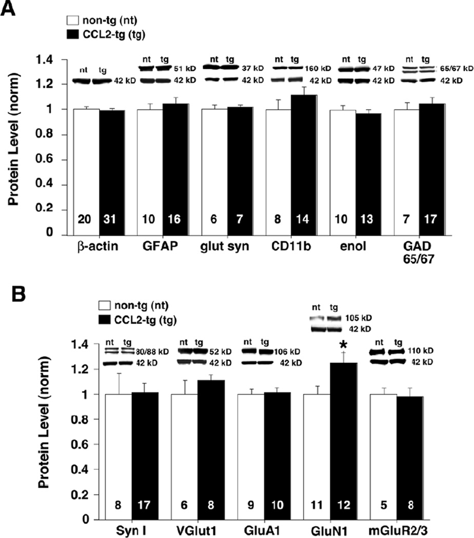
Synaptic function was studied in the hippocampus from young adult CCL2-tg and non-tg mice. The elevated levels of CCL2 in this transgenic model are produced by altered expression in astrocytes, a cell type that is closely associated with synapses in the CNS and a primary source for CCL2 under physiological conditions and after alcohol exposure. CCL2 expression is under control of the GFAP promoter and presumably starts during the postnatal period when GFAP expression is initiated (Lewis and Cowan, 1985; Landry et al., 1990). For each animal studied, one hippocampus was used for the electrophysiological studies and the second was used to assess the general status of the hippocampal tissue by ELISA and Western blot. Analysis of hippocampal lysates by ELISA, to determine the protein levels of CCL2, revealed a 6-fold increase in levels of CCL2 protein in the hippocampus of CCL2-tg mice (1191±105 pg/ml, n=26) compared to their non-tg littermates (196±29 pg/ml, n=19). Analysis of hippocampal lysates by Western blot, to determine if elevated levels of CCL2 altered the expression of important cellular and synaptic proteins, revealed no significant difference for most proteins measured. These proteins included β-actin, a structural protein found in all cells; GFAP, an astrocyte structural protein; CD11b, a microglial protein; enolase, a neuron specific protein; and GAD65/67, the synthetic enzyme for GABA expressed by inhibitory interneurons (Fig. 1A). The levels of synaptic proteins including synapsin I, a protein involved in transmitter release; Vglut1, a protein involved in uptake of the transmitter glutamate into synaptic vesicles; GluA1, a subunit of the AMPA subtype of glutamate receptor; and mGluR2/3, a G-protein coupled glutamate receptor, were also comparable in the CCL2-tg and non-tg hippocampus (Fig. 1B). However, the level of the GluN1 subunit of the NMDA subtype of glutamate receptor was significantly increased in CCL2-tg hippocampus (Fig. 1B; p=0.018, one-group t-test). Taken together, these results show that exposure to elevated levels of CCL2 does not produce a general disruption of the hippocampus as determined by levels of protein expression, but does specifically alter the level of at least one important synaptic protein, GluN1.
Fig. 1.
Protein expression in CCL2-tg and non-tg hippocampus. (A-B) Levels of cellular (A) and synaptic (B) proteins determined by Western blot analysis in CCL2-tg and non-tg hippocampus. Graphs show the mean normalized values. The number of animals studied for each protein is marked in the corresponding bar. Inserts above the graphs show representative Western blots. * Indicates a significant difference from non-tg hippocampus (unpaired t-test).
3.2 Acute ethanol alters synaptic transmission in both CCL2-tg and non-tg hippocampus
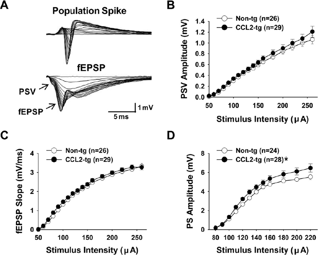
The effect of acute ethanol on synaptic transmission in CCL2-tg and non-tg hippocampus was studied at the Schaffer collateral to CA1 pyramidal neuron synapse using extracellular field potential recordings. Stimulation of the Schaffer collaterals elicited three synaptic events: a presynaptic volley (PSV), a field excitatory postsynaptic potential (fEPSP), and a somatic population spike (PS; Fig. 2A). The PSV represents the summation of action potentials that occur at the synaptic terminals when Schaffer collateral afferents are electrically stimulated. The fEPSP represents the summation of excitatory postsynaptic responses in the dendrites of CA1 pyramidal neurons activated by synaptic transmission. The PS reflects the summation of action potentials in the somatic region of CA1 pyramidal neurons evoked by the dendritic excitatory postsynaptic responses. The PSV and fEPSP were recorded in the dendritic region and the PS was simultaneously recorded from the CA1 pyramidal cell layer. To characterize synaptic transmission, I/O relationships for these synaptic events were constructed for stimulus intensities ranging from subthreshold (i.e., no fEPSP) to the maximum field response for the PS.
Fig. 2.
Input-output (I/O) curves measured in the CA1 region of hippocampal slices from CCL2-tg and non-tg mice. (A) Representative recordings of data for I/O relationship. (B-D) I/O curves constructed from the mean values of the presynaptic volley (PSV) amplitude (B), fEPSP slope (C), and population spike (PS) amplitude (D). No differences were observed in the amplitude of the PSV or the slope of the fEPSP for CCL2-tg hippocampal slices compared with non-tg hippocampal slices. The PS amplitude was significantly larger in CCL2-tg hippocampal slices compared to non-tg hippocampal slices (p=0.024, repeated measures ANOVA). Data are derived primarily from ACSF only exposed slices shown in Figure 3.
I/O relationships for CCL2-tg and non-tg hippocampus were compared in the absence and presence of ethanol to determine whether chronic in vivo exposure to elevated levels of CCL2 produced neuroadaptive changes in synaptic transmission that altered the effects of acute ethanol. Acute ethanol was tested at two concentrations, 20 mM ethanol (100 mg% ethanol) and 60 mM ethanol (300 mg% ethanol). Both concentrations are relevant to alcohol consumption in the human population. The slices were exposed to ethanol for at least 20 minutes prior to performing the I/O protocol.
Under baseline conditions, no differences in the I/O relationship for the PSV or the fEPSP slope were observed between CCL2-tg and non-tg hippocampal slices (Fig. 2B and 2C). However, there was a significant increase in the I/O relationship for the PS amplitude at strong stimulation intensities in CCL2-tg hippocampal slices compared to non-tg hippocampal slices (Fig. 2D; p=0.024, repeated measures ANOVA). These results indicate that chronic in vivo exposure to CCL2 produces neuroadaptive changes that enhance excitability of the pyramidal neuron somata without altering the dendritic synaptic response (fEPSP) or PSV.
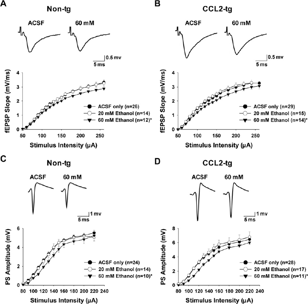
Acute ethanol had no effect on the I/O relationship for the PSV amplitude in either the CCL2-tg or non-tg hippocampus at both concentrations tested (data not shown). Acute ethanol at 20 mM had no effect on the I/O relationship for the fEPSP slope or PS in either CCL2-tg or non-tg hippocampus. However, acute ethanol at 60 mM significantly decreased the I/O relationship for the fEPSP slope in both non-tg (Fig. 3A; p=0.033, repeated measures ANOVA) and CCL2-tg (Fig. 3B; p=0.048, repeated measures ANOVA) hippocampal slices to a similar extent. The I/O relationship for the PS amplitude was also significantly decreased by acute ethanol at 60 mM in both non-tg (Fig. 3C; p=0.003, repeated measures ANOVA) and CCL2-tg (Fig. 3D; p=0.001, repeated measures ANOVA) hippocampal slices to a similar extent. The reduction in the PS by 60 mM acute ethanol in the non-tg and CCL2-tg hippocampus is likely to be a consequence of the reduced fEPSP, which evokes the PS, produced by ethanol. These data indicate that chronic in vivo exposure to elevated levels of CCL2 does not alter the actions of acute ethanol on basal hippocampal synaptic transmission when comparing the I/O relationships.
Fig. 3.
Affect of acute ethanol on input-output (I/O) curves measured in the CA1 region of hippocampal slices from CCL2-tg and non-tg mice. (A-B) I/O curves constructed from the mean values of the fEPSP slope in the presence of 20 mM (open circles) and 60 mM ethanol (closed triangles) in non-tg (A) and CCL2-tg (B) hippocampal slices. 20 mM acute ethanol had no effect on the I/O relationship for the fEPSP slope in either the non-tg (A) or CCL2-tg (B) hippocampal slices. 60 mM acute ethanol significantly decreased the I/O relationship for the fEPSP slope in both non-tg (p=0.033) and CCL2-tg hippocampal slices (p=0.048). (C-D) I/O curves constructed from the mean values of the population spike (PS) amplitude in the presence of 20 mM (open circles) and 60 mM ethanol (closed triangles) in non-tg hippocampal slices. 20 mM acute ethanol had no effect on the I/O relationship for the PS in either the non-tg (C) or CCL2-tg (D) hippocampal slices. 60 mM acute ethanol significantly decreased the I/O relationship for the PS in both non-tg (p=0.003) and CCL2-tg (p=0.001) hippocampal slices. Representative recordings of data for the I/O relationship are shown above each graph. Statistical analysis was determined using repeated measures ANOVA. * Indicates a significant difference between ethanol and ACSF treated hippocampal slices.
3.3 Effects of acute ethanol on paired-pulse facilitation and paired-pulse inhibition
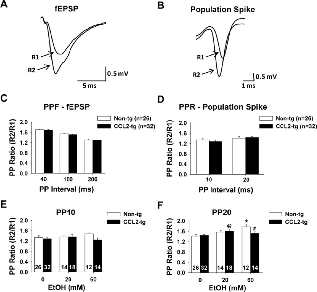
To determine whether chronic in vivo exposure to elevated levels of CCL2 produced neuroadaptive changes that altered the effects of ethanol on short-term synaptic plasticity induced by low-frequency stimulation, paired-pulse facilitation (PPF) and paired-pulse inhibition (PPI) were measured (Fig. 4). PPF is a presynaptic enhancement of transmitter release revealed by the relative magnitude of synaptic responses to a pair of stimuli delivered at short intervals. PPI reflects the strength of inhibitory influences in the somatic region of CA1 neurons. The inhibitory influences involve recurrent inhibitory circuits in the vicinity of the pyramidal cell layer in combination with intrinsic excitability of the pyramidal cells resulting from various ion channels expressed in the cell bodies.
Fig. 4.
Short-term plasticity in CCL2-tg and non-tg hippocampal slices in the absence and presence of acute ethanol. (A-B) Representative traces illustrating paired-pulse facilitation (PPF) of the fEPSP (A) and the PP ratio for the PS (B). Traces R1 and R2 are superimposed responses to paired stimuli separated by 40 ms for the fEPSP and 10 ms for the PS. (C) Summarized results for PPF at 40, 100, and 200 ms paired-pulse intervals. No differences in PPF of the fEPSP slopes were observed for CCL2-tg hippocampal slices compared to non-tg hippocampal slices. (D) Summarized results for the PP ratio at 10 and 20 ms paired-pulse intervals. No differences in the PP ratio for the PS were observed in the CCL2-tg hippocampal slices compared to non-tg hippocampal slices. (E-F) Summarized results for 10 ms (E) and 20 ms (F) paired-pulse intervals for the PS in the presence of 20 mM and 60 mM acute ethanol. Acute ethanol application did not affect the PP ratio of the PS in either CCL2-tg hippocampal slices or non-tg hippocampal slices at the 10 ms interval (E). 20 mM acute ethanol application produced a significant increase in the paired-pulse ratio of the PS in the CCL2-tg hippocampal slices at the 20 ms interval compared with ACSF control CCL2-tg hippocampal slices that were not exposed to ethanol (@, p=0.017). 60 mM acute ethanol application produced a significant increase in the paired-pulse ratio in non-tg hippocampal slices compared with ACSF control non-tg hippocampal slices (*, p=0.004) an effect that was not observed in CCL2-tg hippocampal slices. Thus, there was a significant difference between genotypes in the presence of 60 mM acute ethanol (#, p=0.048). Statistical analysis was determined using the unpaired t-test. The number of slices studied is marked in the corresponding bar.
PPF of the fEPSP slopes was observed at all three interstimulus intervals tested (40, 100, and 200 ms) in both CCL2-tg and non-tg hippocampus and there was no difference in PPF between the CCL2-tg and non-tg hippocampal slices at the three interstimulus intervals (Fig. 4C). Exposure to either 20 or 60 mM ethanol did not produce consistent effects on PPF in CCL2-tg or non-tg hippocampal slices (data not shown). These results indicate that the probability of neurotransmitter release was comparable between the CCL2-tg and non-tg hippocampus in the presence or absence of ethanol.
In both CCL2-tg and non-tg hippocampus, the inhibitory influences at the somatic region were not strong at the stimulus intensity used and facilitation was observed rather than inhibition, presumably due to the angle that the slices were cut. However, no differences were observed in the PP ratio for the PS between CCL2-tg and non-tg hippocampal slices at both interstimulus intervals tested (10 and 20 ms) (Fig. 4D). Acute ethanol at 20 mM and 60 mM did not alter the PP ratio for the PS in either CCL2-tg or non-tg hippocampal slices at the 10 ms interstimulus interval (Fig. 4E). However, at the 20 ms interstimulus interval, a dose dependent increase in the PP ratio (i.e., less inhibition) for the PS was observed in non-tg hippocampal slices compared with baseline (ACSF) controls, which was significant at 60 mM ethanol (Fig. 4F; p=0.004, unpaired t-test). This effect of 60 mM ethanol was not observed for the PS in CCL2-tg hippocampus and the PP ratio was significantly larger in non-tg hippocampus than the CCL2-tg hippocampus in the presence of 60 mM ethanol at the 20 ms interstimulus interval (Fig. 4F; p=0.048, unpaired t-test). A small increase in the PP ratio was observed in CCL2-tg hippocampal slices in the presence of 20 mM ethanol at the 20 ms interstimulus interval compared with ACSF controls (Fig. 4F; p=0.017, unpaired t-test). These results indicate a net increase in somatic excitability in the non-tg hippocampus in the presence of 60 mM acute ethanol, an effect of ethanol that the CCL2-tg hippocampus was resistant to.
3.4 Chronic in vivo overexpression of CCL2 blocks the effects of acute ethanol on hippocampal synaptic plasticity
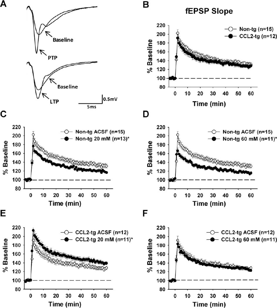
To determine whether chronic in vivo exposure to elevated levels of CCL2 altered the effect of acute ethanol on activity-dependent synaptic plasticity induced by high-frequency stimulation, we compared the ability of an induction stimulation paradigm (TBS) to induce synaptic plasticity at the Schaffer collateral to CA1 pyramidal neuron synapse under baseline conditions and in the presence of ethanol. TBS was used as the induction protocol, because it is thought to reflect normal types of pathway activation (Larson et al., 1986; Otto et al., 1991). TBS induces two forms of short-term plasticity of the fEPSP, post-tetanic potentiation (PTP) and short-term potentiation (STP), in addition to long-term potentiation (LTP) of the fEPSP. All three forms of synaptic plasticity are characterized by an increase in synaptic efficacy and are thought to be cellular mechanisms involved in learning and memory (Lynch, 2004; Martin et al., 2000). PTP was reflected by the initial peak in the magnitude of the fEPSP slope occurring after the termination of the induction protocol (Fig. 5A). The subsequent declining phase of the fEPSP slope following PTP is referred to as STP and lasts ~30 minutes. Both PTP and STP, shortterm enhancements of synaptic strength, are thought to play a role in short-term memory (Erickson et al., 2010). PTP is thought to result from a presynaptic enhancement of synaptic vesicle release that lasts for seconds to minutes after high-frequency stimulation. The mechanisms underlying STP are still not fully understood, but both presynaptic and postsynaptic sites appear to be involved (Erickson et al., 2010). LTP is a long-term change in synaptic strength that is indicated by a stable fEPSP slope that occurs ~50–60 minutes after the induction protocol (Fig. 5A). LTP is mediated primarily by postsynaptic mechanisms involving alterations in expression and/or function of AMPA glutamate receptors (Miyamoto, 2006).
Fig. 5.
Synaptic plasticity measurements following TBS in hippocampal slices from CCL2-tg and non-tg mice in the absence and presence of acute ethanol. (A) Representative dendritic fEPSP traces illustrating post-tetanic potentiation (PTP) 1–3 minutes following TBS and longterm potentiation (LTP) 50–60 minutes following TBS compared to baseline traces recorded prior to TBS. (B) Synaptic plasticity measurements in hippocampal slices from CCL2-tg and non-tg mice expressed as percent of baseline fEPSP slope. High-frequency stimulation (TBS) was used to elicit synaptic plasticity and occurs at time zero. No differences in synaptic plasticity were observed between CCL2-tg and non-tg hippocampal slices (p=0.968). (C-F) Synaptic plasticity measurements in the presence and absence of acute 20 mM and 60 mM ethanol in non-tg (C and D) and CCL2-tg (E and F) hippocampal slices. A decrease in synaptic plasticity was observed in the presence of 20 mM (C, p < 0.0001) and 60 mM (D, p < 0.0001) ethanol in non-tg hippocampal slices when compared to the ACSF treated non-tg hippocampal slices. An enhancement in synaptic plasticity was observed in the presence of 20 mM ethanol (E) in CCL2-tg hippocampal slices when compared to the ACSF treated CCL2-tg hippocampal slices (p < 0.0001). There was no effect of acute 60 mM ethanol on synaptic plasticity in the CCL2-tg hippocampal slices (F) when compared to the ACSF treated CCL2-tg hippocampal slices (p=0.292). Statistical analysis was determined using repeated measures ANOVA. * Indicates a significant difference between ethanol and ACSF treated hippocampal slices.
TBS produced all three forms of synaptic plasticity in the fEPSP slope in both CCL2-tg and non-tg hippocampal slices, as evidenced by a larger fEPSP slope at all time periods following TBS stimulation. The magnitude of the enhancement of the fEPSP slope during PTP, STP, and LTP was similar in CCL2-tg and non-tg hippocampal slices in the absence of ethanol (Fig. 5B). However, differences in synaptic plasticity of the fEPSP slope were revealed in the presence of ethanol. Application of acute 20 mM and 60 mM ethanol to hippocampal slices from non-tg mice significantly reduced the TBS-induced enhancement of the fEPSP slope during PTP, STP, and LTP in a dose-dependent manner compared with vehicle (ACSF) treated non-tg hippocampal slices (Fig. 5C and D; p<0.0001, repeated measures ANOVA). These results are consistent with the known depressive actions of acute ethanol on hippocampal synaptic plasticity (Blitzer et al., 1990; Givens and McMahon, 1995; Izumi et al., 2005; McCool, 2011; Pyapali et al., 1999; Schummers and Browning, 2001; Sinclair and Lo, 1986). Surprisingly, when 20 mM ethanol was applied to hippocampal slices from CCL2-tg mice the enhancement of the fEPSP slope during PTP, STP, and LTP was increased compared with vehicle treated CCL2-tg hippocampal slices (Fig. 5E; p<0.0001, repeated measures ANOVA), an effect of ethanol opposite to that observed in the non-tg hippocampus. In addition, there was no effect of 60 mM ethanol on the TBS-induced enhancement of the fEPSP slope during PTP, STP, and LTP in CCL2-tg hippocampal slices when compared with the vehicle CCL2-tg hippocampal slices (Fig. 5F; p=0.292, repeated measures ANOVA), whereas for the non-tg hippocampal slices a depression of the fEPSP slope was produced by 60 mM ethanol. These data demonstrate that chronic in vivo expression of elevated levels of CCL2 was able to block the depressing effects of acute ethanol on hippocampal synaptic plasticity induced by highfrequency synaptic activity.
3.5 Chronic in vivo overexpression of CCL2 blocks the effects of acute ethanol on cued and contextual fear conditioning
The ability of elevated levels of CCL2 to block the ethanol-induced depression of synaptic plasticity induced by TBS raises the possibility that CCL2 could also block the behavioral effects of ethanol. To test this possibility, we determined whether chronic in vivo CCL2 overexpression altered the effect of acute ethanol on cued and contextual fear conditioning. In cued and contextual fear conditioning, animals learn to associate either a cue (a tone) or the context (environment) with an aversive stimulus, in our case a foot shock. The contextual portion of the task is hippocampal-dependent, whereas the cued conditioning is hippocampal-independent (Logue et al., 1997; Phillips and Ledoux, 1992). Freezing behavior in the context and cued tests (relative to the same context prior to shock and an altered context prior to tone, respectively) are indicative of the formation of an association between the particular stimulus (either the environment or the tone) and the shock; i.e. that learning has occurred. Previous studies have demonstrated that ethanol severely impairs contextual learning, while disrupting cued learning to a lesser extent (Celerier et al., 2000; Gould, 2003; Gulick and Gould, 2007; Melia et al., 1996).
Consistent with the electrophysiological studies that showed only minor differences in hippocampal synaptic function in CCL2-tg mice, we found no significant effects of genotype (non-tg vs. CCL2-tg) in either the context (F(1,68)=0.258, p=0.613) or conditioned stimulus (F(1,68)=0.957, p=0.332) tests. Nor did we see any significant effects of sex on any portion of this test or any effects of genotype or group on freezing behavior in the habituation test and precue portion of the CS+ test. However, ethanol pretreatment (1 g/kg) significantly impaired contextual conditioning (p=0.016) in non-tg mice when compared with control non-tg mice that were injected with saline (Fig. 6A). In comparison, ethanol pretreatment had no effect (p=0.494) on contextual conditioning in CCL2-tg mice (Fig. 6A). A significant decrease in freezing time was also observed during the CS+ test with ethanol pretreatment in non-tg mice when compared to control non-tg mice injected with saline (p=0.037), an effect that was not observed in CCL2-tg mice (Fig. 6B). These results support our previous findings that CCL2 is able to block the depressing effects of acute ethanol and further suggest that overexpression of CCL2 may have a neuroprotective effect against ethanol impairments on hippocampal learning.
Fig. 6.
Effect of acute ethanol on cued and contextual fear conditioning. For fear conditioning, mice were treated with ethanol (1 g/kg) or saline prior to the conditioning trial. (A) Freezing responses during habituation and during exposure to contextual cues. (B) Freezing responses before the cue was activated (pre-cue) and during cue exposure in the conditioned stimulus (CS+ test). Data are expressed as mean ± SEM of the time (s) spent freezing. Non-tg mice treated with ethanol showed significantly less freezing behavior during both the context test (p=0.007) and CS+ test (p=0.037) compared to control non-tg mice treated with saline. The number of animals studied for each test is marked in the corresponding bar. * Indicates a significant difference between ethanol and saline treated mice of the same genotype.
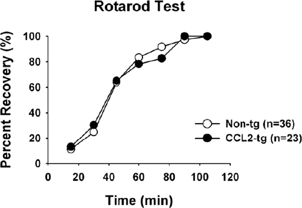
Rotarod performance, which is considered primarily a cerebellar mediated behavior, was also measured in CCL2-tg and non-tg mice. There was no difference between CCL2-tg and non-tg mice in the impairing effects of acute ethanol on performance in the rotarod test (Fig. 7). In addition, the blood ethanol levels at the time of recovery did not differ between CCL2-tg (261.6 ± 18.4 mg%) and non-tg (280.1 ± 14.4 mg%) mice. These data indicate that the protective effect of CCL2 against ethanol-induced behavior impairment is more evident in hippocampal-dependent behaviors than cerebellar-dependent behaviors, at least for the behavioral tests we performed.
Fig. 7.
Ethanol recovery assessed with rotarod testing. Rotarod test recovery times following 2 g/kg ethanol. There was no significant difference between non-tg and CCL2-tg mice in neither this behavioral effect of ethanol, nor the blood ethanol levels determined at the time of recovery.
4. Discussion
The studies described here show that chronic in vivo exposure to elevated levels of CCL2 produces neuroadaptive effects that can block the ability of ethanol to depress synaptic plasticity at the Schaffer collateral to CA1 pyramidal neuron synapse in the hippocampus. Several forms of synaptic plasticity were resistant to the effects of acute ethanol in the CCL2-tg hippocampus including the PP ratio for the PS, PTP, STP, and LTP. Because the generation of these forms of synaptic plasticity involves both presynaptic and postsynaptic mechanisms, the neuroadaptive effects of chronic in vivo exposure to CCL2 are likely to involve multiple sites. A general alteration in hippocampal synaptic function does not appear to be involved, because the effects of ethanol on baseline synaptic transmission and PPF were comparable in CCL2-tg and non-tg hippocampus. Moreover, our Western blot studies revealed only one alteration in the level of cellular and synaptic hippocampal proteins measured, an increase in the level of GluN1, a required subunit of NMDAR. Activation of NMDAR is essential for the induction of LTP (Lynch, 2004) and is a site of ethanol action (Allgaier, 2002; Lovinger et al., 1989; Woodward, 2000). Therefore, the increased levels of GluN1 could play a role in the resistance of CCL2-tg hippocampus to the ethanol-induced depression of LTP observed in the non-tg hippocampus. Other NMDAR subunits were not studied but could also contribute to our results. For example, recent studies show that ethanol-induced phosphorylation of GluN2B can counteract the depressive actions of ethanol on NMDAR-mediated responses (Yaka et al., 2003). Future studies will address this possibility.
Although ethanol has been reported to increase levels of CCL2 in the brain, it is unlikely that the effects we observed were due to acute ethanol-induced CCL2 expression and release. Acute application of CCL2 has been reported to enhance hippocampal neuronal excitability and synaptic transmission (Zhou et al., 2011), whereas in our studies 20 mM ethanol did not produce any changes in synaptic transmission in both CCL2-tg and non-tg slices, while 60 mM ethanol produced a similar decrease in PS amplitude and fEPSP slope in both CCL2-tg and non-tg slices. Importantly, studies byQin et al. (2008) showed that a single high dose of ethanol produced a significant increase in mRNA but not in CCL2 protein in the brain.
The CCL2-tg mice used in our studies (C57BL/6J background) were generated initially from CCL2-tg mice on a SJL background. Previous studies of CCL2-tg (SJL) mice showed that mice less than six months of age appeared normal and were free of obvious neurological impairment, although at older ages (greater than 7 months of age) several neurological impairments such as reduced grooming, postural changes, difficult in righting reflex, and limb weakness were noted (Huang et al., 2005). Studies in our laboratory of hippocampal slices from CCL2-tg (SJL) mice at 7–9 months of age showed that the fEPSP slope and the magnitude of the PSV and PS were significantly smaller in the CCL2-tg (SJL) hippocampus compared with the non-tg hippocampus (Nelson et al., 2011). In addition, both PTP and STP were significantly larger in CCL2-tg (SJL) hippocampus (no difference in LTP) compared to non-tg hippocampus (Nelson et al., 2011). These changes in synaptic transmission and plasticity were not observed in our studies of young adult CCL2-tg mice on the C57BL/6J background. Differences in the age of the animals studied and the higher expression of CCL2 (approximately 2 fold) in the CCL2-tg (SJL) hippocampus are likely to be contributing factors to the differences in results between the two studies.
Ethanol has been shown to modulate both short and long-term changes in synaptic strength in the hippocampus. Ethanol inhibits NMDAR function (Lovinger et al., 1989) and can enhance GABAergic synaptic transmission (Weiner and Valenzuela, 2006), both of which cause a decrease in the extent of LTP. Surprisingly, in our study application of 20 mM acute ethanol caused an enhancement rather than a decrease in synaptic plasticity in CCL2-tg hippocampus. Even in the presence of a higher concentration of ethanol, 60 mM, no difference in synaptic plasticity was observed in CCL2-tg hippocampus compared to vehicle treated (ACSF) CCL2-tg hippocampus, suggesting that CCL2 produces neuroadaptive changes that block the depressing effects of ethanol on hippocampal synaptic plasticity.
In addition to the electrophysiological data, the present study demonstrated that elevated levels of CCL2 can block the ethanol-induced deficits in fear conditioning. Freezing behavior in the context and cued tests is indicative of the formation of an association between the particular stimulus (either the environment or the tone) and the shock. Cue-associated fear memory depends on the amygdala, whereas association of environmental cues with the footshock (contextual fear memory) also involves the hippocampus (Logue et al., 1997; Phillips and Ledoux, 1992). Ethanol administration disrupted both cued and contextual fear conditioning in non-tg mice, whereas CCL2-tg mice did not show any significant deficits following ethanol administration. While not significant, the freezing levels were lower in CCL2-tg than non-tg mice following injection of saline, leaving less room for a further decrease by ethanol in the CCL2-tg mice. However, a potential floor effect cannot be responsible for these results as we see much lower freezing levels during different portions of this test (for example the habituation and precue trials). These behavioral data are consistent with our electrophysiology data and support the idea that CCL2 is able to protect the hippocampus from ethanol induced learning deficits. Thus, ethanol induced increases in CCL2 may have a neuroprotective effect against the detrimental effects of ethanol on hippocampal function.
However, other studies have suggested a detrimental interaction between CCL2 and ethanol. For example, in hippocampal-entorhinal cortex brain slice cultures CCL2 potentiated ethanol-increased glutamate toxicity (Zou and Crews, 2010). In our studies we saw no evidence of interactions between CCL2 and ethanol that were detrimental to synaptic function. In the study by Zou and Crews (2010) the cultures were treated with ethanol for 72 hours, which reflected a more chronic ethanol exposure than used in our studies, and higher concentrations of ethanol were tested. Thus, the neuroprotective vs. neurotoxic abilities of CCL2 are likely to depend on context, such as ethanol dose and duration of exposure.
In addition to alcohol use disorders, elevated CNS expression of CCL2 has been shown to occur in several neurological disorders including multiple sclerosis (Mahad and Ransohoff, 2003; McManus et al., 1998; Simpson et al., 1998), Alzheimer’s disease (Ishizuka et al., 1997; Sokolova et al., 2009), HIV-associated encephalitis (Sanders et al., 1998), and ischemic brain injury (Losy and Zaremba, 2001). Correlative studies suggest that elevated levels of CCL2 may play a role in the cognitive dysfunction associated with several of these disorders. For example, the increased levels of CCL2 in the CSF of older Alzheimer’s patients correlate with the severity of cognitive deficits (Galimberti et al., 2006). In addition, studies of bigenic mice constructed by crossing an Aβ deposition mouse model (Tg2576) of Alzheimer’s disease with a CCL2 overexpressing mouse showed enhanced Aβ oligomer formation, which led to greater deficits in hippocampal synaptic transmission and spatial memory (Kiyota et al., 2009).
Studies also show that CCL2 can be neuroprotective. For example, CCL2 protected against NMDA induced toxicity in mixed cortical cultures and reduced the toxic consequences of oxygen-glucose deprivation in cortical neurons (Bruno et al., 2000; Madrigal et al., 2009). CCL2 also protected mixed cultures of human neurons and astrocytes against NMDA and HIV-tat induced apoptosis (Eugenin et al., 2003). In addition, CCL2 has been described to mediate neuroprotection induced by neurotransmitters, such as noradrenaline (Madrigal et al., 2009, 2010). Our data showing that chronic in vivo elevated levels of CCL2 provide a neuroprotective effect against the depressing effects of pharmacologically relevant concentrations of ethanol on hippocampal synaptic plasticity and hippocampal dependent learning support the ability of CCL2 to serve a neuroprotective role. Thus, the ethanol-induced production of CCL2 may reflect a response of the innate immune system of the CNS designed to protect the CNS from the detrimental effects of ethanol. The success of this protective mechanism is likely to depend on a variety of factors including the extent of ethanol abuse. Excessive ethanol use may cause a dysregulation of the protective mechanism that could compound the detrimental effects of ethanol.
Highlights.
Synaptic transmission in CCL2-transgenic mice is similar to non-transgenic mice.
CCL2 protects against the depressing effects of ethanol on synaptic plasticity.
CCL2 protects against ethanol induced learning impairments in fear conditioning.
Acknowledgments
Supported by NIAAA Grant AA019261 and the Integrated Neuroscience Initiative on Alcoholism (INAI)-West grant AA020893. Jennifer G. Bray was supported by a National Research Service Award, FAA020441. We thank Floriska Chizer-Byas for administrative assistance and Christine Hao for technical assistance.
Footnotes
Publisher's Disclaimer: This is a PDF file of an unedited manuscript that has been accepted for publication. As a service to our customers we are providing this early version of the manuscript. The manuscript will undergo copyediting, typesetting, and review of the resulting proof before it is published in its final citable form. Please note that during the production process errors may be discovered which could affect the content, and all legal disclaimers that apply to the journal pertain.
References
- Allgaier C. Ethanol sensitivity of NMDA receptors. Neurochem. Int. 2002;41:377–382. doi: 10.1016/s0197-0186(02)00046-3. [DOI] [PubMed] [Google Scholar]
- Ambrosini E, Aloisi F. Chemokines and glial cells: a complex network in the central nervous system. Neurochem. Res. 2004;29:1017–1038. doi: 10.1023/b:nere.0000021246.96864.89. [DOI] [PubMed] [Google Scholar]
- Bertollini C, Ragozzino D, Gross C, Limatola C, Eusebi F. Fractalkine/CX3CL1 depresses central synaptic transmission in mouse hippocampal slices. Neuropharmacology. 2006;51:816–821. doi: 10.1016/j.neuropharm.2006.05.027. [DOI] [PubMed] [Google Scholar]
- Blednov YA, Bergeson SE, Walker D, Ferreira VM, Kuziel WA, Harris RA. Perturbation of chemokine networks by gene deletion alters the reinforcing actions of ethanol. Behav. Brain Res. 2005;165:110–125. doi: 10.1016/j.bbr.2005.06.026. [DOI] [PMC free article] [PubMed] [Google Scholar]
- Blitzer RD, Gil O, Landau EM. Long-term potentiation in rat hippocampus is inhibited by low concentrations of ethanol. Brain Res. 1990;537:203–208. doi: 10.1016/0006-8993(90)90359-j. [DOI] [PubMed] [Google Scholar]
- Bruno V, Copani A, Besong G, Scoto G, Nicoletti F. Neuroprotective activity of chemokines against N-methyl-D-aspartate or beta-amyloid-induced toxicity in culture. Eur. J. Pharmacol. 2000;399:117–121. doi: 10.1016/s0014-2999(00)00367-8. [DOI] [PubMed] [Google Scholar]
- Cardona AE, Li M, Liu L, Savarin C, Ransohoff RM. Chemokines in and out of the central nervous system: much more than chemotaxis and inflammation. J. Leukoc. Biol. 2008;84:587–594. doi: 10.1189/jlb.1107763. [DOI] [PMC free article] [PubMed] [Google Scholar]
- Cartier L, Hartley O, Dubois-Dauphin M, Krause KH. Chemokine receptors in the central nervous system: role in brain inflammation and neurodegenerative diseases. Brain Res. Brain Res. Rev. 2005;48:16–42. doi: 10.1016/j.brainresrev.2004.07.021. [DOI] [PubMed] [Google Scholar]
- Celerier A, Ognard R, Decorte L, Beracochea D. Deficits of spatial and non-spatial memory and of auditory fear conditioning following anterior thalamic lesions in mice: comparison with chronic alcohol consumption. Eur. J. Neurosci. 2000;12:2575–2584. doi: 10.1046/j.1460-9568.2000.00115.x. [DOI] [PubMed] [Google Scholar]
- Cho J, Gruol DL. The chemokine CCL2 activates p38 mitogen-activated protein kinase pathway in cultured rat hippocampal cells. J. Neuroimmunol. 2008;199:94–103. doi: 10.1016/j.jneuroim.2008.05.011. [DOI] [PMC free article] [PubMed] [Google Scholar]
- Conductier G, Blondeau N, Guyon A, Nahon JL, Rovere C. The role of monocyte chemoattractant protein MCP1/CCL2 in neuroinflammatory diseases. J. Neuroimmunol. 2010;224:93–100. doi: 10.1016/j.jneuroim.2010.05.010. [DOI] [PubMed] [Google Scholar]
- Crews FT, Bechara R, Brown LA, Guidot DM, Mandrekar P, Oak S, Qin L, Szabo G, Wheeler M, Zou J. Cytokines and alcohol. Alcohol Clin. Exp. Res. 2006;30:720–730. doi: 10.1111/j.1530-0277.2006.00084.x. [DOI] [PubMed] [Google Scholar]
- de Haas AH, van Weering HR, de Jong EK, Boddeke HW, Biber KP. Neuronal chemokines: versatile messengers in central nervous system cell interaction. Mol. Neurobiol. 2007;36:137–151. doi: 10.1007/s12035-007-0036-8. [DOI] [PMC free article] [PubMed] [Google Scholar]
- Erickson MA, Maramara LA, Lisman J. A single brief burst induces GluR1-dependent associative short-term potentiation: a potential mechanism for short-term memory. J Cogn Neurosci. 2010;22:2530–2540. doi: 10.1162/jocn.2009.21375. [DOI] [PMC free article] [PubMed] [Google Scholar]
- Eugenin EA, D'Aversa TG, Lopez L, Calderon TM, Berman JW. MCP-1 (CCL2) protects human neurons and astrocytes from NMDA or HIV-tat-induced apoptosis. J. Neurochem. 2003;85:1299–1311. doi: 10.1046/j.1471-4159.2003.01775.x. [DOI] [PubMed] [Google Scholar]
- Flora G, Pu H, Lee YW, Ravikumar R, Nath A, Hennig B, Toborek M. Proinflammatory synergism of ethanol and HIV-1 Tat protein in brain tissue. Exp. Neurol. 2005;191:2–12. doi: 10.1016/j.expneurol.2004.06.007. [DOI] [PubMed] [Google Scholar]
- Flugel A, Hager G, Horvat A, Spitzer C, Singer GM, Graeber MB, Kreutzberg GW, Schwaiger FW. Neuronal MCP-1 expression in response to remote nerve injury. J. Cereb. Blood Flow Metab. 2001;21:69–76. doi: 10.1097/00004647-200101000-00009. [DOI] [PubMed] [Google Scholar]
- Galimberti D, Schoonenboom N, Scheltens P, Fenoglio C, Bouwman F, Venturelli E, Guidi I, Blankenstein MA, Bresolin N, Scarpini E. Intrathecal chemokine synthesis in mild cognitive impairment and Alzheimer disease. Arch. Neurol. 2006;63:538–543. doi: 10.1001/archneur.63.4.538. [DOI] [PubMed] [Google Scholar]
- Givens B, McMahon K. Ethanol suppresses the induction of long-term potentiation in vivo. Brain Res. 1995;688:27–33. doi: 10.1016/0006-8993(95)00499-g. [DOI] [PubMed] [Google Scholar]
- Gould TJ. Ethanol disrupts fear conditioning in C57BL/6 mice. J. Psychopharmacol. 2003;17:77–81. doi: 10.1177/0269881103017001702. [DOI] [PubMed] [Google Scholar]
- Gulick D, Gould TJ. Acute ethanol has biphasic effects on short- and long-term memory in both foreground and background contextual fear conditioning in C57BL/6 mice. Alcohol Clin. Exp. Res. 2007;31:1528–1537. doi: 10.1111/j.1530-0277.2007.00458.x. [DOI] [PMC free article] [PubMed] [Google Scholar]
- Harrison JK, Jiang Y, Chen S, Xia Y, Maciejewski D, McNamara RK, Streit WJ, Salafranca MN, Adhikari S, Thompson DA, Botti P, Bacon KB, Feng L. Role for neuronally derived fractalkine in mediating interactions between neurons and CX3CR1-expressing microglia. Proc. Natl. Acad. Sci. U.S.A. 1998;95:10896–10901. doi: 10.1073/pnas.95.18.10896. [DOI] [PMC free article] [PubMed] [Google Scholar]
- He J, Crews FT. Increased MCP-1 and microglia in various regions of the human alcoholic brain. Exp. Neurol. 2008;210:349–358. doi: 10.1016/j.expneurol.2007.11.017. [DOI] [PMC free article] [PubMed] [Google Scholar]
- Huang D, Tani M, Wang J, Han Y, He TT, Weaver J, Charo IF, Tuohy VK, Rollins BJ, Ransohoff RM. Pertussis toxin-induced reversible encephalopathy dependent on monocyte chemoattractant protein-1 overexpression in mice. J. Neurosci. 2002;22:10633–10642. doi: 10.1523/JNEUROSCI.22-24-10633.2002. [DOI] [PMC free article] [PubMed] [Google Scholar]
- Huang D, Wujek J, Kidd G, He TT, Cardona A, Sasse ME, Stein EJ, Kish J, Tani M, Charo IF, Proudfoot AE, Rollins BJ, Handel T, Ransohoff RM. Chronic expression of monocyte chemoattractant protein-1 in the central nervous system causes delayed encephalopathy and impaired microglial function in mice. FASEB J. 2005;19:761–772. doi: 10.1096/fj.04-3104com. [DOI] [PubMed] [Google Scholar]
- Ishizuka K, Kimura T, Igata-yi R, Katsuragi S, Takamatsu J, Miyakawa T. Identification of monocyte chemoattractant protein-1 in senile plaques and reactive microglia of Alzheimer's disease. Psychiatry Clin. Neurosci. 1997;51:135–138. doi: 10.1111/j.1440-1819.1997.tb02375.x. [DOI] [PubMed] [Google Scholar]
- Izumi Y, Nagashima K, Murayama K, Zorumski CF. Acute effects of ethanol on hippocampal long-term potentiation and long-term depression are mediated by different mechanisms. Neuroscience. 2005;136:509–517. doi: 10.1016/j.neuroscience.2005.08.002. [DOI] [PubMed] [Google Scholar]
- Jung H, Toth PT, White FA, Miller RJ. Monocyte chemoattractant protein-1 functions as a neuromodulator in dorsal root ganglia neurons. J. Neurochem. 2008;104:254–263. doi: 10.1111/j.1471-4159.2007.04969.x. [DOI] [PMC free article] [PubMed] [Google Scholar]
- Kiyota T, Yamamoto M, Xiong H, Lambert MP, Klein WL, Gendelman HE, Ransohoff RM, Ikezu T. CCL2 accelerates microglia-mediated Abeta oligomer formation and progression of neurocognitive dysfunction. PLoS. One. 2009;4:e6197. doi: 10.1371/journal.pone.0006197. [DOI] [PMC free article] [PubMed] [Google Scholar]
- Landry CF, Ivy GO, Brown IR. Developmental expression of glial fibrillary acidic protein mRNA in the rat brain analyzed by in situ hybridization. J. Neurosci. Res. 1990;25:194–203. doi: 10.1002/jnr.490250207. [DOI] [PubMed] [Google Scholar]
- Larson J, Wong D, Lynch G. Patterned stimulation at the theta frequency is optimal for the induction of hippocampal long-term potentiation. Brain Res. 1986;368:347–350. doi: 10.1016/0006-8993(86)90579-2. [DOI] [PubMed] [Google Scholar]
- Lauro C, Di AS, Cipriani R, Sobrero F, Antonilli L, Brusadin V, Ragozzino D, Limatola C. Activity of adenosine receptors type 1 Is required for CX3CL1-mediated neuroprotection and neuromodulation in hippocampal neurons. J. Immunol. 2008;180:7590–7596. doi: 10.4049/jimmunol.180.11.7590. [DOI] [PubMed] [Google Scholar]
- Lewis SA, Cowan NJ. Temporal expression of mouse glial fibrillary acidic protein mRNA studied by a rapid in situ hybridization procedure. J. Neurochem. 1985;45:913–919. doi: 10.1111/j.1471-4159.1985.tb04080.x. [DOI] [PubMed] [Google Scholar]
- Limatola C, Giovannelli A, Maggi L, Ragozzino D, Castellani L, Ciotti MT, Vacca F, Mercanti D, Santoni A, Eusebi F. SDF-1alpha-mediated modulation of synaptic transmission in rat cerebellum. Eur. J. Neurosci. 2000;12:2497–2504. doi: 10.1046/j.1460-9568.2000.00139.x. [DOI] [PubMed] [Google Scholar]
- Losy J, Zaremba J. Monocyte chemoattractant protein-1 is increased in the cerebrospinal fluid of patients with ischemic stroke. Stroke. 2001;32:2695–2696. doi: 10.1161/hs1101.097380. [DOI] [PubMed] [Google Scholar]
- Logue SF, Paylor R, Wehner JM. Hippocampal lesions cause learning deficits in inbred mice in the Morris water maze and conditioned-fear task. Behav. Neurosci. 1997;111:104–113. doi: 10.1037//0735-7044.111.1.104. [DOI] [PubMed] [Google Scholar]
- Lovinger DM, White G, Weight FF. Ethanol inhibits NMDA-activated ion current in hippocampal neurons. Science. 1989;243:1721–1724. doi: 10.1126/science.2467382. [DOI] [PubMed] [Google Scholar]
- Lynch MA. Long-term potentiation and memory. Physiol Rev. 2004;84:87–136. doi: 10.1152/physrev.00014.2003. [DOI] [PubMed] [Google Scholar]
- Madrigal JL, Leza JC, Polak P, Kalinin S, Feinstein DL. Astrocyte-derived MCP-1 mediates neuroprotective effects of noradrenaline. J. Neurosci. 2009;29:263–267. doi: 10.1523/JNEUROSCI.4926-08.2009. [DOI] [PMC free article] [PubMed] [Google Scholar]
- Madrigal JL, Garcia-Bueno B, Hinojosa AE, Polak P, Feinstein DL, Leza JC. Regulation of MCP-1 production in brain by stress and noradrenaline-modulating drugs. J Neurochem. 2010;113:543–551. doi: 10.1111/j.1471-4159.2010.06623.x. [DOI] [PubMed] [Google Scholar]
- Mahad DJ, Ransohoff RM. The role of MCP-1 (CCL2) and CCR2 in multiple sclerosis and experimental autoimmune encephalomyelitis (EAE) Semin. Immunol. 2003;15:23–32. doi: 10.1016/s1044-5323(02)00125-2. [DOI] [PubMed] [Google Scholar]
- Martin SJ, Grimwood PD, Morris RG. Synaptic plasticity and memory: an evaluation of the hypothesis. Annu. Rev. Neurosci. 2000;23:649–711. doi: 10.1146/annurev.neuro.23.1.649. [DOI] [PubMed] [Google Scholar]
- Matthews DB, Silvers JR. The use of acute ethanol administration as a tool to investigate multiple memory systems. Neurobiol. Learn. Mem. 2004;82:299–308. doi: 10.1016/j.nlm.2004.06.007. [DOI] [PubMed] [Google Scholar]
- McCool BA. Ethanol modulation of synaptic plasticity. Neuropharmacology. 2011;61:1097–1108. doi: 10.1016/j.neuropharm.2010.12.028. [DOI] [PMC free article] [PubMed] [Google Scholar]
- McManus C, Berman JW, Brett FM, Staunton H, Farrell M, Brosnan CF. MCP-1, MCP-2 and MCP-3 expression in multiple sclerosis lesions: an immunohistochemical and in situ hybridization study. J. Neuroimmunol. 1998;86:20–29. doi: 10.1016/s0165-5728(98)00002-2. [DOI] [PubMed] [Google Scholar]
- Melia KR, Ryabinin AE, Corodimas KP, Wilson MC, LeDoux JE. Hippocampal-dependent learning and experience-dependent activation of the hippocampus are preferentially disrupted by ethanol. Neuroscience. 1996;74:313–322. doi: 10.1016/0306-4522(96)00138-8. [DOI] [PubMed] [Google Scholar]
- Meng SZ, Oka A, Takashima S. Developmental expression of monocyte chemoattractant protein-1 in the human cerebellum and brainstem. Brain Dev. 1999;21:30–35. doi: 10.1016/s0387-7604(98)00065-5. [DOI] [PubMed] [Google Scholar]
- Miyamoto E. Molecular mechanism of neuronal plasticity: induction and maintenance of long-term potentiation in the hippocampus. J. Pharmacol. Sci. 2006;100:433–442. doi: 10.1254/jphs.cpj06007x. [DOI] [PubMed] [Google Scholar]
- Nelson TE, Hao C, Manos J, Ransohoff RM, Gruol DL. Altered hippocampal synaptic transmission in transgenic mice with astrocyte-targeted enhanced CCL2 expression. Brain Behav. Immun. 2011;25(Suppl. 1):S106–S119. doi: 10.1016/j.bbi.2011.02.013. [DOI] [PMC free article] [PubMed] [Google Scholar]
- Otto T, Eichenbaum H, Wiener SI, Wible CG. Learning-related patterns of CA1 spike trains parallel stimulation parameters optimal for inducing hippocampal long-term potentiation. Hippocampus. 1991;1:181–192. doi: 10.1002/hipo.450010206. [DOI] [PubMed] [Google Scholar]
- Phillips RG, LeDoux JE. Differential contribution of amygdala and hippocampus to cued and contextual fear conditioning. Behav. Neurosci. 1992;106:274–285. doi: 10.1037//0735-7044.106.2.274. [DOI] [PubMed] [Google Scholar]
- Pyapali GK, Turner DA, Wilson WA, Swartzwelder HS. Age and dose-dependent effects of ethanol on the induction of hippocampal long-term potentiation. Alcohol. 1999;19:107–111. doi: 10.1016/s0741-8329(99)00021-x. [DOI] [PubMed] [Google Scholar]
- Qin L, He J, Hanes RN, Pluzarev O, Hong JS, Crews FT. Increased systemic and brain cytokine production and neuroinflammation by endotoxin following ethanol treatment. J. Neuroinflammation. 2008;5:10. doi: 10.1186/1742-2094-5-10. [DOI] [PMC free article] [PubMed] [Google Scholar]
- Ransohoff RM. Chemokines and chemokine receptors: standing at the crossroads of immunobiology and neurobiology. Immunity. 2009;31:711–721. doi: 10.1016/j.immuni.2009.09.010. [DOI] [PMC free article] [PubMed] [Google Scholar]
- Roberts AJ, Krucker T, Levy CL, Slanina KA, Sutcliffe JG, Hedlund PB. Mice lacking 5-HT receptors show specific impairments in contextual learning. Eur. J. Neurosci. 2004;19:1913–1922. doi: 10.1111/j.1460-9568.2004.03288.x. [DOI] [PubMed] [Google Scholar]
- Rock RB, Hu S, Sheng WS, Peterson PK. Morphine stimulates CCL2 production by human neurons. J. Neuroinflammation. 2006;3:32. doi: 10.1186/1742-2094-3-32. [DOI] [PMC free article] [PubMed] [Google Scholar]
- Rollins BJ. Chemokines. Blood. 1997;90:909–928. [PubMed] [Google Scholar]
- Rossi D, Zlotnik A. The biology of chemokines and their receptors. Annu. Rev. Immunol. 2000;18:217–242. doi: 10.1146/annurev.immunol.18.1.217. [DOI] [PubMed] [Google Scholar]
- Sánchez-Alcañiz JA, Haege S, Mueller W, Pla R, Mackay F, Schulz S, Lopez-Bendito G, Stumm R, Marin O. Cxcr7 controls neuronal migration by regulating chemokine responsiveness. Neuron. 2011;69:77–90. doi: 10.1016/j.neuron.2010.12.006. [DOI] [PubMed] [Google Scholar]
- Sanders VJ, Pittman CA, White MG, Wang G, Wiley CA, Achim CL. Chemokines and receptors in HIV encephalitis. AIDS. 1998;12:1021–1026. [PubMed] [Google Scholar]
- Savarin-Vuaillat C, Ransohoff RM. Chemokines and chemokine receptors in neurological disease: raise, retain, or reduce? Neurotherapeutics. 2007;4:590–601. doi: 10.1016/j.nurt.2007.07.004. [DOI] [PMC free article] [PubMed] [Google Scholar]
- Schummers J, Browning MD. Evidence for a role for GABA(A) and NMDA receptors in ethanol inhibition of long-term potentiation. Brain Res. Mol. Brain Res. 2001;94:9–14. doi: 10.1016/s0169-328x(01)00161-9. [DOI] [PubMed] [Google Scholar]
- Semple BD, Kossmann T, Morganti-Kossmann MC. Role of chemokines in CNS health and pathology: a focus on the CCL2/CCR2 and CXCL8/CXCR2 networks. Journal of Cerebral Blood Flow and Metabolism. 2010;30:459–473. doi: 10.1038/jcbfm.2009.240. [DOI] [PMC free article] [PubMed] [Google Scholar]
- Simpson JE, Newcombe J, Cuzner ML, Woodroofe MN. Expression of monocyte chemoattractant protein-1 and other beta-chemokines by resident glia and inflammatory cells in multiple sclerosis lesions. J. Neuroimmunol. 1998;84:238–249. doi: 10.1016/s0165-5728(97)00208-7. [DOI] [PubMed] [Google Scholar]
- Sinclair JG, Lo GF. Ethanol blocks tetanic and calcium-induced long-term potentiation in the hippocampal slice. Gen. Pharmacol. 1986;17:231–233. doi: 10.1016/0306-3623(86)90144-8. [DOI] [PubMed] [Google Scholar]
- Sokolova A, Hill MD, Rahimi F, Warden LA, Halliday GM, Shepherd CE. Monocyte chemoattractant protein-1 plays a dominant role in the chronic inflammation observed in Alzheimer's disease. Brain Pathol. 2009;19:392–398. doi: 10.1111/j.1750-3639.2008.00188.x. [DOI] [PMC free article] [PubMed] [Google Scholar]
- Ubogu EE, Cossoy MB, Ransohoff RM. The expression and function of chemokines involved in CNS inflammation. Trends Pharmacol. Sci. 2006;27:48–55. doi: 10.1016/j.tips.2005.11.002. [DOI] [PubMed] [Google Scholar]
- van Gassen KL, Netzeband JG, de Graan PN, Gruol DL. The chemokine CCL2 modulates Ca2+ dynamics and electrophysiological properties of cultured cerebellar Purkinje neurons. Eur. J. Neurosci. 2005;21:2949–2957. doi: 10.1111/j.1460-9568.2005.04113.x. [DOI] [PubMed] [Google Scholar]
- Vlkolinský R, Siggins GR, Campbell IL, Krucker T. Acute exposure to CXC chemokine ligand 10, but not its chronic astroglial production, alters synaptic plasticity in mouse hippocampal slices. J. Neuroimmunol. 2004;150:37–47. doi: 10.1016/j.jneuroim.2004.01.011. [DOI] [PubMed] [Google Scholar]
- Weiner JL, Valenzuela CF. Ethanol modulation of GABAergic transmission: the view from the slice. Pharmacol. Ther. 2006;111:533–554. doi: 10.1016/j.pharmthera.2005.11.002. [DOI] [PubMed] [Google Scholar]
- Woodward JJ. Ethanol and NMDA receptor signaling. Crit Rev. Neurobiol. 2000;14:69–89. doi: 10.1080/08913810008443548. [DOI] [PubMed] [Google Scholar]
- Xiong H, Boyle J, Winkelbauer M, Gorantla S, Zheng J, Ghorpade A, Persidsky Y, Carlson KA, Gendelman HE. Inhibition of long-term potentiation by interleukin-8: implications for human immunodeficiency virus-1-associated dementia. J. Neurosci. Res. 2003;71:600–607. doi: 10.1002/jnr.10503. [DOI] [PubMed] [Google Scholar]
- Yaka R, Phamluong K, Ron D. Scaffolding of Fyn kinase to the NMDA receptor determines brain region sensitivity to ethanol. J. Neurosci. 2003;23:3623–3632. doi: 10.1523/JNEUROSCI.23-09-03623.2003. [DOI] [PMC free article] [PubMed] [Google Scholar]
- Zhou Y, Tang H, Liu J, Dong J, Xiong H. Chemokine CCL2 modulation of neuronal excitability and synaptic transmission in rat hippocampal slices. J. Neurochem. 2011;116:406–414. doi: 10.1111/j.1471-4159.2010.07121.x. [DOI] [PMC free article] [PubMed] [Google Scholar]
- Zou J, Crews F. Induction of innate immune gene expression cascades in brain slice cultures by ethanol: key role of NF-kappaB and proinflammatory cytokines. Alcohol Clin. Exp. Res. 2010;34:777–789. doi: 10.1111/j.1530-0277.2010.01150.x. [DOI] [PubMed] [Google Scholar]


