Abstract
Right-sided aortic arch with aberrant left subclavian artery is an uncommon anomaly. We describe a case of Kommerell’s diverticulum involving the distal portion of a right-sided aortic arch and the origin of an aberrant left subclavian artery in a 74-year-old man with hoarseness. The patient underwent successful endovascular repair of the aneurysm with use of a Gore TAG thoracic endoprosthesis and coil embolization of the left subclavian artery. Postoperative computed tomography showed complete exclusion of the lesion, without endoleaks. Endovascular repair is feasible and can be effective in such cases.
Keywords: endovascular repair, Kommerell’s diverticulum, right-sided aortic arch
Right-sided aortic arch is an anatomical variant occurring in approximately 0.1% of the general population.1) About 50% of people with this variant also have an aberrant left subclavian artery (ALSA).2, 3) These anomalies may be either isolated or coexistent with congenital heart disease.4) We describe what we believe is the first case of endovascular treatment of a Kommerell’s diverticulum involving a right-sided aortic arch and ALSA.
Case
A 74-year-old man with hypertension and a history of smoking had recent development of hoarseness. The patient had previously undergone surgical treatment of rectal cancer. His family history was unremarkable. A physical examination showed that his blood pressure was 136/86 mmHg in both arms and his heart rate was regular and 72 beats per minute. There were no carotid bruits or other abnormal results on either the physical examination or laboratory studies.
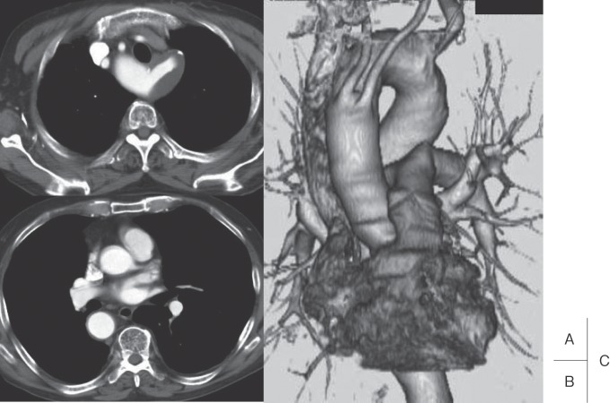
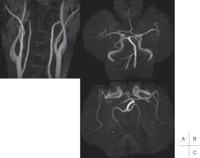
Computed tomography (CT) scanning with 3-mm sections showed a right-sided aortic arch and a right descending thoracic aorta with an ALSA (Fig. 1A, B). A 42-mm aneurysm involved the distal portion of the arch and the origin of the ALSA (Kommerell’s diverticulum). The trachea and esophagus were located at the anterior portion of the arch. Three-dimensional CT scanning showed the proximal-to-distal order of the branches of the aortic arch to be as follows: left common carotid artery, right common carotid artery, right subclavian artery, and ALSA (Fig. 1C). Magnetic resonance angiography found no carotid artery disease on either side, a complete circle of Willis, and bilateral vertebral circulation (Fig. 2). An endovascular approach was chosen to repair the aneurysm because the patient’s anatomical characteristics would have increased the risk of complications associated with thoracotomy.
Fig. 1.
Preoperative computed tomographic (CT) findings. A, B: CT scans show a right-sided aortic arch, a right descending thoracic aorta with an aberrant left subclavian artery (ALSA), and a 42-mm aneurysm involving the distal arch and origin of the ALSA, C: Three-dimensional CT scan shows the proximal-to-distal order of the aortic arch branches to be as follows: left common carotid artery, right common carotid artery, right subclavian artery, and ALSA.
Fig. 2.
Preoperative magnetic resonance angiography (MRA) findings. A, B, C: MRA images show no carotid artery disease on either side, a complete circle of Willis, and bilateral vertebral circulation.
The endovascular procedure was performed with the patient under general anesthesia in an operating room equipped with an angiographic system and prepared for an emergency conversion to open surgery. Access to the right femoral artery for angiography was obtained percutaneously. The left femoral artery was surgically exposed. After administration of heparin (5000 U), a 300-cm guidewire (Radifocus, Terumo, Tokyo, Japan) was introduced through the right brachial artery, advanced across the aortic arch through the brachiocephalic artery, and inserted downstream of the femoral artery. The guidewire was kept under continuous tension by applying traction at both ends of the brachial and femoral access with the aim of overcoming any tortuosity in the aortofemoral route during advancement of the catheter. A 24F introducer sheath with a silicon pinch valve (WL Gore & Associates, Flagstaff, AZ, USA) was advanced into the abdominal aorta. Under fluoroscopic guidance, a self-expanding, 37 × 150-mm Gore TAG thoracic endoprosthesis (WL Gore & Associates) was advanced and deployed at the level of the ALSA coverage.
After deployment of the device, angiography showed a type 1 proximal endoleak caused by insufficient length of the stent-graft proximally. Therefore, a second device (37 × 100 mm) was inserted, overlapping the first one. Angioplasty using a Gore Tri-Lobe balloon (WL Gore & Associates) was employed to seal the endoprosthesis against the aneurysm neck and at the overlap between the devices. Percutaneous coil embolization of the proximal segment of the ALSA was then performed with a 7 Tornado coil (diameter, 10 mm; Cook, Bloomington, IN, USA) to ensure complete exclusion of the aneurysm.
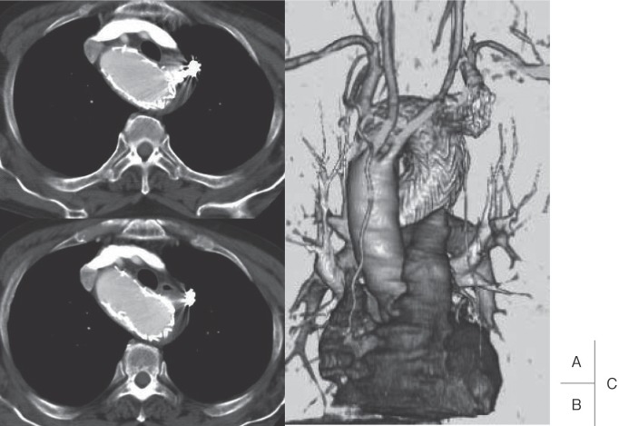
The patient’s post-procedure course was uneventful. A blood pressure gradient of 50 mmHg was observed in both arms, but neither arm claudication nor subclavian steal syndrome occurred. He was discharged from the hospital on the eighth day after treatment. CT scanning confirmed that the stent-graft was positioned correctly and that the aneurysm had been successfully excluded, without endoleaks (Fig. 3).
Fig. 3.
Post-procedure operative computed tomographic (CT) findings. A, B: CT scans, and C: three-dimensional CT scan show correct stent-graft position and successful exclusion of the aneurysm, without endoleaks.
Discussion
In a person with a right-sided aortic arch and ALSA, the left common carotid artery is the first branch arising from the aortic arch, followed by the right common carotid artery, right subclavian artery, and ALSA.5) Aortic aneurysms associated with a right-sided aortic arch are especially rare. Kommerell’s diverticulum, an aneurysm located at the base of the ALSA, is known to cause tracheal compression or dysphagia. In our patient, compression by the lesion had caused hoarseness.
Aberrant subclavian arteries are more likely to dilate than nonaberrant vessels, and rupture of an aneurysm in an aberrant subclavian artery has a 100% mortality rate.6) Therefore, Myers and Gomes6) recommended aggressive surgery for even very small aneurysms in patients with this anomaly. Mid-sternotomy or right thoracotomy is the preferred surgical approach, and cardiopulmonary bypass and deep hypothermia with circulatory arrest are commonly used during the open repair.7, 8) Rates of morbidity and mortality are high.
The goal of thoracic endografting is to provide a method for excluding thoracic aneurysms that is safer than traditional open surgery.9) The TAG device is currently the only commercially available endoprosthesis approved by the US Food and Drug Administration for use in the treatment of thoracic aortic aneurysms. In our case, two TAG devices were employed successfully with ALSA coverage to exclude the aneurysm, and the patient had no complications. Cinà et al.10) commented that reconstruction of the ALSA should be attempted in patients with Kommerell’s diverticulum and a right-sided aortic arch because this may prevent arm claudication in younger patients and subclavian steal syndrome in older patients. In our patient, we used ALSA coverage with coil embolization to achieve good vertebrobasilar circulation. For Kommerell’s diverticula larger than the one in our case, however, complete exclusion may not be achievable with coil embolization alone and surgical treatment may be necessary.
In conclusion, our case shows that endovascular repair is feasible for a Kommerell’s diverticulum involving a right-sided aortic arch and ALSA. Careful follow-up is essential in patients who have undergone this procedure to ensure that such problems as new aneurysms, stent-graft migration, and endoleaks are treated promptly. Formal studies of this endovascular application are needed to determine whether it would have a reduced risk of complications and death compared with surgical treatment.
References
- Drnovsek V, Weber ED, Snow RD. Stenotic origin of an aberrant left subclavian artery from a right-sided aortic arch. A case report. Angiology. 1996; 47: 523–9 [DOI] [PubMed] [Google Scholar]
- Minato N, Rikitake K, Murayama J, Ohnishi H, Takarabe K. Surgery of the dissecting aneurysm involving a right aortic arch. J Cardiovasc Surg (Torino). 1999; 40: 121–5 [PubMed] [Google Scholar]
- Shuford WH, Sybers RG, Gordon IJ, Baron MG, Carson GC. Circumflex retroesophageal right aortic arch simulating mediastinal tumor or dissecting aneurysm. AJR Am J Roentgenol. 1986; 146: 491–6 [DOI] [PubMed] [Google Scholar]
- Felson B, Palayew MJ. The two types of right aortic arch. Radiology. 1963; 81: 745–59 [DOI] [PubMed] [Google Scholar]
- Stewart JR, Kincaid OW, Titus JL. Right aortic arch: plain film diagnosis and significance. Am J Roentgenol Radium Ther Nucl Med. 1966; 97: 377–89 [DOI] [PubMed] [Google Scholar]
- Myers JL, Gomes MN. Management of aberrant subclavian artery aneurysms. J Cardiovasc Surg (Torino). 2000; 41: 607–12 [PubMed] [Google Scholar]
- Kieffer E, Bahnini A, Koskas F. Aberrant subclavian artery: surgical treatment in thirty-three adult patients. J Vasc Surg. 1994; 19: 100–11 [DOI] [PubMed] [Google Scholar]
- Tsukube T, Ataka K, Sakata M, Wakita N, Okita Y. Surgical treatment of an aneurysm in the right aortic arch with aberrant left subclavian artery. Ann Thorac Surg. 2001; 71: 1710–1 [DOI] [PubMed] [Google Scholar]
- Makaroun MS, Dillavou ED, Wheatley GH, Cambria RP; Gore TAG investigators Five-year results of endovascular treatment with the Gore TAG device compared with open repair of thoracic aortic aneurysms. J Vasc Surg. 2008; 47: 912–8 [DOI] [PubMed] [Google Scholar]
- Cinà CS, Arena GO, Bruin G, Clase CM. Kommerell’s diverticulum and aneurysmal right-sided aortic arch: a case report and review of the literature. J Vasc Surg. 2000; 32: 1208–14 [DOI] [PubMed] [Google Scholar]


