Abstract
The development of a ganglion in the hip joint is a rare cause of lower limb swelling. We herein describe a case of a ganglion of the hip with compression of the femoral vein that produced signs and symptoms that mimicked a deep vein thrombosis. Needle aspiration of the ganglion was easily performed, and swelling of the left lower limb promptly improved. Intensive follow-up of this case was important because the recurrence rate of ganglions after needle aspiration is high.
Keywords: ganglion, deep vein thrombosis, pseudothrombophlebitis
Introduction
Synovial/ganglion cysts of the hip joint are rare. Compression of the femoral vein by a synovial/ganglion cyst of the hip joint result in leg swelling resembling that caused by deep vein thrombosis.1) We herein present a case of lower limb swelling due to compression of the femoral vein by a ganglion of the hip joint.
Case Report
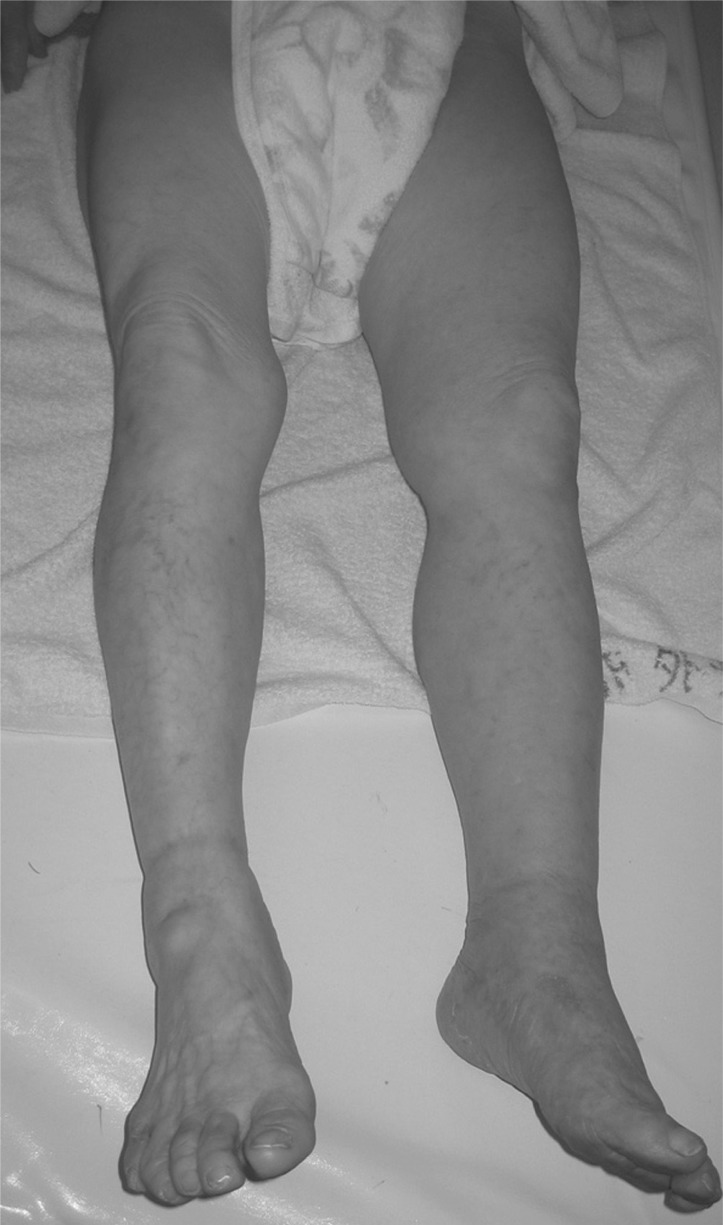
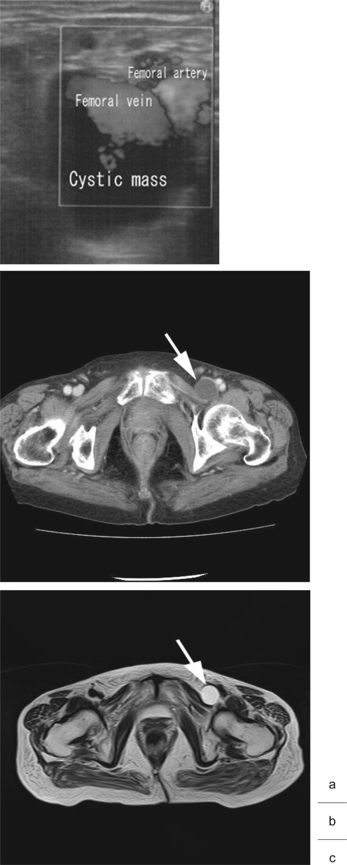
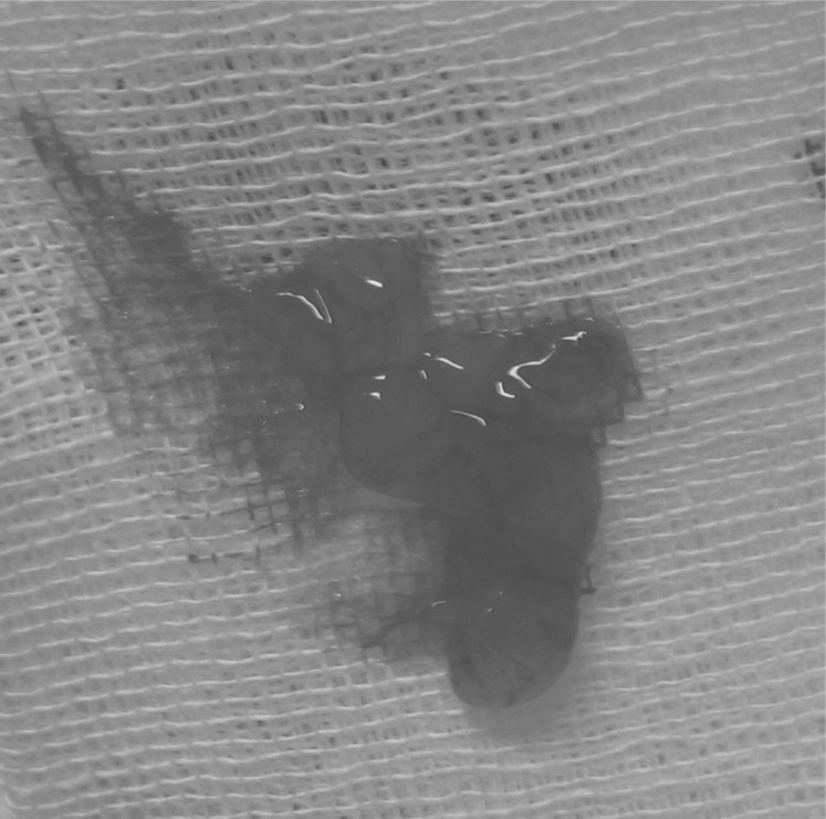
An 85 year-old female came to our hospital with a three-month history of swelling and edema of the left leg. The left thigh circumference was measured to be 4 cm greater, and the left calf circumference was measured to be 2 cm greater than that of the right leg. The grayish-purple color of the left lower limb suggested venous stasis; however, the patient did not complain of either pain or tenderness in the left leg (Fig. 1). Ultrasonography (U.S.) was performed. No venous thrombi were seen; however, U.S. showed a cystic lesion posterior to the left femoral vein, measuring approximately 3 cm in diameter. The left femoral vein curved anteriorly and was severely compressed by the lesion. A computer tomography (CT) scan and magnetic resonance imaging (MRI) of the left hip region revealed, both in high-T2- and low-T1-weighted signal intensity, a cystic lesion 3 cm in diameter arising from the anterio-medial aspect of the acetabulum (Fig. 2). Needle aspiration of the cyst was easily performed with a 21-gauge short needle under U.S. guidance. Approximately 2 mL of clear, jelly-like fluid was yielded (Fig. 3). The size of the cyst had decreased, and the swelling of the left lower limb promptly improved; therefore, the patient did not require surgery for the ganglion cyst of the hip joint. No recurrence of swelling of the left lower limb was observed up to two months after the needle aspiration.
Fig. 1.
Lower limbs of the patient at the first visit.
The left lower limbs were swollen and the skin was grayish-purple, suggesting venous hemostasis.
Fig. 2.
US, CT, and MRI findings.
US (a), CT (b), and MRI (c) each showed a cystic mass (arrow) arising from the left hip joint compressing the femoral vein.
Fig. 3.
The content of the cystic mass obtained by needle aspiration. Clear, jelly-like fluid was yielded.
Discussion
The development of cystic lesions around the joints is a common clinical problem. Histologically, there are two types of cysts; ganglia and synovial cysts. Synovial cysts usually demonstrate a communication with the adjacent facet joint and have a lining of synovial cells seen on a histologic analysis. Ganglion cysts are thought to be the result of myxomatous degeneration of certain fibrous tissue structures, and they do not have a lining of synovial cells on a histologic analysis.2) Because symptoms of and therapy for para-articular cysts do not depend on the histologic composition of the cyst wall, differentiation of ganglia and synovial cysts may be more of academic interest than of clinical importance. Moreover, distinguishing between ganglia and synovial cysts using imaging is not always possible, so that the terms ganglion and synovial cyst are often used interchangeably.2) Synovial/ ganglion cysts can develop on any joint lining or tendon sheath, though, they usually occur at the wrist, ankle, or knee. Synovial/ganglion cysts of the hip joint are not common and are usually accompanied by hip disorders such as rheumatoid arthritis, osteoarthritis, or trauma. The development of an inguinal mass and the presence of groin or thigh pain form the usual clinical presentation of synovial/ganglion cysts. However, compressive symptoms caused by synovial/ganglion cysts of the hip joint are unusual. Compression of the femoral or iliac vein causes leg swelling resembling that caused by deep vein thrombosis,1,3–5) called pseudothrombophlebitis, and sometimes induces deep vein thrombosis,6) called pseudo-pseudothrombophlebitis. Compression of the femoral artery causes leg ischemia,7) and compression of the femoral nerve causes femoral nerve palsy.8)
In 2006, Colasanti et al. reported their case and summarized 27 previously reported cases of lower limb swelling caused by synovial cysts of the hip joint. The mean age of these patients was 62 (35–80) years, and 60% of the patients were female. In 55% of the cases, the joint cysts were accompanied by hip disorders: rheumatoid arthritis or osteoarthritis. Surgery was the treatment of choice in 70% of the cases, and the remaining cases were treated with needle aspiration. Lower limb swelling recurred in 37% of the patients initially treated with needle aspiration, whereas recurrence was noted in only one patient treated surgically.5)
Radiographs are useful to observe the presence of hip disorders. Arthrography can be used to demonstrate the communication of a cyst with the joint cavity; however, cysts may fail to fill when the communication is very narrow or when the cyst is filled with highly viscous fluid. Ultrasonography is one of the most useful imaging modalities; it facilitates diagnosis and is not invasive. However, ultrasonography does not show subtle sites of joint communication and has a limited ability to evaluate associated intra-articular abnormalities. Furthermore, cysts containing debris or hyperplastic synovium may simulate solid mass lesions on an ultrasound examination. A computer tomography (CT) scan is an excellent tool to assess abnormalities of calcified tissues because of its high spatial resolution. Nonetheless, its value for assessing soft-tissue contrast is still limited. Para-articular cysts have lower attenuation than muscle and higher attenuation than fat. Rim enhancement is seen after administration of intravenous contrast. Magnetic resonance imaging (MRI) is superior to all other imaging modalities for investigating soft-tissue abnormalities, particularly for demonstrating the location and extent of lesions. It offers superior soft-tissue contrast and multiplanar capabilities and is noninvasive. It demonstrates the exact location and extent of a lesion, shows the relationship of a lesion to the joint and surrounding structures, and with the aid of intravenous contrast, shows rim enhancement that indicates the cystic nature of a lesion. In addition, MRI is very accurate in depicting associated joint disorders such as meniscal tears, ligamentous abnormalities, and degenerative or inflammatory changes.2)
Treatment of synovial/ganglion cysts depends on their size, location and symptoms. Asymptomatic synovial/ ganglion cysts are often treated with observation. Once a synovial/ganglion cyst of the hip joint causes compressive symptoms, the ideal treatment is surgical excision because the recurrence rate for cysts treated with needle aspiration is high. However, needle aspiration is easier to perform and is less invasive than surgery, and the contents of a cyst can aid diagnosis.1)
In the present case, we treated the cystic mass with needle aspiration only; therefore, no histological diagnosis was done. The CT and MRI revealed no direct communication of the cystic mass and the hip joint, and the CT and MRI findings were similar to that found in cases of ganglia of the hip joint previously reported by Gale et al.6) and Bhan et al.4) Therefore, we diagnosed the cystic mass as a ganglion. Because the patient’s leg swelling promptly improved, she did not request surgery. Surgery has been recommended for the treatment of synovial cysts because of frequent recurrence after needle aspiration. However, according to Savarese et al.,9) surgical excision should be reserved for patients with recurrent cysts or for those who do not respond to needle aspiration. Ideally, surgery was indicated in this case, and a careful follow-up was needed because of the high possibility of recurrence
Conclusions
We herein report a rare case of leg swelling due to femoral vein compression by a ganglion of the hip joint. Ganglia of the hip joint should be taken into consideration in the differential diagnosis of deep vein thrombosis. Needle aspiration is one of the more useful and less invasive options to treat ganglia; however, the recurrence rate following this procedure is high.
Disclosure Statement
We declare no conflicts of interest. references
References
- Sugiura M, Komiyama T, Akagi D, et al. Compression of the iliac vein by a synovial cyst. Ann Vasc Surg 2004; 18: 369-71 [DOI] [PubMed] [Google Scholar]
- Steiner E, Steinbach LS, Schnarkowski P, et al. Ganglia and cysts around joints. Radiol Clin North Am 1996; 34: 395-425 xi-xii [PubMed] [Google Scholar]
- Endo M, Sato H, Murakami S, et al. A case of pseudothrombophlebitis due to inguinal synovial cyst. Am Surg 1990; 56: 533-4 [PubMed] [Google Scholar]
- Bhan C, Corfield L. A case of unilateral lower limb swelling secondary to a ganglion cyst. Eur J Vasc Endovasc Surg 2007; 33: 371-2 [DOI] [PubMed] [Google Scholar]
- Colasanti M, Sapienza P, Moroni E, et al. An unusual case of synovial cyst of the hip joint presenting as femoral vein compression and severe lower limb edema. Eur J Vasc Endovasc Surg 2006; 32: 468-70 [DOI] [PubMed] [Google Scholar]
- Gale SS, Fine M, Dosick SM, et al. Deep vein obstruction and leg swelling caused by femoral ganglion. J Vasc Surg 1990; 12: 594-5 [PubMed] [Google Scholar]
- Beardsmore D, Spark JI, MacAdam R, et al. A psoas ganglion causing obstruction of the iliofemoral arteries. Eur J Vasc Endovasc Surg 2000; 19: 554-5 [DOI] [PubMed] [Google Scholar]
- Kalacı A, Dogramaci Y, Sevinç TT, et al. Femoral nerve compression secondary to a ganglion cyst arising from a hip joint: a case report and review of the literature. J Med Case Reports 2009; 3: 33. [DOI] [PMC free article] [PubMed] [Google Scholar]
- Savarese RP, Kaplan SM, Calligaro KD, et al. Iliopectineal bursitis: an unusual cause of iliofemoral vein compression. J Vasc Surg 1991; 13: 725-7 [PubMed] [Google Scholar]


