Abstract
Aneurysms of the intrathoracic subclavian artery are extremely rare. A 74 year-old man was referred to our hospital with an abnormal chest X-ray film. Contrast computed tomography revealed an intrathoracic left subclavian artery aneurysm. Via left 4th posterolateral thoracotomy, the aneurysm was exposed under systemic deep hypothermia and circulatory arrest. The distal arch was replaced with a 26 mm single-branched graft and the left subclavian artery was reconstructed with a 10 mm graft.
Keywords: subclavian artery, aneurysm, surgery
Introduction
Subclavian artery aneurysm (SAA) is a rare peripheral aneurysm. The most common causes of true SAA are atherosclerosis, thoracic outlet syndrome, infection, trauma, and congenital arterial anomalies such as Marfan syndrome or von Recklinghausen’s disease.1) These aneurysms can undergo rupture, thrombosis, or embolism, and can cause symptoms by local compression. Here, we present our experience with an aneurysm of the intrathoracic left subclavian artery.
Case Report
A 74 year-old man was referred to our institution after an abnormality was detected on his chest X-ray film(Fig. 1). Physical examination was unremarkable. The blood pressure was normal and equal in both upper extremities (120/80 mmHg). He had no history of trauma, infection, congenital arterial anomalies, or systemic vasculitis. Computed tomography (CT) demonstrated an aneurysm that measured 3.9 cm on the proximal part of the left subclavian artery. There were no other aneurysms. CT also revealed dilatation of the ostium of the left subclavian artery (Fig. 2). The aneurysm increased from 3.9 cm to 4.9 cm in diameter after 11 months on repeat CT (Fig. 3). The patient was anxious about the aneurysm and requested graft replacement.
Fig. 1.
Preoperative chest X-ray film shows a left mediastinal mass.
Fig. 2.
Contrast-enhanced CT scan showing the dilated ostium of the left subclavian artery.
Fig. 3.
Contrast-enhanced CT scan showing an aneurysm of the left subclavian artery.
The operation was done via left posterolateral thoracotomy through the 4th intercostal space. After systemic heparinization, the right femoral artery and femoral vein were cannulated, and cardiopulmonary bypass was established. The aneurysm was exposed under systemic deep hypothermia and circulatory arrest. The distal arch was replaced with a 26 mm single-branched graft (Triplex, Terumo Co., Tokyo, Japan) and the left subclavian artery was reconstructed with a 10 mm graft. Operative findings confirmed that the aneurysm was due to atherosclerosis and not trauma.
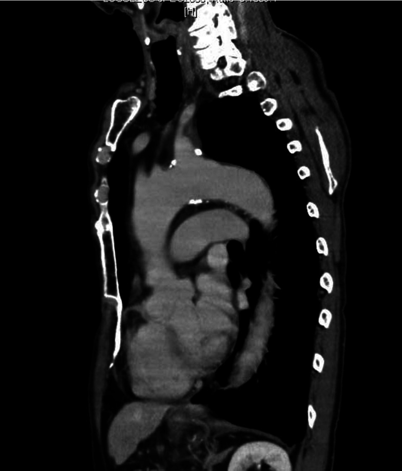
His postoperative course was uneventful, and follow-up CT confirmed a good result (Fig. 4).
Fig. 4.
Multislice CT at 2 months postoperatively.
Discussion
Aneurysms of the subclavian artery are infrequent, accounting for less than 1% of atherosclerotic aneurysms in a large series.1) The most common etiology of aneurysms at this location is atherosclerosis, followed by infection, Marfan syndrome, and thoracic outlet syndrome.2)
SAA can sometimes be completely asymptomatic, as in the present case. When symptoms occur, chest pain or shoulder pain is most common. SAA can undergo rupture, thrombosis, or embolism, and can cause symptoms due to local compression.
The natural history of SAA is unknown. However, a high incidence of death from rupture and exsanguination has been reported regardless of the size of the aneurysm.3) Therefore, an aggressive approach must be taken when dealing with SAA. It has been reported that the operative mortality rate for SAA repair is 6%, with a morbidity rate of 25%.4) In patients with atherosclerotic aneurysms, it is important to rule out other aneurysms elsewhere in the body since the incidence of associated aneurysms ranges from 33% to 47% and a significant number of late deaths from rupture of such aneurysms has been noted.4)
In many patients with a proximal left SAA, the orifice of the subclavian artery is fragile and dilated, so reconstruction by anastomosis or direct closure of the artery with a patch graft is difficult.2) The best approach for proximal left SAA remains controversial. It has been conventional to approach the intrathoracic left subclavian artery through a left 4th intercostal posterolateral thoracotomy in order to obtain proximal control, but median sternotomy has also been employed.5) However, the left subclavian arteries arises from the distal arch of the aorta and lies posteriorly in the thoracic cavity, sometimes making it difficult to obtain distal side of subclavian artery control via an anterior approach, in these cases the posterolateral approach is considered to be useful.
Surgery is the main treatment for SAA, and endoluminal stent grafting may be the method of choice for high-risk surgical candidates.2) On the other hand, endovascular repair of intrathoracic left subclavian artery aneurysm and total endovascular debranching of the aortic arch have recently been reported.6,7) Endovascular aortic repair may be an alternative to conventional surgical repair for patients with left SAA.
In conclusion, we reported a 74 year-old man in whom we treated an atherosclerotic aneurysm of the intrathoracic left subclavian artery.
References
- 1.Dent TL, Lindenauer SM, Ernst CB.Multiple arteriosclerotic arterial aneurysms. Arch Surg 1972; 105: 338-44 10.1001/archsurg.1972.04180080184031 [DOI] [PubMed] [Google Scholar]
- 2.Dougherty MJ, Calligaro KD, Savarese RP.Atherosclerotic aneurysm of the intrathoracic subclavian artery: a case report and review of the literature. J Vasc Surg 1995; 21: 521-9 10.1016/S0741-5214(95)70297-0 [DOI] [PubMed] [Google Scholar]
- 3.Argotte AF, Giron F, Bilfinger TV. Bilateral subclavian artery aneurysms with pseudocoarctation of the aorta. Case report and review of the literature. J Cardiovasc Surg (Torino) 1998; 39: 747-50 [PubMed] [Google Scholar]
- 4.Coselli JS, Crawford ES. Surgical treatment of aneurysms of the intrathoracic segment of the subclavian artery. Chest 1987; 91: 704-8 10.1378/chest.91.5.704 [DOI] [PubMed] [Google Scholar]
- 5.Kawaguchi S, Watanabe M, Hachimaru T.Atherosclerotic pseudoaneurysm of the left subclavian artery: a case report. Ann Thorac Cardiovasc Surg 2010; 16: 376-9 [PubMed] [Google Scholar]
- 6.Veraldi GF, Furlan F, Tasselli S.Endovascular repair of intrathoracic left subclavian artery aneurysm with stent grafts: report of a case and review of the literature. Chir Ital 2005; 57: 355-9 [PubMed] [Google Scholar]
- 7.Cires G, Noll RE, Jr, Albuquerque FC., JrEndovascular debranching of the aortic arch during thoracic endograft repair. J Vasc Surg 2011; 53: 1485-91 Epub 2011 Apr 16. 10.1016/j.jvs.2011.01.053 [DOI] [PubMed] [Google Scholar]


