Abstract
A 7-year-old, 46-kg spayed female rottweiler dog was presented with sudden onset of disorientation, bilateral convergent strabismus, and enophthalmos. Diagnostic workup revealed hypothyroid-associated cranial neuropathy. Symptoms abated considerably upon treatment with levothyroxine-sodium (T4) at an initial dose of 800 μg/kg body weight (BW), PO, q12h, which was reduced 3 days later to 600 μg/kg BW, q12h due to severe agitation and panting. Two weeks later the dosage of the levothyroxine-sodium (T4) was reduced to 400 μg/kg BW in the morning and 600 μg/kg BW in the evening. Eight weeks after the initial presentation, the dog had recovered with only mild convergent strabismus in the right eye. This is the first case report of suspected hypothyroid-associated neuropathy resulting in these symptoms.
Résumé
Neuropathie hypothyroïdienne chez une chienne rottweiler. Une chienne rottweiler de 7 ans, pesant 46 kilogrammes, est présentée pour désorientation, strabisme convergent et enophtalmie d’apparition brutale. Les examens complémentaires révèlent une neuropathie hypothyroïdienne affectant les nerfs crâniens. Le traitement, levothyroxine sodium initialment à la dose 800 μg per os, deux fois par jour, reduit à 600 μg deux fois par jour en raison de apgitation et haleter, permet considerablement l’amélioration des symptômes. Le propriétaire ait conjeillié à diminuer la dose à 400 μg le matin et 600 μg dans la soirée. La chienne recupérait avec solement daix strabisme convergent dans l’oeil drait 8 semaines après la presentation initiale. Ceci est le premier cas rapporté de suppose neuropathie thyroïdienne présentant cette association de symptômes.
(Traduit par les auteurs)
The most frequently described neurological signs associated with hypothyroidism in dogs are head tilt, ataxia, circling, and strabismus. Hypothyroidism has also been associated with peripheral vestibular disease (1). This may manifest as vestibular symptoms or signs of keratoconjunctivitis sicca, if the facial nerve is affected (2). Dogs with more severe polyneuropathy may have more generalized signs, such as flaccid limb paresis (3). Dogs may suffer from hypothyroidism in the absence of common signs such as weight gain and lethargy, making diagnosis of the condition challenging (4).
The pathogenesis of hypothyroid-associated peripheral neuropathy is assumed to be the result of a disturbance in axonal transport, caused by a deficit in energy metabolism (4). Some authors blame a myxomatous compression of cranial nerves, exiting through their respective skull foraminae, for the acute onset of clinical symptoms (3). Discrepancy among different pathological pathways is evidence that the true pathogenesis is still unknown and has yet to be elucidated.
This article describes the clinical findings, diagnostic workup, treatment, and outcome of suspected hypothyroid-associated cranial neuropathy in a 7-year-old female spayed rottweiler dog.
Case description
A 7-year-old, 46-kg spayed female rottweiler dog was presented to the Clinic for Small Animal Surgery, Dentistry and Ophthalmology of the Veterinary University Vienna with the primary complaint of sudden onset blindness of 3 days duration. On further questioning, the owners described progressive lethargy and weight gain over a period of several weeks that was not monitored on a scale. The dog had been treated with nonsteroidal anti-inflammatory drugs and a vitamin K preparation by the referring veterinarian; the dosage could not be determined retrospectively. Food and water consumption, as well as urination and defecation were unaltered. The dog had a previous history of orthopedic disease of the hind limbs of half a year duration, for which a complete orthopedic examination had not been performed and thus accurate diagnosis was unavailable. The dog had received no medications prior to the acute onset of clinical symptoms; however, consumption of toxic substances could not be ruled out, since the dog had been walked by the owners a few hours prior to the onset of clinical symptoms.
On initial examination the dog showed severe signs of disorientation, when entering the examination room, with its head and neck in dorsiflexion and a markedly careful gait. However the dog was still able to navigate through the examination room with obstacles deliberately placed in front of it.
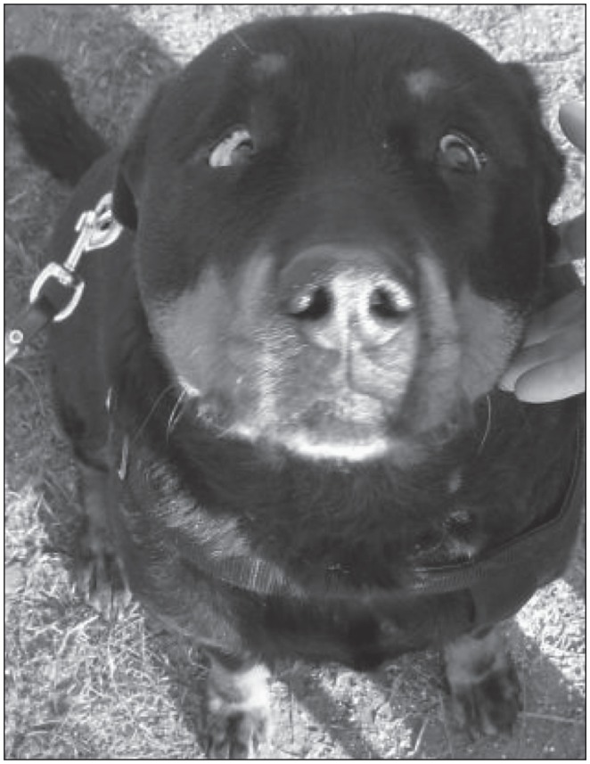
Complete ophthalmic examination using slitlamp biomicroscopy (Kowa SL 15; Kowa, Tokyo, Japan) and indirect ophthalmoscopy (Heine Omega 2C; Heine Optotechnik GmbH & Co KG, Hersching, Germany) revealed bilateral convergent strabismus and enophthalmos (Figure 1). Bilateral resting mydriasis was present; however, pupillary light reflex was prompt and incomplete using a focal light source (Ophthalmic examination lamp; Heine, Herrsching, Germany). Dazzle reflex and menace response were sluggish in both eyes. After the bright light stimulus was applied to induce the dazzle reflex, a horizontal pendular nystagmus was evoked. Schirmer Tear Test I (Schirmer-Tränentest; Essex Pharma GmbH, Munich, Germany) exceeded 15 mm in 60 s bilaterally. Eyelid and corneal sensation were evaluated and found to be normal. Further examination using Melan -100 (Melan -100; Biomed Vision Technologies Ames, Iowa, USA) revealed prompt and incomplete pupillary reaction in both eyes with red and blue light sources. Melan -100 is routinely used in cases of dilated pupils, in order to distinguish between a malfunction of photoreceptors and ganglionic cells. Photoreceptors react to red light, thus resulting in pupillary contraction, whereas ganglionic cells selectively respond to blue light.
Figure 1.
Clinical appearance on initial examination. Note obvious convergent strabismus and marked enophthalmos.
After induced mydriasis (Mydriaticum “Agepha,” Vienna, Austria), biomicroscopy and indirect ophthalmoscopy revealed bilateral posterior focal cortical polar cataract and normal fundi. Electroretinography (ERG) was not performed due to the fact that the dog was not blind but disoriented.
Based on the findings of the ophthalmic examination the initial complaint of blindness could not be confirmed, since the dog still managed to avoid obstacles. The neurolocalization based on ophthalmic examination was within the central nervous system (CNS). An ophthalmic manifestation of systemic disease was suspected, and the dog was referred to the Clinic for Small Animal Internal Medicine for a complete neurologic examination. Complete neurological examination revealed no further abnormalities of cranial nerves, spinal reflexes, or postural reactions, other than already described in the ophthalmic examination. A positional nystagmus following passive horizontal and vertical movement of the head was evident in addition to the light induced horizontal nystagmus.
Differential diagnoses for the observed abnormalities included incipient tetanus, lymphoma of the CNS, and granulomatous meningoencephalitis (GME). For further investigation of the underlying disease, laboratory analyses such as a complete blood (cell) count (CBC) and a biochemistry panel were performed. On gross examination, high grade hyperlipidemia and moderate hemolysis of serum and plasma were present. Serum triglycerides and cholesterol were not measured.
Results of the blood test revealed mild anemia [RBC 5.12 × 10⁶/μL; reference interval (RI): 5.5 to 8.0 × 10⁶/μL], low hematocrit (30.9%; RI: 37% to 55%), slight increase in mean corpuscular hemoglobin concentration (MCHC) (395 g/L; RI: 310 to 340 g/L) due to hemolysis, mild thrombocytosis (586 × 103/μL; RI: 150 to 500 × 103/μL), and mild hyperglycemia (7.5 mmol/L; RI: 2.8 to 5.0 mmol/L). Based on the age, posture, lethargy, and the presence of lipemic serum and mild anemia, thyroid stimulating hormone (TSH) and thyroxine (T4) were measured, for assessment of thyroid function. The results revealed marked increase in canine TSH (0.89 ng/mL; RI: < 0.6 ng/mL) and a T4 value below the reportable range of the assay (< 0.50 μg/L).
Contrast computed tomography (CT) of the head and neck was performed under general anesthesia to exclude inflammatory or neoplastic disease of the CNS. Pre-anesthetic medication was intravenous midazolam (Mayrhofer; Mayrhofer Pharmazeutika GmbH & Co Kg, Leonding, Austria), 0.2 mg/kg body weight (BW) and butorphanol (Butomidor 10 mg/mL, Richterpharma, Wels, Austria), 0.2 mg/kg BW. Anesthesia was induced with propofol and maintained with isoflurane (initial end-tidal concentration of 1%). An atlanto-occipital cerebrospinal fluid (CSF) sample was collected and submitted to cytological and biochemical examination. Cytology revealed no abnormalities of the CSF, the biochemical analysis showed a mild increase in glucose (4.1 mmol/L). Total protein, CSF lactate dehydrogenase (LDH), and creatine kinase (CK) were within reference intervals. Computed tomography scans revealed unremarkable CNS, bullae, and lymph nodes of the neck region. Central nervous system neoplasia or diffuse inflammatory diseases were excluded based on the CT findings and the results of the CSF analysis.
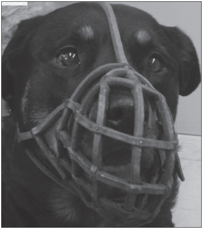
A diagnosis of hypothyroidism-associated neuropathy was made. The dog was put on a course of levothyroxine-sodium (T4) (Forthyron®400; VANA GmbH, Vienna, Austria), 800 μg (18 μg/kg BW), PO, q12h, until re-evaluation after 2 wk. However, the owners reported severe agitation and panting 3 days into the course of treatment, hence the dosage was reduced to 600 μg/kg BW, q12h, which resulted in amelioration of the neurologic signs. Re-evaluation at the Clinic for Small Animal Internal Medicine 2 wk later revealed marked abatement of clinical symptoms. The dog’s general condition was excellent; she was bright, alert, and responsive. Signs of strabismus and enophthalmus had improved considerably (Figure 2), disorientation and nystagmus were no longer present.
Figure 2.
Clinical appearance 3 months after initial examination. Note signs of strabismus and enophthalmos had abated significantly.
The owners were advised to reduce the dosage of the levothyroxin-sodium (T4) to 400 μg/kg BW in the morning and 600 μg/kg BW in the evening, until re-examination after 6 wk. Upon re-evaluation, the dog was in excellent health, clinical symptoms had completely resolved, apart from a mild degree of convergent strabismus of the right eye. The owners were instructed to maintain the dog on medication. Follow-up 18 mo after the initial presentation revealed that the dog had died due to unrelated causes. Unfortunately, necropsy was not performed.
Discussion
When confronted with a patient presented for sudden disorientation, the clinician is inclined to keep all possible differential diagnoses in mind. The most common reasons for vision impairment are opacity of ocular media, retinal disease, such as chorioretinitis, retinal detachment, retinal degeneration, or optic nerve disease (5). A number of intracranial diseases may cause acute blindness, including diseases of the optic chiasm to the visual cortex (5). In this case disorientation was evidently not caused by a disease of the eyes, but rather the effects of the significant convergent strabismus, resulting in marked loss of visual acuity and depth perception. The reduced menace response and dazzle reflex may also be the result of an impairment of the processing of nerve fiber projections, rather than a mechanical block of the visual pathway.
Eye movement is performed by the extraocular muscles, innervated by the oculomotor, trochlear, and abducent nerves. In order to achieve synergistic or antagonistic movement of both eyes, constant feedback by the upper motor neuron (UMN) from the cerebral cortex and brain stem vestibular reflexes is imperative. Abnormalities of muscle groups or innervation result in a fixed globe position, strabismus, or nystagmus (6). Vestibular strabismus may be observed in peripheral or central vestibular lesions and is characterized by ventral deviation of the globe on the side of the lesion, when the head and neck is in extension (7). In this case bilateral convergent strabismus was unrelated to the position of the head. There was no further evidence of vestibular disease, other than a positional nystagmus throughout the neurologic examination. Possible explanations for this finding are bilateral neuropathy of the abducent nerve or dysfunction of the lateral rectus muscle; however, both of these explanations are unlikely, since abducent nerve neuropathy would not result in enophthalmos. Furthermore, horizontal nystagmus would be rendered impossible, if myopathy or atrophy of the lateral rectus muscle was present.
Nystagmus is an involuntary and rhythmic movement of the eyes. It may be pendular (equal movement speed in both directions) or jerk (fast and slow phases), depending on the site of the disease. Nystagmus may be characterized by its direction, as horizontal, vertical, or rotary. Pendular nystagmus is often seen in patients suffering from diseases of the cerebellum or in congenital defects of the visual pathway in Siamese, Birman, and Himalayan cats (7). Siamese and Himalayan cats commonly reveal convergent strabismus and nystagmus, due to a misdirection of axonal projections from the retina, resulting in loss of binocular vision, decrease in visual acuity, and miswiring of central visual pathways (8).
In this case positional nystagmus was noted on passive movement of the head. Unlike in Siamese cats, a congenital defect is highly unlikely, since the dog had never suffered from these symptoms before. However it is possible that both strabismus and nystagmus may have been the result of a temporary misdirection in axonal function, as an effect of hypothyroidism. The final noteworthy feature of this patient was a marked enophthalmos. Enophthalmos may be caused by an abnormally sized eye or a relatively too large orbit. Active or passive retraction of the eye, as an effect of reflectory contraction of the retractor bulbi muscle or space occupying alterations of the orbit respectively, may result in enophthalmos. Finally intra- or periorbital atrophy and sympathicolysis, as seen in Horner’s syndrome, commonly results in this clinical feature (9).
The cause of enophthalmos in this patient remains unclear, since the position of the globes returned to the initial state on re-presentation, rendering atrophy of periocular structures unlikely. Furthermore, no other signs of sympathicolysis were noted — the pupils were dilated and ptosis was not present. Finally, active retraction of the globes remains unlikely, since no cause of pain of ocular structures was found. A possible explanation for enophthalmos in this case is a temporary misdirection in axonal function of the abducent nerves to the retractor bulbi muscles.
The reason for dorsiflexion of the head and neck remains speculation, since there was no anatomical reason for this posture. However, a possible explanation for this symptom is a compensation for the dog to look straight ahead. This explanation would also concur with the careful gait at initial presentation.
Hypothyroidism is widely associated with neuromuscular dysfunction in human and canine patients (10). Clinical signs result from a decreased production of thyroxine and triiodothyronine by the thyroid gland. The most common form of hypothyroidism is acquired primary hypothyroidism, due to lymphocytic thyroiditis or idiopathic thyroid atrophy. Antithyroglobulin antibodies have turned out to be sensitive markers for thyroid inflammation.
Hypothyroidism may occur in any dog; however, some breeds, such as the golden retriever or the doberman pinscher are reported to be at higher risk than others. Furthermore, middle aged, spayed female and neutered male dogs have a higher chance of developing clinical signs (11).
Panciera (2) evaluated 66 cases of hypothyroidism in a period of 5 y. In that report he confirmed the strong association of neutering/spaying and the presence of clinical signs. Symptoms of hypothyoroidism include lethargy, weight gain, alterations of the hair coat quality or color, alopecia, and hyperkeratosis. Poor wound healing, ceruminous otitis, and myxedema have also been reported in affected animals (11). In Panciera’s retrospective study the most common clinical findings were obesity, seborrhoea, and alopecia with 41%, 39%, and 26% frequency, respectively (2). Apart from these nonspecific symptoms several publications report on neuromuscular diseases caused by hypothyroidism.
Higgins et al (12) reported on 10 dogs with hypothyroid-associated central vestibular disease in a period of 6 y. All 10 dogs were aged between 5 and 10 y and were referred for progressive neurologic disease. Five dogs had lesions localized in the myelencephalic region and 5 in the vestibulocerebellar pathways. The only consistent general symptom of hypothyroidism was hypercholesterolemia in 7 of the 10 dogs. However, all animals had total thyroxine and free thyroxine concentrations below reference ranges. Intracranial imaging studies using pre- and post-contrast CT images, as well as 1.5 Tesla MRI, revealed lesions consistent with infarctions in merely 3 of 8 animals; the rest showed no abnormalities. The diagnosis was confirmed by an excellent response to levothyroxine therapy.
Jaggy et al (1) evaluated 29 dogs with neurologic manifestations of hypothyroidism. The most common signs were lower motor neuron dysfunction, peripheral vestibular deficits, megaesophagus, and laryngeal paralysis, in descending order. The study group included mainly older, large breed dogs; however, no gender or breed predisposition was found in that study. The diagnosis was based on the results of neurologic examination, EMG abnormalities, high serum cholesterol concentrations, low response to thyroid stimulating hormone, and good response to therapy with thyroxine.
In several reports on hypothyroidism-induced peripheral neuropathies in the dog, facial nerve paralysis and ataxia are the most common clinical signs (13,14). Other sources report on cranial nerves V, VII, and VIII being commonly involved individually or in combination in hypothyroidism-associated peripheral nerve disease (6).
To the authors’ knowledge, there is no evidence as to which nerve fiber types (sensory or motor), are more prone to failure in hypothyroidism-induced neuropathy. Two major pathomechanisms are suspected for the development of this disease. One proposes that decreased mitochondrial ATPase activity impedes the axonal transport of nerves, leading to axonal degeneration and nerve dysfunction. The other suggests that accumulation of acid mucopolysaccharides in the endoneurium and perineurium of peripheral nerves may impair normal nerve function (6).
Rossmeisl (3) suspects a myxomatous compression of the nerves through their respective foraminae to result in a mechanical disturbance of nerves; however, this theory seems unlikely to apply to this particular case, since no compression was evident on CT. The abducent nerve arises from the nucleus motorius n. abducentis and is incorporated in a common dura-sheath with the oculomotor and ophthalmic nerves, passing through the orbital fissure (15). Myxomatous compression of this nerve would also result in oculomotor and ophthalmic nerve deficits, which was clearly not the case. The theory of myxomatous compression of cranial nerves in veterinary medicine is derived from human studies, in which mononeuropathies are attributed to mucinous deposits in soft tissue structures, thus causing compressive neuropathy. However this pathomechanism remains unproven in animal patients (10).
What still remains unclear about this case is the obvious abducent nerve dysfunction consistent with bilateral nasal strabismus and the presence of bilateral enophthalmos. An explanation described in literature for this particular feature is type II fiber atrophy of the retractor bulbi muscle. This theory is based on the histologic evaluation of muscle biopsies from patients with hypothyroidism-induced peripheral neuropathy (10). This may also explain why strabismus may persist despite successful treatment of hypothyroidism (4). However, muscle atrophy as an explanation for enophthalmos in this case is improbable, since clinical signs had abated within a few weeks.
The most likely explanation for symptoms in this case is failure or misdirection of central axonal transport, due to or in addition to one of the aforementioned reasons. The location of the lesion in the central nervous system cannot be determined precisely by diagnostic imaging, unlike in human medicine where SPECT — single photon emission computed tomography-scans are routinely used to display brain functions (16).
Adequate treatment of canine hypothyroidism consists of synthetic sodium L-thyroxine. Both liquid formulations and tablets are available. L-thyroxine has a serum half-life of 12 to 16 h. A dosage of 20 μg/kg BW, PO, q12h, which may be reduced to q24h, is recommended. In cases of poor suitability for the patient, especially in cases of congestive heart failure, the dosage may be reduced by half. Synthetic T3 administration is indicated if the dog does not respond to T4 supplementation. Improvement of clinical signs should occur within 4 to 6 wk after initial therapy; however, a positional strabismus may persist (11). In this case the patient started to improve within 2 wk of initial presentation. Hypothyroidism-associated neuropathy has often been reported, but, to the authors’ knowledge, this is the first report on hypothyroidism-induced axonopathy in the CNS with effects on the peripheral nervous system. The exact pathomechanism in this case remains mere speculation, since the dog was lost to long-term follow-up and postmortem necropsy. Clinical trials may be needed in order to elucidate the poorly understood pathological pathway of this disease.
Acknowledgments
The authors thank Ilse Schwendenwein (Department for Pathobiology), for interpretation of blood and CSF results, and Michaela Gumpenberger (Department for Companion Animals and Horses) for CT scan interpretation. CVJ
Footnotes
Use of this article is limited to a single copy for personal study. Anyone interested in obtaining reprints should contact the CVMA office (hbroughton@cvma-acmv.org) for additional copies or permission to use this material elsewhere.
References
- 1.Jaggy A, Oliver JE, Ferguson DC, Mahaffey EA, Glaus T., Jr Neurological manifestations of hypothyroidism: A retrospective study of 29 dogs. J Vet Intern Med. 1994;8:328–336. doi: 10.1111/j.1939-1676.1994.tb03245.x. [DOI] [PubMed] [Google Scholar]
- 2.Panciera DL. Hypothyroidism in dogs: 66 cases (1987–1992) J Am Vet Med Assoc. 1994;204:761–767. [PubMed] [Google Scholar]
- 3.Rossmeisl JH. Vestibular disease in dogs and cats. Vet Clin North Am Small Anim Pract. 2010;40:81–100. doi: 10.1016/j.cvsm.2009.09.007. [DOI] [PubMed] [Google Scholar]
- 4.Munana KR. Head tilt and nystagmus. In: Olby NJ, Platt SR, editors. BSAVA Manual of Canine and Feline Neurology. 3rd ed. Quedgeley, Gloucester, UK: British Small Animal Veterinary Association; 2004. pp. 155–171. [Google Scholar]
- 5.Hamilton HL, McLaughlin SA. Differential diagnosis of blindness. In: Bonagura JD, Twedt DC, Kirk RW, editors. Kirk’s Current Veterinary Therapy XIV. St. Louis, Missouri: Elsevier Saunders; 2009. pp. 1163–1167. [Google Scholar]
- 6.Dewey CW. Disorders of the peripheral nervous system: Mononeuropathies and polyneuropathies. In: Dewey CW, editor. A Practical Guide to Canine and Feline Neurology. 1st ed. Ames, Iowa: Iowa State University Press; 2003. pp. 367–412. [Google Scholar]
- 7.Coates JR, O’Brien DP. Brain diseases. In: Ettinger SJ, editor. Textbook of Veterinary Internal Medicine: Diseases of the Dog and Cat. 27th ed. Philadelphia, Pennsylvania: Saunders; 2010. pp. 668–726. [Google Scholar]
- 8.Johnson BW. Congenitally abnormal visual pathways of Siamese cats. Comp Contin Educ Pract Vet. 1991;13:374–378. [Google Scholar]
- 9.Walde I, Nell B, Schäffer EH, Köstlin RG. 4 Augapfel und Augenhöhle (Orbita) In: Walde I, Nell B, Schäffer EH, Köstlin RG, editors. Augenheilkunde: Lehrbuch und Atlas; Hund, Katze, Kaninchen und Meerschweinchen. 3rd ed. Stuttgart, Germany: Schattauer; 2008. pp. 133–164. [Google Scholar]
- 10.Mariane CL. Peripheral nerve disorders. In: Ettinger SJ, editor. Textbook of Veterinary Internal Medicine: Diseases of the Dog and Cat. 27th ed. Philadelphia, Pennsylvania: Saunders; 2010. pp. 834–843. [Google Scholar]
- 11.Scott-Moncrieff JC. Hypothyroidism. In: Bonagura JD, Twedt DC, Kirk RW, editors. Kirk’s Current Veterinary Therapy XIV. St. Louis, Missouri: Elsevier, Saunders; 2009. pp. 185–191. [Google Scholar]
- 12.Higgins MA. Hypothyroid-associated central vestibular disease in 10 dogs: 1999–2005. J Vet Intern Med. 2006;20:1363–1369. doi: 10.1892/0891-6640(2006)20[1363:hcvdid]2.0.co;2. [DOI] [PubMed] [Google Scholar]
- 13.Bichsel P, Jacobs G, Oliver JE. Neurologic manifestations associated with hypothyroidism in four dogs. J Am Vet Med Assoc. 1988;192:1745–1747. [PubMed] [Google Scholar]
- 14.Kern TJ. Facial neuropathy in dogs and cats: 95 cases (1975–1985) J Am Vet Med Assoc. 1987;191:1604–1609. [PubMed] [Google Scholar]
- 15.Böhme G, Nickel R, Schummer A, et al. Peripheres nervensystem. In: Böhme G, Nickel R, Schummer A, et al., editors. Lehrbuch der Anatomie der Haustiere 4 Nervensystem, Sinnesorgane, endokrine Drüsen. 3rd ed. Berlin, Germany: Parey; 1992. pp. 228–384. [Google Scholar]
- 16.Warwick JM. Imaging of brain function using SPECT. Metab Brain Dis. 2004;19:113–123. doi: 10.1023/b:mebr.0000027422.48744.a3. [DOI] [PubMed] [Google Scholar]


