Abstract
Exhaled breath nitric oxide (NO) is an accepted asthma biomarker. Lung concentrations of NO and its amino acid precursor, L-arginine, are regulated by the relative expressions of the NO synthase (NOS) and arginase isoforms. Increased expression of arginase I and NOS2 occurs in murine models of allergic asthma and in biopsies of asthmatic airways. Although clinical trials involving the inhibition of NO-producing enzymes have shown mixed results, small molecule arginase inhibitors have shown potential as a therapeutic intervention in animal and cell culture models. Their transition to clinical trials is hampered by concerns regarding their safety and potential toxicity. In this review, we discuss the paradigm of arginase and NOS competition for their substrate L-arginine in the asthmatic airway. We address the functional role of L-arginine in inflammation and the potential role of arginase inhibitors as therapeutics.
Keywords: nitric oxide, L-arginine, arginase, nor-NOHA, nitrosation, nitric oxide synthase
INTRODUCTION
Asthma is a common disease characterized by a syndrome of persistent airway inflammation and reversible airway obstruction. Intermittent obstruction of the airways results from influx of inflammatory cells, increased mucus secretion, edema, and airway smooth muscle constriction. Chronic inflammation leads to long term remodeling of the lung including mucus cell hyperplasia and metaplasia[1], smooth muscle hyperplasia[1],[2], and increased basement membrane thickness from accumulation of collagens in the submucosal and reticular basement membrane[3]. The airway remodeling and resultant reduction in overall lung function can become irreversible.
Current methods of diagnosing asthma and assessing patient response to therapy are inexact and include measuring lung function with spirometry and assessing noninvasive exhaled breath biomarkers[4] and expectorated sputum samples. One biomarker present in higher concentrations in the exhaled breath of asthmatics, exhaled nitric oxide (NO), has been positively correlated with lung inflammation severity. However, clinical trials with inhibitors targeting the NO producing enzymes have produced mixed results[5],[6], indicating that the role of NO during asthma exacerbation or mediation is much more complex than previously thought.
Derived primarily from the metabolism of L-arginine by the NO synthase (NOS) family of enzymes, NO is essential in preserving normal lung function. The NO diffusion gradient ensures sufficient blood oxygenation by dilating vascular smooth muscle at regions of hypoxia, thereby maintaining proper ventilation-perfusion matching[7]–[9]. NO also regulates the ciliary beat frequency[10] of columnar epithelial cells in the airway that clear potentially obstructive agents, including foreign materials and mucus from the upper conducting airways. As an inhibitory non-adrenergic non-cholinergic (iNANC) signaling molecule[11],[12], NO controls smooth muscle tone in the airways by activating the soluble guanylate cyclase in the smooth muscle[13]. NO modulates inflammation by affecting leukocyte adhesion to the endothelium[14],[15] and vascular permeability[16] and also is an integral part of the immune system anti-microbial arsenal, reacting with other reactive species to form potent oxidant molecules[17],[18].
Thus, despite the correlation of increased exhaled NO with inflammatory severity in the lung[4],[19], reducing the overall production of NO by inhibiting NOS enzymes would undoubtedly also affect NO-dependent regulation of normal lung function. The variability in outcomes using NOS inhibitors in animal models of allergic inflammation supports the conclusion that not all sources of NO are equal (See Mathrani, et al. 2007 for review of NOS inhibition in allergic asthma models[20]). Focusing entirely on regulating a measurable parameter, exhaled NO, does not take into account the sources of NO production or the delicate balance of NO in the lung as a whole. The more telling question may be whether there is “good NO” and “bad NO”, what their cellular sources are, and what changes occur in the lung during allergic inflammation that affect both “good” and “bad” NO.
THE FUNCTIONAL ROLE OF NO AND ITS PRESURSOR, L-ARGININE
Nitric oxide: function and form interdependence
The NO molecule is a neutral-charged free radical with a short half life in biological fluids (<1 ms) due to its reactivity with surrounding proteins, free radical species, and reducing molecules of the intra- and extracellular compartments like glutathione. NO is primarily derived from the enzymatic conversion of the amino acid L-arginine and molecular oxygen into NO and citrulline by the NOS family of enzymes.
The NOS enzyme family is comprised of three isoforms, NOS1, NOS2 and NOS3, which vary in their regulatory mechanism and tissue expression patterns[21]. NOS1 and NOS3 are constitutive NOS enzymes that require intracellular calcium/calmodulin binding for activation. In addition to a calcium concentration dependence, NOS3 activity is also regulated by multi-site phosphorylation of serine and threonine residues[22]. NOS2, the inducible NOS, is predominantly regulated at the transcriptional level. Due to its high affinity for calmodulin, NOS2 activity is relatively independent of intracellular calcium fluxes but requires binding of transcriptional activators nuclear factor-kappa B(NF-κB), activator protein-1 (AP-1) or signal transducers and activators of transcription 1α STAT1α[23]–[25] for expression. The NOS2 isoform can be rapidly induced by pro-inflammatory cytokines, resulting in heightened levels of NOS2 protein expression and NO production; thus, NOS2 can become the major source of NO under inflammatory conditions.
The three NOS isoforms are differentially expressed in numerous resident and inflammatory cell types in the lung and can vary in both expression and activity under normal and proinflammatory conditions. NOS1 is expressed mainly in airway epithelial cells[26] while NOS3 is expressed in the airway epithelium and vascular endothelium[27]. NOS1 and NOS3 are both expressed under basal conditions and contribute to the baseline concentrations of exhaled NO. NOS2 is expressed at low to undetectable levels under non-inflammatory conditions but can be expressed at high levels in the airway epithelium, airway smooth muscle, inflammatory cells and alveolar type 2 cells under inflammatory conditions. NOS2 is thought to contribute to the increase in exhaled NO observed in asthmatics and animal models of allergic inflammation. Despite tight regulatory controls over the constitutive NOS1 and NOS3 isoforms, NOS2 isoform expression can change depending on surrounding NO concentration and cytokine expression[28]. As a result, NO production by the different enzymatic isoforms can vary significantly depending on the surrounding conditions and have sweeping effects on lung function.
The rate of clearance of NO also depends on numerous factors. Accumulation in protected cellular compartments, including the plasma membrane, lipophilic protein folds and interstitial spaces (the inner mitochondrial space or vesicles) can increase the half-life of the molecule[29]. Reaction of NO with glutathione, forming S-nitrosoglutathione (GSNO), or with albumin or hemoglobin can convert NO into a more stable intermediate, giving NO the capacity to have functional activity far removed from its temporal and positional origin. The oxidization products of NO, nitrate and nitrite, are more stable than NO and can serve as a substrate pool for NO under hypoxic conditions by enzymatic conversion using xanthine oxidoreductase[30] or by non-enzymatic reduction via electron and proton transfer reactions with both free and protein-associated heme[31],[32]. Excessive nitrate and nitrite can be filtered from the plasma and excreted in the urine or exhaled from the lung directly as either NO or as one of its many oxidation products.
L–arginine and inflammation
NO production can be greatly increased under inflammatory conditions but, like other enzymatic reactions, is limited by the amount of active enzyme present, the concentration of the enzymatic cofactor, tetrahydrobiopterin (BH4), and of the substrate, L-arginine. L-arginine is a semi-essential amino acid that serves as a substrate for numerous enzymatic pathways and a precursor for protein synthesis[33]. In the body, circulating L-arginine concentration in the plasma is the sum of the dynamic interconversion of L-arginine downstream metabolites, protein synthesis and degradation, dietary intake and excretion. L-Arginine is first absorbed in the gut through the epithelium of the small intestine where it is converted into L-citrulline and then enters the circulation. Synthesis of L-arginine from L-citrulline occurs mainly in the kidney by the concerted enzymatic activities of the argininosuccinate synthase (AS) and argininosuccinate lyase (AL), although certain cell types, including alveolar macrophages, retain the capacity to regenerate L-arginine by this pathway.
L-arginine incorporated into proteins can be posttranslationally modified by methyltransferases, yielding asymmetric dimethylarginine (ADMA), symmetric dimethylarginine (SDMA) and N-monomethylarginine (L-NMMA). After protein degradation, these methylated products are released back into the free amino acid pool[34],[35] where they can competitively inhibit NOS activity and compete with L-arginine for transmembrane transport by the cationic amino acid transporter (CAT). The dimethylarginine molecules can also be enzymatically converted back into L-arginine by dimethylarginine dimethylaminohydrolase (DDAH) or excreted in the urine[36].
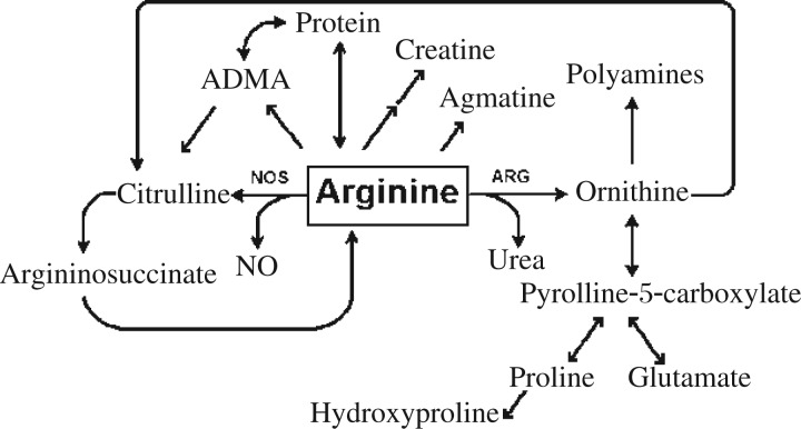
L-arginine is also metabolized by several different metabolic pathways, resulting in the formation of downstream products such as creatine, agmatine, glutamate, proline, polyamines, ornithine and NO (Fig. 1). These metabolic products can affect the development of or perpetuate the asthmatic phenotype. Creatine has been linked to increased airway hyperreactivity as measured by Penh and increased airway eosinophilia, though only at high supplemented dosages[37]. Agmatine is a weak NOS inhibitor[38] and glutamine is a precursor for γ-aminobutyric acid (GABA) and glutathione, which increases mucus production in the airway epithelium or act as an antioxidant, respectively[39],[40]. Proline is a precursor for collagen, a component of the basement membrane that becomes thickened during asthmatic airway remodeling[41],[42]. Polyamines are important regulators of the cell cycle, proliferation, differentiation and apoptosis[41]–[43].
Fig. 1. Schema of major L-arginine metabolic pathways.
With systemic or chronic inflammation, the rate of L-arginine reconversion may become insufficient to maintain normal plasma concentration[44]–[46]. In this case, total L-arginine catabolic and anabolic reaction rates have become unbalanced, producing an overall shift toward L-arginine catabolism. This L-arginine flux imbalance may occur throughout the whole body, reducing overall circulating L-arginine, be limited to a tissue microenvironment where the diffusion of L-arginine from the circulating plasma to the target tissues becomes the limiting factor, or occur within specific cellular compartments where diffusion and transmembrane transport rates may affect L-arginine supply[47]–[49].
Reduced L-arginine content has been detected in the airway compartment of mice exposed to ovalbumin (OVA) to mimic allergic airway disease[50],[51]. In moderate asthmatic subjects, circulating plasma L-arginine is reduced compared to control subjects (45 µmol/L L-arginine compared to control values of 94 µmol/L). The decrease in plasma L-arginine in asthmatics coincides with a 3-fold increase in plasma arginase activity[52] and occurs concurrently with changes in the dynamics of L-arginine's primary metabolic pathways, the NOS pathway and the arginase pathway.
Expression of arginase I (Arg1) increases in murine models of allergic asthma induced by either OVA, A. fumigatis exposure[53]–[56], or house dust mite allergen (HDMA)[57] (up to an 8-fold increase in total Arg1 expression in the airways of mice exposed to OVA). The increase in Arg1 expression in mice directly correlates with increased arginase activity[56]. This change in L-arginine metabolism is thought to control NOS-dependent production of NO by the depletion of their common substrate, L-arginine.
L-arginine depletion: enzyme uncoupling
The depletion of L-arginine in the airways can have consequences in addition to reducing total NO production. Constitutive NOS isoforms perform a two step conversion of L-arginine and oxygen to L-citrulline and NO. In the absence of sufficient L-arginine (under 100 µmol/L) and/or BH4, NOS activity can produce superoxide (•O2—). This is termed “uncoupling” as the oxygenase domain is uncoupled from the reductase domain of the enzyme[58]. Uncoupling results in the constitutively active NOS enzymes producing a combination of NO and superoxide[59]–[61], which can combine to form peroxynitrite.
The uncoupling phenomenon only occurs at low BH4 concentration in the inducible NOS isoform[62]. Examination of the catalytic mechanism of the NOS2 enzyme identified the flavin-binding reductase domain, not the oxygenase domain, of NOS2 as the source of superoxide production[63]. This difference in catalytic mechanism in the NOS2 isoform, as contrasted with the constitutive NOS1 and NOS3 isoforms, may exist because the antimicrobial action of NO depends on its reaction with the free radical species superoxide to form the more potent oxidant peroxynitrite.
Arginase: a competitor for L-arginine
The arginase enzymes are homotrimeric metalloenzymes stabilized in conformation by two Mn2+ ions per monomeric structure. There are two isoforms of arginase, Arg1 and arginase 2 (Arg2). Arg1 is often referred to as liver arginase, especially in older texts, as it is found at high levels in the liver as part of the urea cycle of enzymes, but it is also expressed in bone marrow-derived cells[64], such as neutrophils, macrophages[65] and red blood cells in humans, as well as in fibroblasts[66], epithelial and endothelial cells[67] upon activation by proinflammatory cytokines or hypoxia. Arg2 is found ubiquitously at low levels in mitochondria and at high levels in the kidney.
Although Arg1 and NOS2 are expressed in the airway and inflammatory cells of the lung, and in some cases within the same cell, there is controversy regarding the ability of arginase to compete with NOS for L-arginine due to kinetic properties of the two enzymes. Kinetic assays using isolated arginase have indicated the maximum rate of hydrolysis of L-arginine into L-ornithine and urea at a Km of 1 mmol/L and a Vmax of 4,380 µmol/(min·mg)[68]. Based solely upon the calculated Km values, (Arg: 1 mmol/L, and NOS: 10 µmol/L), the arginase enzymes do not appear capable of competing with the NOS enzymes at L-arginine concentrations below 100 µmol/L. However, the enzymes' kinetic parameters were determined in a closed system with isolated enzymes and do not take into account enzyme coupling, non-freely diffusible substrate pools, intracellular localization of the enzymes and substrate transporter expression and activity, diffusion gradients, and potential sequestration[69]–[72].
In vitro studies in isolated endothelial cells note that increasing the extracellular arginine concentration increases NO production despite average intracellular L-arginine concentrations than would be considered saturating for the enzyme based upon its kinetics. Colocalization of cationic amino acid transporters (CAT) 1 and 2B, and L-arginine recycling enzymes, AS and AL, with NOS3 make the NOS3 enzyme dependent upon extracellular L-arginine concentration, the concentration of other cationic amino acids that can compete for transport via CAT, and/or the rates of L-arginine re-synthesis[73],[74].
In the 1990s, research began to focus on the potential for arginase activity to regulate airway reactivity. In a series of experiments utilizing ex vivo airway preparations from allergen-exposed guinea pigs, de Boer et al. observed the dependence of airway hyperreactivity on NO deficiency[75],[76] and that this deficiency was L-arginine concentration dependent[77]. By analyzing NO production in the presence and absence of the arginase inhibitor Nω-hydroxy-nor-arginine (nor-NOHA), researchers showed that the deficiency of NO caused by arginase activity was the causative agent for the development of airway hyperreactivity[78]–[81]. Although this mechanism was previously identified in cultured macrophages[82],[83] and endothelial cells[84], the identification of this phenomenon in the resident airway cells of the lung had a significant impact on the understanding of NO function in the airways.
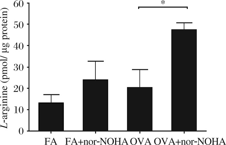
Recently, we have found that systemic treatment of mice with a competitive inhibitor of arginase in a model of allergen-induced airway inflammation significantly increased the amount of NO produced, as measured by the nitrite + nitrate (NOx) concentration in lung lavage supernatant[50]. We also verified that arginase inhibition increases L-arginine content in the conducting airways of C57BL/6 mice exposed to OVA (Fig. 2). Examination of Arg1 expression in OVA-exposed C57BL/6 and congenic NOS2 knockout mice[1] demonstrated that Arg1 content in the airway compartment may be regulated by NOS2 activity. Expression levels of Arg1 also vary between BALB/c mice and C57BL/6 strains[50],[56], but these strain differences may be linked to variability in NF-κB signaling which is altered in the two strains[85] and in the NOS2 knockout mice. Given the apparent linked regulation of Arg1 and NOS, targeted therapeutic interventions at one of these enzymes will be needed to recognize and account for the effects on the other.
Fig. 2. L-arginine content in microdissected airways of BALB/c mice sensitized and exposed to ovalbumin (OVA) for two weeks.
Mice exposed to ovalbumin and treated with the arginase inhibitor Nω-hydroxy-nor-L-arginine (nor-NOHA) had higher levels of L-arginine in their dissected airways compared to mice exposed to ovalbumin alone. FA: filtered air. *P < 0.05.
L-arginine and arginase: potential therapeutic targets
In addition to asthma, arginase activity has been shown to correlate with the disease severity in several lung diseases including COPD, cystic fibrosis and sickle cell anemia[86],[87]. To better understand the role of arginase activity in perpetuating and/or potentiating inflammatory lung disease, arginase inhibitors have been used extensively in animal models of allergen-induced inflammatory lung disease using tracheal explants and in cell culture. In these models, the inhibition of arginases increases the production of NO, the product of the competing NOS pathway.
The liver and kidneys contribute a significant portion of the body's overall arginase activity and prolonged treatment with arginase inhibitors may inadvertently target these highly perfused organ systems causing metabolic imbalance. Tissue-specific targeting of arginase inhibitors would limit systemic effects, but can be difficult to achieve due to the hydrophilic nature of the currently available drugs, which are mainly structural analogues of arginine[88].
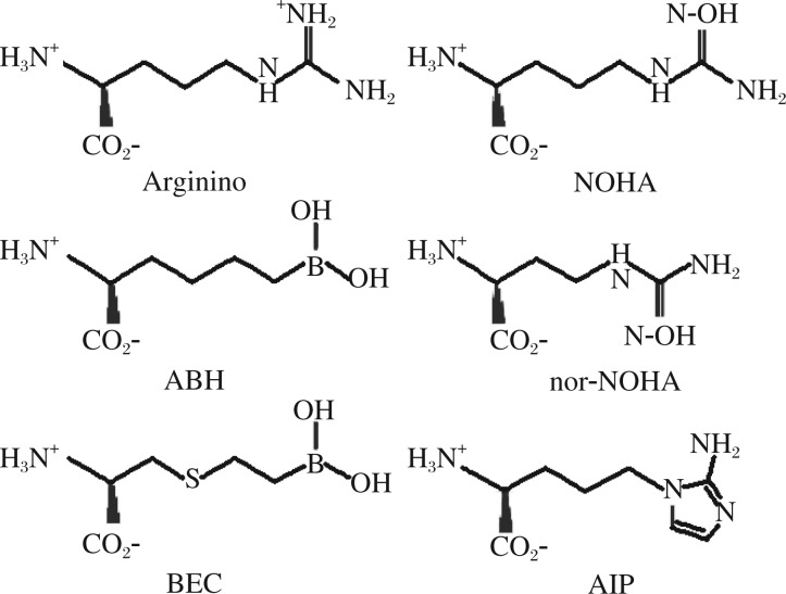
There are two basic subsets of arginase inhibitors: reversible and irreversible. The reversible inhibitors can be further divided into boronic acid-based inhibitors and non-boronic acid-based inhibitors. Most reversible inhibitors are L-arginine molecular analogs, which mimic the transition-state structure of L-arginine during the arginase hydrolysis reaction (Fig. 3). The inhibitors are structurally similar to the molecule, Nω-hydroxy-L-arginine (NOHA), an intermediate in the synthesis of NO by the NOS family of enzymes. Although NOHA can act as an arginase inhibitor and a substrate for NOS, transition state inhibitors, such as nor-NOHA and N(G)-hydroxy-L-arginine (L-NOHA) cannot be converted to NO by the NOS enzymes or inhibit NOS enzyme activity. In addition to the NOHA-based molecules, there is a small group of amino acid sulfonamides that can inhibit arginase activity[89]. These compounds are also transition state mimetics with an activated guanidino group that bridges the binuclear manganese cluster, but with a lower binding efficiency than the boronic and NOHA-based inhibitors.
Fig. 3. Key arginase inhibitors of therapeutic interest in asthma.
Adapted from a research of Morris SM Jr. [88]
2-(S)-amino-5-(2-aminoimidazol-1-yl)pentanoic acid (A1P) is a recently developed arginase inhibitor that utilizes a 2-aminoimidazole moiety in place of a guanidine side chain (Fig. 3). This newly developed arginase inhibitor has a Ki of 4 µmol/L and has recently been used in a murine model of allergic inflammation to significantly reduce airway hyperresponsiveness[90]. Much like the non-boronic acid based inhibitors, the boron-based arginase inhibitors are also transition state inhibitors but with a significantly higher potency, ≥5-fold lower IC50 and 5-fold lower Ki, and cannot serve as a substrate for NOS enzymes[91],[92]. Boron-based inhibitors, 2(S)-amino-6-boronohexonic acid (ABH) or S-(2-boronoethyl)-L-cysteine (BEC), have been used extensively in animal models of allergic asthma with considerable success[79],[93]–[95].
The irreversible arginase inhibitor (+)-S-2-amino-6-iodoacetamidohexanoic acid (2-AIHA) is also a potent inhibitor of the enzyme. 2-AIHA is an Nω-derivative of (+)-lysine with significant toxicity issues, including liver and kidney damage in mice[96].
Several other classes of drugs can also affect arginase indirectly by acting as a general anti-inflammatory agent: NSAIDs, corticosteroids, and statins – the latter two of which block Arg1 induction in murine and human tissues[66],[97],[98]
Several investigators have taken the information from previously described in vitro and ex vivo studies to examine animal models of allergic airway disease in order to better understand the complex relationship between NO, airway hyperreactivity and airway inflammation. Experiments utilizing arginase inhibitors and Arg1 RNAi have indicated that arginase inhibition can decrease airway inflammation and reduce airway hyperreactivity[56],[93],[94],[99]. However, a study by Ckless et al.[100] noted an increase in airway inflammation and hyperresponsiveness, with significant increases in nitrotyrosine and S-nirosylation, indicating an increase in NO-based oxidative products. In this study, mice were treated with the inhibitor using a single intratracheal dose 2 h after the final (3rd) allergen exposure. Although oxidative products increased with a single dose, there were reductions in IL-4; thus, the increase in oxidative stress products may have been due to preestablished uncoupling of the NOS enzymes in the lung.
Supplementation of L-arginine or manipulation of NOS expression has shown similar effects on airway hyperreactivity and inflammation, further supporting the theory that increasing NOS substrate concentration, either by manipulation of competing metabolic pathways or increasing substrate concentration, directly impacts upon the severity of the allergic response[93],[101]. For example, the deletion of the NOS2 isoform increases the influx of inflammatory cells and airway hyperreactivity, whereas overexpression of NOS3 reduces these parameters[102]–[104]. It is interesting that both Ten Broeke et al[103] and Kobayashi et al[104] found that a strategy of overexpressing NOS3 in the vascular and airway epithelium of mice to increase NO levels decreased allergic airway inflammation, chemokine expression, and airway hyperrsponsiveness. Providing substrate to the “right” NOS isoform is appealing therapeutically, but clearly a challenge. For example, we found that supplementation of asthmatic patients with relatively high doses of L-arginine (5-8 g/day) in order to increase substrate availability to the NOS enzymes gives unpredictable results. Some asthmatic subjects boost exhaled NO levels, while others have a significant increase in the downstream products of arginase, as measured in serum. Furthermore, supplementation in asthmatic patients may cause short term increases in plasma ADMA[105], which may result from increased efflux of intracellular ADMA into the plasma compartment. While the strategy of L-arginine supplementation is appealing because it is inexpensive and readily available, it will be important to discover a biomarker or NOS/arginase genotype that predicts some response to therapy.
The use of arginase inhibitors and of L-arginine administration as a therapeutic intervention is not limited to models of allergic asthma. These approaches have also been used successfully in models of ischemia–reperfusion injury[106]–[109], endothelial dysfunction[110]–[112], and Leishmania infection[113],[114], all of which share the commonality of arginase depletion of L-arginine.
CONCLUSION
The current paradigm on the interaction between arginase and NOS enzymes in allergic airway inflammation and hyperreactivity is that increased NOS and arginase activity results in competition between these two metabolic pathways for the common substrate, L-arginine. The competition between the ARG and NOS does not necessarily occur solely through the ARG1 and NOS2 isoforms, as this has been shown to vary depending on the tissue and disease state of interest[106]–[116]. The increase in L-arginine metabolism that accompanies allergic inflammation can lead to the depletion of L-arginine, which may be limited to the lung tissue or cause depletion of circulating plasma L-arginine. Supplementation of L-arginine and/or arginase inhibition can reduce the impact of the arginase metabolic pathways on NO production, reducing the severity of airway inflammation and the development of airway hyperreactivity.
Footnotes
The study was supported by the following grants: National Institute of Environmental Health Sciences funded training program in Environmental Health Sciences (No. T32 ES007058-33) to Jennifer M. Bratt, CTSC K12 Award (No. UL1RR024146) and KL2RR024144 to Amir A. Zeki, and the American Asthma Foundation to Nicholas J. Kenyon.
References
- 1.Siddiqui S, Martin JG. Structural aspects of airway remodeling in asthma. Curr Allergy Asthma Rep. 2008;8:540–7. doi: 10.1007/s11882-008-0098-3. [DOI] [PubMed] [Google Scholar]
- 2.Bentley JK, Hershenson MB. Airway smooth muscle growth in asthma: proliferation, hypertrophy, and migration. Proc Am Thorac Soc. 2008;5:89–96. doi: 10.1513/pats.200705-063VS. [DOI] [PMC free article] [PubMed] [Google Scholar]
- 3.Wilson JW, Li X. The measurement of reticular basement membrane and submucosal collagen in the asthmatic airway. Clin Exp Allergy. 1997;27:363–71. [PubMed] [Google Scholar]
- 4.ATS/ERS recommendations for standardized procedures for the online and offline measurement of exhaled lower respiratory nitric oxide and nasal nitric oxide. Am J Respir Crit Care Med. 2005;171:912–30. doi: 10.1164/rccm.200406-710ST. [DOI] [PubMed] [Google Scholar]
- 5.Singh D, Richards D, Knowles RG, Schwartz S, Woodcock A, Langley S, et al. Selective inducible nitric oxide synthase inhibition has no effect on allergen challenge in asthma. Am J Respir Crit Care Med. 2007;176:988–93. doi: 10.1164/rccm.200704-588OC. [DOI] [PubMed] [Google Scholar]
- 6.Mabalirajan U, Ahmad T, Leishangthem GD, Joseph DA, Dinda AK, Agrawal A, et al. Beneficial effects of high dose of L-arginine on airway hyperresponsiveness and airway inflammation in a murine model of asthma. J Allergy Clin Immunol. 2010;125:626–35. doi: 10.1016/j.jaci.2009.10.065. [DOI] [PubMed] [Google Scholar]
- 7.Gaston B, Singel D, Doctor A, Stamler JS. S-nitrosothiol signaling in respiratory biology. Am J Respir Crit Care Med. 2006;173:1186–93. doi: 10.1164/rccm.200510-1584PP. [DOI] [PMC free article] [PubMed] [Google Scholar]
- 8.Ricciardolo FL, Sterk PJ, Gaston B, Folkerts G. Nitric oxide in health and disease of the respiratory system. Physiol Rev. 2004;84:731–65. doi: 10.1152/physrev.00034.2003. [DOI] [PubMed] [Google Scholar]
- 9.Lipton A, Johnson M, Macdonald T, Lieberman M, Gozal D, Gaston B. S-Nitrosothiols signal the ventilatory response to hypoxia. Nature. 2001;413:171–4. doi: 10.1038/35093117. [DOI] [PubMed] [Google Scholar]
- 10.Li D, Shirakami G, Zhan X, Johns RA. Regulation of ciliary beat frequency by the nitric oxide-cyclic guanosine monophosphate signaling pathway in rat airway epithelial cells. Am J Respir Cell Mol Biol. 2000;23:175–81. doi: 10.1165/ajrcmb.23.2.4022. [DOI] [PubMed] [Google Scholar]
- 11.Aizawa H, Takata S, Inoue H, Matsumoto K, Koto H, Hara N. Role of nitric oxide released from iNANC neurons in airway responsiveness in cats. Eur Respir J. 1999;3:775–80. doi: 10.1034/j.1399-3003.1999.13d13.x. [DOI] [PubMed] [Google Scholar]
- 12.Tucker JF, Brave SR, Charalambous L, Hobbs AJ, Gibson A. L-NG-nitroarginine inhibits non-adrenergic, non-cholinergic relaxations of guinea-pig isolated tracheal smooth muscle. Br J Pharmacol. 1990;100:663–4. doi: 10.1111/j.1476-5381.1990.tb14072.x. [DOI] [PMC free article] [PubMed] [Google Scholar]
- 13.Kesler BS, Mazzone SB, Canning BJ. Nitric Oxide–dependent Modulation of Smooth-Muscle Tone by Airway Parasympathetic Nerves. Am J Respir Crit Care Med. 2002;165:481–8. doi: 10.1164/ajrccm.165.4.2004005. [DOI] [PubMed] [Google Scholar]
- 14.Kubes P, Suzuki M, Granger DN. Nitric oxide: an endogenous modulator of leukocyte adhesion. Proc Natl Acad Sci USA. 1991;88:4651–5. doi: 10.1073/pnas.88.11.4651. [DOI] [PMC free article] [PubMed] [Google Scholar]
- 15.De Caterina R, Libby P, Peng HB, Thannickal VJ, Rajavashisth TB, Gimbrone MA, et al. Nitric oxide decreases cytokine-induced endothelial activation. Nitric oxide selectively reduces endothelial expression of adhesion molecules and proinflammatory cytokines. J Clin Invest. 1995;96:60–8. doi: 10.1172/JCI118074. [DOI] [PMC free article] [PubMed] [Google Scholar]
- 16.Bernareggi M, Mitchell JA, Barnes PJ, Belvisi MG. Dual action of nitric oxide on airway plasma leakage. Am J Respir Crit Care Med. 1997;155:869–74. doi: 10.1164/ajrccm.155.3.9117019. [DOI] [PubMed] [Google Scholar]
- 17.Schmidt HW, Walter U. NO at work. Cell. 1994;78:919–25. doi: 10.1016/0092-8674(94)90267-4. [DOI] [PubMed] [Google Scholar]
- 18.MacMicking JD, Nathan C, Hom G, Chartrain N, Fletcher DS, Trumbauer M, et al. Altered responses to bacterial infection and endotoxic shock in mice lacking inducible nitric oxide synthase. Cell. 1995;81:641–50. doi: 10.1016/0092-8674(95)90085-3. [DOI] [PubMed] [Google Scholar]
- 19.Shaw DE, Berry MA, Thomas M, Green RH, Brightling CE, Wardlaw AJ, et al. The use of exhaled nitric oxide to guide asthma management: a randomized controlled trial. Am J Respir Crit Care Med. 2007;176:231–7. doi: 10.1164/rccm.200610-1427OC. [DOI] [PubMed] [Google Scholar]
- 20.Mathrani VC, Kenyon NJ, Zeki A, Last JA. Mouse models of asthma: can they give us mechanistic insights into the role of nitric oxide? Curr Med Chem. 2007;14:2204–13. doi: 10.2174/092986707781389628. [DOI] [PubMed] [Google Scholar]
- 21.Vercelli D. Arginase: marker, effector, or candidate gene for asthma? J Clin Invest. 2003;111:1815–7. doi: 10.1172/JCI18908. [DOI] [PMC free article] [PubMed] [Google Scholar]
- 22.Kukreja RC, Xi L. eNOS phosphorylation: a pivotal molecular switch in vasodilation and cardioprotection? J Mol Cell Cardiol. 2007;42:280–2. doi: 10.1016/j.yjmcc.2006.10.011. [DOI] [PMC free article] [PubMed] [Google Scholar]
- 23.Guo Z, Shao L, Du Q, Park KS, Geller DA. Identification of a classic cytokine-induced enhancer upstream in the human iNOS promoter. FASEB J. 2007;21:535–42. doi: 10.1096/fj.06-6739com. [DOI] [PubMed] [Google Scholar]
- 24.Marks-Konczalik J, Chu SC, Moss J. Cytokine-mediated transcriptional induction of the human inducible nitric oxide synthase gene requires both activator protein 1 and nuclear factor kappaB-binding sites. J Biol Chem. 1998;273:22201–8. doi: 10.1074/jbc.273.35.22201. [DOI] [PubMed] [Google Scholar]
- 25.Chu SC, Marks-Konczalik J, Wu HP, Banks TC, Moss J. Analysis of the cytokine-stimulated human inducible nitric oxide synthase (iNOS) gene: characterization of differences between human and mouse iNOS promoters. Biochem Biophys Res Commun. 1998;248:871–8. doi: 10.1006/bbrc.1998.9062. [DOI] [PubMed] [Google Scholar]
- 26.Shan J, Carbonara P, Karp N, Tulic M, Hamid Q, Eidelman DH. Localization and distribution of NOS1 in murine airways. Nitric Oxide. 2007;17:25–32. doi: 10.1016/j.niox.2007.05.001. [DOI] [PubMed] [Google Scholar]
- 27.Asano K, Chee CB, Gaston B, Lilly CM, Gerard C, Drazen JM, et al. Constitutive and inducible nitric oxide synthase gene expression, regulation, and activity in human lung epithelial cells. Proc Natl Acad Sci U S A. 1994;91:10089–93. doi: 10.1073/pnas.91.21.10089. [DOI] [PMC free article] [PubMed] [Google Scholar]
- 28.Bratt JM, Williams K, Rabowsky MF, Last MS, Franzi LM, Last JA, et al. Nitric oxide synthase enzymes in the airways of mice exposed to ovalbumin: NOS2 expression is NOS3 dependent. Mediators Inflamm. 2010 doi: 10.1155/2010/321061. [DOI] [PMC free article] [PubMed] [Google Scholar]
- 29.Liu X, Miller MJ, Joshi MS, Thomas DD, Lancaster JR., Jr Accelerated reaction of nitric oxide with O2 with in hydrophobic interior of biological membranes. Proc Natl Acad Sci U S A. 1998;95:2175–9. doi: 10.1073/pnas.95.5.2175. [DOI] [PMC free article] [PubMed] [Google Scholar]
- 30.Zuckerbraun BS, Shiva S, Ifedigbo E, Mathier MA, Mollen KP, Rao J, et al. Nitrite potently inhibits hypoxic and inflammatory pulmonary arterial hypertension and smooth muscle proliferation via xanthine oxidoreductase-dependent nitric oxide generation. Circulation. 2010;121:98–109. doi: 10.1161/CIRCULATIONAHA.109.891077. [DOI] [PubMed] [Google Scholar]
- 31.Shiva S, Huang Z, Grubina R, Sun J, Ringwood LA, MacArthur PH, et al. Deoxymyoglobin is a nitrite reductase that generates nitric oxide and regulates mitochondrial respiration. Circ Res. 2007;100:654–61. doi: 10.1161/01.RES.0000260171.52224.6b. [DOI] [PubMed] [Google Scholar]
- 32.Gladwin MT, Kim-Shapiro DB. The functional nitrite reductase activity of the heme-globins. Blood. 2008;112:2636–47. doi: 10.1182/blood-2008-01-115261. [DOI] [PMC free article] [PubMed] [Google Scholar]
- 33.Cynober L. Pharmacokinetics of arginine and related amino acids. J Nutr. 2007;137:1646S–9S. doi: 10.1093/jn/137.6.1646S. [DOI] [PubMed] [Google Scholar]
- 34.Rawal N, Rajpurohit R, Lischwe MA, Williams KR, Paik WK, Kim S. Structural specificity of substrate for S-adenosylmethionine:protein arginine N-methyltransferases. Biochim Biophys Acta. 1995;1248:11–8. doi: 10.1016/0167-4838(94)00213-z. [DOI] [PubMed] [Google Scholar]
- 35.Cantoni GL. Biological methylation: Selected aspects. Annu Rev Biochem. 1975;44:435–51. doi: 10.1146/annurev.bi.44.070175.002251. [DOI] [PubMed] [Google Scholar]
- 36.MacAllister RJ, Fickling SA, Whitley GS, Vallance P. Metabolism of methylarginines by human vasculature; implications for the regulation of nitric oxide synthesis. Br J Pharmacol. 1994;112:43–8. doi: 10.1111/j.1476-5381.1994.tb13026.x. [DOI] [PMC free article] [PubMed] [Google Scholar]
- 37.Vieira RP, Duarte AC, Claudino RC, Perini A, Santos AB, Moriya HT, et al. Creatine supplementation exacerbates allergic lung inflammation and airway remodeling in mice. Am J Respir Cell Mol Biol. 2007;37:660–7. doi: 10.1165/rcmb.2007-0108OC. [DOI] [PubMed] [Google Scholar]
- 38.Galea E, Regunathan S, Eliopoulos V, Feinstein DL, Reis DJ. Inhibition of mammalian nitric oxide synthases by agmatine, an endogenous polyamine formed by decarboxylation of arginine. Biochem J. 1996;316:247–9. doi: 10.1042/bj3160247. [DOI] [PMC free article] [PubMed] [Google Scholar]
- 39.Xiang YY, Wang S, Liu M, Hirota JA, Li J, Ju W, et al. A GABAergic system in airway epithelium is essential for mucus overproduction in asthma. Nat Med. 2007;13:862–7. doi: 10.1038/nm1604. [DOI] [PubMed] [Google Scholar]
- 40.Sackesen C, Ercan H, Dizdar E, Soyer O, Gumus P, Tosun BN, et al. A comprehensive evaluation of the enzymatic and nonenzymatic antioxidant systems in childhood asthma. J Allergy Clin Immunol. 2008;122:78–85. doi: 10.1016/j.jaci.2008.03.035. [DOI] [PubMed] [Google Scholar]
- 41.Li H, Meininger CJ, Hawker JR, Jr, Haynes TE, Kepka-Lenhart D, Mistry SK, et al. Regulatory role of arginase I and II in nitric oxide, polyamine, and proline syntheses in endothelial cells. Am J Physiol Endocrinol Metab. 2001;280:E75–82. doi: 10.1152/ajpendo.2001.280.1.E75. [DOI] [PubMed] [Google Scholar]
- 42.Grody WW, Dizikes GJ, Cederbaum SD. Human arginase isozymes. Curr Top Biol Med Res. 1987;13:181–214. [PubMed] [Google Scholar]
- 43.Agostinelli E, Marques MP, Calheiros R, Gil FP, Tempera G, Viceconte N, et al. Polyamines: fundamental characters in chemistry and biology. Amino Acids. 2010;38:393–403. doi: 10.1007/s00726-009-0396-7. [DOI] [PubMed] [Google Scholar]
- 44.Lau T, Owen W, Yu YM, Noviski N, Lyons J, Zurakowski D, et al. Arginine, citrulline, and nitric oxide metabolism in end-stage renal disease patients. J Clin Invest. 2000;105:1217–25. doi: 10.1172/JCI7199. [DOI] [PMC free article] [PubMed] [Google Scholar]
- 45.Kao CC, Bandi V, Guntupalli KK, Wu M, Castillo L, Jahoor F. Arginine, citrulline and nitric oxide metabolism in sepsis. Clin Sci (Lond) 2009;117:23–30. doi: 10.1042/CS20080444. [DOI] [PubMed] [Google Scholar]
- 46.Askanazi J, Carpentier YA, Michelsen CB, Elwyn DH, Furst P, Kantrowitz LR, et al. Muscle and plasma amino acids following injury. Influence of intercurrent infection. Ann Surg. 1980;192:78–85. doi: 10.1097/00000658-198007000-00014. [DOI] [PMC free article] [PubMed] [Google Scholar]
- 47.Shen LJ, Beloussow K, Shen WC. Accessibility of endothelial and inducible nitric oxide synthase to the intracellular citrulline–arginine regeneration pathway. Biochem. Pharmacol. 2005;69:97–104. doi: 10.1016/j.bcp.2004.09.003. [DOI] [PubMed] [Google Scholar]
- 48.Topal G, Brunet A, Walch L, Boucher JL, David-Dufilho M. Mitochondrial arginase II modulates nitric-oxide synthesis through nonfreely exchangeable L-arginine pools in human endothelial cells. J Pharmacol Exp Ther. 2006;318:1368–74. doi: 10.1124/jpet.106.103747. [DOI] [PubMed] [Google Scholar]
- 49.Closs EI, Scheld JS, Sharafi M, Förstermann U. Substrate supply for nitric oxide synthase in macrophages and endothelial cells: role of cationic amino acid transporters. Mol Pharmacol. 2000;57:68–74. [PubMed] [Google Scholar]
- 50.Kenyon NJ, Bratt JM, Linderholm AL, Last MS, Last JA. Arginases I and II in lungs of ovalbumin-sensitized mice exposed to ovalbumin: sources and consequences. Toxicol Appl Pharmacol. 2008;230:269–75. doi: 10.1016/j.taap.2008.03.004. [DOI] [PMC free article] [PubMed] [Google Scholar]
- 51.Bratt JM, Franzi LM, Linderholm AL, O'Roark EM, Kenyon NJ, Last JA. Arginase inhibition in airways from normal and nitric oxide synthase 2-knockout mice exposed to ovalbumin. Toxicol Appl Pharmacol. 2010;242:1–8. doi: 10.1016/j.taap.2009.09.018. [DOI] [PMC free article] [PubMed] [Google Scholar]
- 52.Morris CR, Poljakovic M, Lavrisha L, Machado L, Kuypers FA, Morris SM., Jr Decreased arginine bioavailability and increased serum arginase activity in asthma. Am J Respir Crit Care Med. 2004;170:148–53. doi: 10.1164/rccm.200309-1304OC. [DOI] [PubMed] [Google Scholar]
- 53.King NE, Rothenberg ME, Zimmermann N. Arginine in asthma and lung inflammation. J Nutr. 2004;134:2830S–6S. doi: 10.1093/jn/134.10.2830S. [DOI] [PubMed] [Google Scholar]
- 54.Zimmermann N, King NE, Laporte J, Yang M, Mishra A, Pope SM, et al. Dissection of experimental asthma with DNA microarray analysis identifies arginase in asthma pathogenesis. J Clin Invest. 2003;111:1863–74. doi: 10.1172/JCI17912. [DOI] [PMC free article] [PubMed] [Google Scholar]
- 55.Fajardo I, Svensson L, Bucht A, Pejler G. Increased levels of hypoxia sensitive proteins in allergic airway inflammation. Am J Respir Crit Care Med. 2004;170:477–84. doi: 10.1164/rccm.200402-178OC. [DOI] [PubMed] [Google Scholar]
- 56.Bratt JM, Franzi LM, Linderholm AL, Last MS, Kenyon NJ, Last JA. Arginase enzymes in isolated airways from normal and nitric oxide synthase 2-knockout mice exposed to ovalbumin. Toxicol Appl Pharmacol. 2009;234:273–80. doi: 10.1016/j.taap.2008.10.007. [DOI] [PMC free article] [PubMed] [Google Scholar]
- 57.Takahashi N, Ogino K, Takemoto K, Hamanishi S, Wang DH, Takigawa T, et al. Direct inhibition of arginase attenuated airway allergic reactions and inflammation in a Dermatophagoides farinae-induced NC/Nga mouse model. Am J Physiol Lung Cell Mol Physiol. 2010 doi: 10.1152/ajplung.00216.2009. [DOI] [PubMed] [Google Scholar]
- 58.Xia Y, Dawson VL, Dawson TM, Snyder SH, Zweier JL. Nitric oxide synthase generates superoxide and nitric oxide in arginine-depleted cells leading to peroxynitrite-mediated cellular injury. Proc Natl Acad Sci U S A. 1996;93:6770–4. doi: 10.1073/pnas.93.13.6770. [DOI] [PMC free article] [PubMed] [Google Scholar]
- 59.Xia Y, Tsai AL, Berka V, Zweier JL. Superoxide generation from endothelial nitric-oxide synthase. A Ca2+/calmodulin-dependent and tetrahydrobiopterin regulatory process. J Biol Chem. 1998;273:25804–8. doi: 10.1074/jbc.273.40.25804. [DOI] [PubMed] [Google Scholar]
- 60.Sullivan JC, Pollock JS. Coupled and Uncoupled NOS: Separate But Equal? Uncoupled NOS in Endothelial Cells Is a Critical Pathway for Intracellular Signaling. Circ Res. 2006;98:717. doi: 10.1161/01.RES.0000217594.97174.c2. [DOI] [PubMed] [Google Scholar]
- 61.Yoneyama H, Yamamoto A, Kosaka H. Neuronal nitric oxide synthase generates superoxide from the oxygenase domain. Biochem J. 2001;360:247–53. doi: 10.1042/0264-6021:3600247. [DOI] [PMC free article] [PubMed] [Google Scholar]
- 62.Xia Y, Zweier JL. Superoxide and peroxynitrite generation from inducible nitric oxide synthase in macrophages. Proc Natl Acad Sci U S A. 1997;94:6954–8. doi: 10.1073/pnas.94.13.6954. [DOI] [PMC free article] [PubMed] [Google Scholar]
- 63.Xia Y, Roman LJ, Masters BS, Zweier JL. Inducible nitric-oxide synthase generates superoxide from the reductase domain. J Biol Chem. 1998;273:22635–9. doi: 10.1074/jbc.273.35.22635. [DOI] [PubMed] [Google Scholar]
- 64.Niese KA, Collier AR, Hajek AR, Cederbaum SD, O'Brien WE, Wills-Karp M, et al. Bone marrow cell derived arginase I is the major source of allergen-induced lung arginase but is not required for airway hyperresponsiveness, remodeling and lung inflammatory responses in mice. BMC Immunol. 2009;10:33. doi: 10.1186/1471-2172-10-33. [DOI] [PMC free article] [PubMed] [Google Scholar]
- 65.Klasen S, Hammermann R, Fuhrmann M, Lindemann D, Beck KF, Pfeilschifter J, et al. Glucocorticoids inhibit lipopolysaccharideinduced up-regulation of arginase in rat alveolar macrophages. Br J Pharmacol. 2001;132:1349–57. doi: 10.1038/sj.bjp.0703951. [DOI] [PMC free article] [PubMed] [Google Scholar]
- 66.Lindemann D, Racké K. Glucocorticoid inhibition of interleukin-4 (IL-4) and interleukin-13 (IL-13) induced up-regulation of arginase in rat airway fibroblasts. Naunyn Schmiedebergs Arch Pharmacol. 2003;368:546–50. doi: 10.1007/s00210-003-0839-8. [DOI] [PubMed] [Google Scholar]
- 67.Takemoto K, Ogino K, Shibamori M, Gondo T, Hitomi Y, Takigawa T, et al. Transiently, paralleled upregulation of arginase and nitric oxide synthase and the effect of both enzymes on the pathology of asthma. Am J Physiol Lung Cell Mol Physiol. 2007;293:L1419–26. doi: 10.1152/ajplung.00418.2006. [DOI] [PubMed] [Google Scholar]
- 68.Reczkowski RS, Ash DE. Rat liver arginase: kinetic mechanism, alternate substrates, and inhibitors. Arch Biochem Biophys. 1994;312:31–7. doi: 10.1006/abbi.1994.1276. [DOI] [PubMed] [Google Scholar]
- 69.Shen LJ, Beloussow K, Shen WC. Accessibility of endothelial and inducible nitric oxide synthase to the intracellular citrulline–arginine regeneration pathway. Biochem Pharmacol. 2005;69:97–104. doi: 10.1016/j.bcp.2004.09.003. [DOI] [PubMed] [Google Scholar]
- 70.Topal G, Brunet A, Walch L, Boucher JL, David-Dufilho M. Mitochondrial arginase II modulates nitric-oxide synthesis through nonfreely exchangeable L-arginine pools in human endothelial cells. J Pharmacol Exp Ther. 2006;318:1368–74. doi: 10.1124/jpet.106.103747. [DOI] [PubMed] [Google Scholar]
- 71.Closs EI, Scheld JS, Sharafi M, Förstermann U. Substrate supply for nitric oxide synthase in macrophages and endothelial cells: role of cationic amino acid transporters. Mol Pharmacol. 2000;57:68–74. [PubMed] [Google Scholar]
- 72.Jiang J, George SC. Modeling gas phase nitric oxide release in lung epithelial cells. Nitric Oxide. 2011 doi: 10.1016/j.niox.2011.04.010. [DOI] [PMC free article] [PubMed] [Google Scholar]
- 73.McDonald KK, Zharikov S, Block ER, Kilberg MS. A caveolar complex between the cationic amino acid transporter 1 and endothelial nitric-oxide synthase may explain the arginine paradox. J Biol Chem. 1997;272:31213–6. doi: 10.1074/jbc.272.50.31213. [DOI] [PubMed] [Google Scholar]
- 74.Greene B, Pacitti AJ, Souba WW. Characterization of L-arginine transport by pulmonary artery endothelial cells. Am J Physiol. 1993;264:L351–6. doi: 10.1152/ajplung.1993.264.4.L351. [DOI] [PubMed] [Google Scholar]
- 75.de Boer J, Meurs H, Coers W, Koopal M, Bottone AE, Visser AC, et al. Deficiency of nitric oxide in allergen-induced airway hyperreactivity to contractile agonists after the early asthmatic reaction: an ex vivo study. Br J Pharmacol. 1996;119:1109–16. doi: 10.1111/j.1476-5381.1996.tb16011.x. [DOI] [PMC free article] [PubMed] [Google Scholar]
- 76.Ricciardolo FL, Di Maria GU, Mistretta A, Sapienza MA, Geppetti P. Impairment of bronchoprotection by nitric oxide in severe asthma. Lancet. 1997;350:1297–8. doi: 10.1016/s0140-6736(05)62474-9. [DOI] [PubMed] [Google Scholar]
- 77.de Boer J, Duyvendak M, Schuurman FE, Pouw FM, Zaagsma J, Meurs H. Role of L-arginine in the deficiency of nitric oxide and airway hyperreactivity after the allergen-induced early asthmatic reaction in guineapigs. Br J Pharmacol. 1999;128:1114–20. doi: 10.1038/sj.bjp.0702882. [DOI] [PMC free article] [PubMed] [Google Scholar]
- 78.Meurs H, Hamer MA, Pethe S, Vadon-Le Goff S, Boucher JL, Zaagsma J. Modulation of cholinergic airway reactivity and nitric oxide production by endogenous arginase activity. Br J Pharmacol. 2000;130:1793–8. doi: 10.1038/sj.bjp.0703488. [DOI] [PMC free article] [PubMed] [Google Scholar]
- 79.Maarsingh H, Tio MA, Zaagsma J, Meurs H. Arginase attenuates inhibitory nonadrenergic noncholinergic nerve-induced nitric oxide generation and airway smooth muscle relaxation. Respir Res. 2005;6:23. doi: 10.1186/1465-9921-6-23. [DOI] [PMC free article] [PubMed] [Google Scholar]
- 80.Maarsingh H, Leusink J, Bos IS, Zaagsma J, Meurs H. Arginase strongly impairs neuronal nitric oxide-mediated airway smooth muscle relaxation in allergic asthma. Respir Res. 2006;7:6. doi: 10.1186/1465-9921-7-6. [DOI] [PMC free article] [PubMed] [Google Scholar]
- 81.Maarsingh H, Leusink J, Zaagsma J, Meurs H. Role of the L-citrulline/ L-arginine cycle in iNANC nerve-mediated nitric oxide production and airway smooth muscle relaxation in allergic asthma. Eur J Pharmacol. 2006;546:171–6. doi: 10.1016/j.ejphar.2006.07.041. [DOI] [PubMed] [Google Scholar]
- 82.Hrabák A, Temesi A, Csuka I, Antoni F. Inverse relation in the de novo arginase synthesis and nitric oxide production in murine and rat peritoneal macrophages in long-term cultures in vitro. Comp Biochem Physiol B. 1992;103:839–45. doi: 10.1016/0305-0491(92)90202-3. [DOI] [PubMed] [Google Scholar]
- 83.Gotoh T, Mori M. Arginase II downregulates nitric oxide (NO) production and prevents NO-mediated apoptosis in murine macrophage-derived RAW 264.7 cells. J Cell Biol. 1999;144:427–34. doi: 10.1083/jcb.144.3.427. [DOI] [PMC free article] [PubMed] [Google Scholar]
- 84.Chicoine LG, Paffett ML, Young TL, Nelin LD. Arginase inhibition increases nitric oxide production in bovine pulmonary arterial endothelial cells. Am J Physiol Lung Cell Mol Physiol. 2004;287:L60–8. doi: 10.1152/ajplung.00194.2003. [DOI] [PubMed] [Google Scholar]
- 85.Alcorn JF, Ckless K, Brown AL, Guala AS, Kolls JK, Poynter ME, et al. Strain-dependent activation of NF-kappaB in the airway epithelium and its role in allergic airway inflammation. Am J Physiol Lung Cell Mol Physiol. 2010;298:L57–66. doi: 10.1152/ajplung.00037.2009. [DOI] [PMC free article] [PubMed] [Google Scholar]
- 86.Maarsingh H, Pera T, Meurs H. Arginase and pulmonary diseases. Naunyn Schmiedebergs Arch Pharmacol. 2008;378:171–84. doi: 10.1007/s00210-008-0286-7. [DOI] [PMC free article] [PubMed] [Google Scholar]
- 87.Morris CR. Asthma management: reinventing the wheel in sickle cell disease. Am J Hematol. 2009;84:234–41. doi: 10.1002/ajh.21359. [DOI] [PubMed] [Google Scholar]
- 88.Morris SM., Jr Recent advances in arginine metabolism: Roles and regulation of the arginases. Br J Pharmacol. 2009;157:922–30. doi: 10.1111/j.1476-5381.2009.00278.x. [DOI] [PMC free article] [PubMed] [Google Scholar]
- 89.Cama E, Shin H, Christianson DW. Design of Amino Acid Sulfonamides as Transition-State Analogue Inhibitors of Arginase. J Am Chem Soc. 2003;125:13052–7. doi: 10.1021/ja036365b. [DOI] [PubMed] [Google Scholar]
- 90.Ilies M, Di Costanzo L, North ML, Scott JA, Christianson DW. 2-Aminoimidazole Amino Acids as Inhibitors of the Binuclear Manganese Metalloenzyme Human Arginase I. J Med Chem. 2010;53:4266–76. doi: 10.1021/jm100306a. [DOI] [PMC free article] [PubMed] [Google Scholar]
- 91.Colleluori DM, Ash DE. Classical and Slow-Binding Inhibitors of Human Type II Arginase. Biochemistry. 2001;40:9356–62. doi: 10.1021/bi010783g. [DOI] [PubMed] [Google Scholar]
- 92.Kim NN, Cox JD, Baggio RF, Emig FA, Mistry SK, Harper SL, et al. Probing erectile function: S-(2-boronoethyl)-l-cysteine binds to arginase as a transition state analogue and enhances smooth muscle relaxation in human penile corpus cavernosum. Biochemistry. 2001;40:2678–88. doi: 10.1021/bi002317h. [DOI] [PubMed] [Google Scholar]
- 93.Maarsingh H, Zuidhof AB, Bos IS, van Duin M, Boucher JL, Zaagsma J, et al. Arginase inhibition protects against allergen-induced airway obstruction, hyperresponsiveness, and inflammation. Am J Respir Crit Care Med. 2008;178:565–73. doi: 10.1164/rccm.200710-1588OC. [DOI] [PubMed] [Google Scholar]
- 94.North ML, Khanna N, Marsden PA, Grasemann H, Scott JA. Functionally important role for arginase 1 in the airway hyperresponsiveness of asthma. Am J Physiol Lung Cell Mol Physiol. 2009;296:L911–20. doi: 10.1152/ajplung.00025.2009. [DOI] [PubMed] [Google Scholar]
- 95.Maarsingh H, Dekkers BG, Zuidhof AB, Bos IS, Menzen MH, Klein T, et al. Increased arginase activity contributes to airway remodelling in chronic allergic asthma. Eur Respir J. 2011 doi: 10.1183/09031936.00057710. [DOI] [PubMed] [Google Scholar]
- 96.Trujillo-Ferrara J, Koizumia G, Munoz O, Joseph-Nathan P, Yañez R. Antitumor effect and toxicity of two new active-site-directed irreversible ornithine decarboxylase and extrahepatic arginase inhibitors. Cancer Letters. 1992;67:193–7. doi: 10.1016/0304-3835(92)90143-j. [DOI] [PubMed] [Google Scholar]
- 97.Mayorek N, Naftali-Shani N, Grunewald M. Diclofenac inhibits tumor growth in a murine model of pancreatic cancer by modulation of VEGF levels and arginase activity. PLoS One. 2010;5:e12715. doi: 10.1371/journal.pone.0012715. [DOI] [PMC free article] [PubMed] [Google Scholar]
- 98.Zeki AA, Bratt JM, Rabowsky M, Last JA, Kenyon NJ. Simvastatin inhibits goblet cell hyperplasia and lung arginase in a mouse model of allergic asthma: a novel treatment for airway remodeling? Transl Res. 2010;156:335–49. doi: 10.1016/j.trsl.2010.09.003. [DOI] [PMC free article] [PubMed] [Google Scholar]
- 99.Yang M, Rangasamy D, Matthaei KI, Frew AJ, Zimmmermann N, Mahalingam S, et al. Inhibition of arginase I activity by RNA interference attenuates IL-13-induced airways hyperresponsiveness. J Immunol. 2006;177:5595–603. doi: 10.4049/jimmunol.177.8.5595. [DOI] [PubMed] [Google Scholar]
- 100.Ckless K, Lampert A, Reiss J, Kasahara D, Poynter ME, Irvin CG, et al. Inhibition of arginase activity enhances inflammation in mice with allergic airway disease, in association with increases in protein S-nitrosylation and tyrosine nitration. J Immunol. 2008;181:4255–64. doi: 10.4049/jimmunol.181.6.4255. [DOI] [PMC free article] [PubMed] [Google Scholar]
- 101.Mabalirajan U, Ahmad T, Leishangthem GD, Joseph DA, Dinda AK, Agrawal A, et al. Beneficial effects of high dose of L-arginine on airway hyperresponsiveness and airway inflammation in a murine model of asthma. J Allergy Clin Immunol. 2010;125:626–35. doi: 10.1016/j.jaci.2009.10.065. [DOI] [PubMed] [Google Scholar]
- 102.Bratt JM, Williams K, Rabowsky MF, Last MS, Franzi LM, Last JA, et al. Nitric oxide synthase enzymes in the airways of mice exposed to ovalbumin: NOS2 expression is NOS3 dependent. Mediators Inflamm. 2010 doi: 10.1155/2010/321061. [DOI] [PMC free article] [PubMed] [Google Scholar]
- 103.Ten Broeke R, De Crom R, Van Haperen R, Verweij V, Leusink-Muis T, Van Ark I, et al. Overexpression of endothelial nitric oxide synthase suppresses features of allergic asthma in mice. Respir Res. 2006;5:58. doi: 10.1186/1465-9921-7-58. [DOI] [PMC free article] [PubMed] [Google Scholar]
- 104.Kobayashi K, Nishimura Y, Yamashita T, Sataouchi M, Yokoyama M. The effect of overexpression of endothelial nitric oxide synthase on eosinophilic lung inflammation in a murine model. Int Immunopharmacol. 2006;6:1040–52. doi: 10.1016/j.intimp.2005.09.016. [DOI] [PubMed] [Google Scholar]
- 105.Kenyon NJ, Last M, Bratt JM, Kwan VW, O'Roark E, Linderholm A. L-Arginine Supplementation and Metabolism in Asthma. Pharmaceuticals. 2011;4:187–201. [Google Scholar]
- 106.Huk I, Nanobashvili J, Neumayer C, Punz A, Mueller M, Afkhampour K, et al. L-arginine treatment alters the kinetics of nitric oxide and superoxide release and reduces ischemia/reperfusion injury in skeletal muscle. Circulation. 1997;96:667–75. doi: 10.1161/01.cir.96.2.667. [DOI] [PubMed] [Google Scholar]
- 107.Jeyabalan G, Klune JR, Nakao A, Martik N, Wu G, Tsung A, et al. Arginase blockade protects against hepatic damage in warm ischemia-reperfusion. Nitric Oxide. 2008;19:29–35. doi: 10.1016/j.niox.2008.04.002. [DOI] [PMC free article] [PubMed] [Google Scholar]
- 108.Reid KM, Tsung A, Kaizu T, Jeyabalan G, Ikeda A, Shao L, et al. Liver I/R injury is improved by the arginase inhibitor, N(omega)-hydroxy-nor- L-arginine (nor-NOHA) Am J Physiol Gastrointest Liver Physiol. 2007;292:G512–7. doi: 10.1152/ajpgi.00227.2006. [DOI] [PubMed] [Google Scholar]
- 109.Engelman DT, Watanabe M, Maulik N, Cordis GA, Engelman RM, Rousou, et al. L-arginine reduces endothelial inflammation and myocardial stunning during ischemia/reperfusion. Ann Thorac Surg. 1995;60:1275–81. doi: 10.1016/0003-4975(95)00614-Q. [DOI] [PubMed] [Google Scholar]
- 110.Kim JH, Bugaj LJ, Oh YJ, Bivalacqua TJ, Ryoo S, Soucy KG, et al. Arginase inhibition restores NOS coupling and reverses endothelial dysfunction and vascular stiffness in old rats. J Appl Physiol. 2009;107:1249–57. doi: 10.1152/japplphysiol.91393.2008. [DOI] [PMC free article] [PubMed] [Google Scholar]
- 111.Bagnost T, Berthelot A, Bouhaddi M, Laurant P, André C, Guillaume Y, et al. Treatment with the arginase inhibitor N(omega)-hydroxy-nor- L-arginine improves vascular function and lowers blood pressure in adult spontaneously hypertensive rat. J Hypertens. 2008;26:1110–8. doi: 10.1097/HJH.0b013e3282fcc357. [DOI] [PubMed] [Google Scholar]
- 112.Berkowitz DE, White R, Li D, Minhas KM, Cernetich A, Kim S, et al. Arginase reciprocally regulates nitric oxide synthase activity and contributes to endothelial dysfunction in aging blood vessels. Circulation. 2003;108:2000–6. doi: 10.1161/01.CIR.0000092948.04444.C7. [DOI] [PubMed] [Google Scholar]
- 113.Iniesta V, Gómez-Nieto LC, Corraliza I. The inhibition of arginase by N-(omega)-hydroxy- L-arginine controls the growth of Leishmania inside macrophages. J Exp Med. 2001;193:777–84. doi: 10.1084/jem.193.6.777. [DOI] [PMC free article] [PubMed] [Google Scholar]
- 114.Iniesta V, Gómez-Nieto LC, Molano I, Mohedano A, Carcelén J, Mirón C, et al. Arginase I induction in macrophages, triggered by Th2-type cytokines, supports the growth of intracellular Leishmania parasites. Parasite Immunol. 2002;24:113–8. doi: 10.1046/j.1365-3024.2002.00444.x. [DOI] [PubMed] [Google Scholar]
- 115.Bivalacqua TJ, Burnett AL, Hellstrom WJ, Champion HC. Overexpression of arginase in the aged mouse penis impairs erectile function and decreases eNOS activity: influence of in vivo gene therapy of anti-arginase. Am J Physiol Heart Circ Physiol. 2007;292:H1340–51. doi: 10.1152/ajpheart.00121.2005. [DOI] [PubMed] [Google Scholar]
- 116.Lim HK, Lim HK, Ryoo S, Benjo A, Shuleri K, Miriel V, et al. Mitochondrial arginase II constrains endothelial NOS-3 activity. Am J Physiol Heart Circ Physiol. 2007;293:H3317–24. doi: 10.1152/ajpheart.00700.2007. [DOI] [PubMed] [Google Scholar]


