Figure 1.

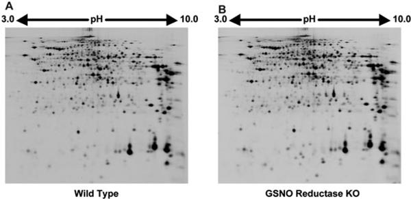
Representative 2D images of proteins in the liver extracts of wild-type (A) and GSNOR−/− mice (B) following LPS-treatment. Approximately 4000 protein spots were visualized by laser scanning.

Representative 2D images of proteins in the liver extracts of wild-type (A) and GSNOR−/− mice (B) following LPS-treatment. Approximately 4000 protein spots were visualized by laser scanning.