
Fig. 1.
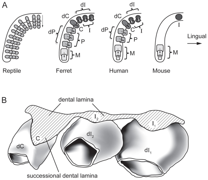
Variations in tooth replacement in reptiles and mammals, and serial addition of molars in mammals. (A) Schematics of dentitions of different vertebrates showing the left half of the lower jaws. Replacement teeth form at the lingual side of the dental arch. Mammalian molars are added serially in the posterior direction. Most reptiles have a homodont dentition, which is continuously replaced. Humans and ferrets represent typical mammals with a heterodont dentition composed of incisors, one canine, premolars and molars, and all teeth except molars are replaced once. Mice have one continuously growing incisor, a toothless diastema region and three molars. Mouse teeth are not replaced. (B) Reconstruction of human deciduous tooth germs illustrating their connection by the continuous dental lamina and the initiation of replacement tooth formation by budding of the successional dental lamina. Lingual view of anterior tooth germs in the lower jaw. A similar dental lamina is present in most other mammals and squamate reptiles. C, permanent canine; dC, deciduous canine; dI, deciduous incisor; dP, deciduous premolar; I, permanent incisor; M, molar; P, permanent premolar.