Abstract
The mitogen-activated protein kinases (MAPKs; also known as ERKs) are key intracellular signaling molecules that are ubiquitously expressed in tissues and were assumed to be functionally equivalent. Here, we use the mouse lens as a model system to investigate whether MAPK1 plays a specific role during development. MAPK3 is known to be dispensable for lens development. We demonstrate that, although MAPK1 is uniformly expressed in the lens epithelium, its deletion significantly reduces cell proliferation in the peripheral region, an area referred to as the lens germinative zone in which most active cell division occurs during normal lens development. By contrast, cell proliferation in the central region is minimally affected by MAPK1 deletion. Cell cycle regulators, including cyclin D1 and survivin, are downregulated in the germinative zone of the MAPK1-deficient lens. Interestingly, loss of MAPK1 subsequently induces upregulation of phosphorylated MAPK3 (pMAPK3) levels in the lens epithelium; however, this increase in pMAPK3 is not sufficient to restore cell proliferation in the germinative zone. Additionally, MAPK1 plays an essential role in epithelial cell survival but is dispensable for fiber cell differentiation during lens development. Our data indicate that MAPK1/3 control cell proliferation in the lens epithelium in a spatially defined manner; MAPK1 plays a unique role in establishing the highly mitotic zone in the peripheral region, whereas the two MAPKs share a redundant role in controlling cell proliferation in the central region of the lens epithelium.
Keywords: MAPK1, ERK, Lens, Proliferation, Survival, Mouse
INTRODUCTION
MAPK1 and MAPK3 (also known as ERK2 and ERK1, respectively) are members of the mitogen-activated protein kinase family. The classic MAPK1/3 activation pathway involves the sequential activation of the serine/threonine kinase Raf, a dual-specificity MAPK kinase (MAPKK or MEK) and then the MAPK (Sebolt-Leopold and Herrera, 2004). Activation of the Raf-MAPKK-MAPK kinase pathway transmits signals to both cytoplasmic signaling complexes and nuclear transcription factors, including protein kinases and phosphatases, signaling effectors, transcriptional regulators and cytoskeletal proteins (Yoon and Seger, 2006). As such, MAPK1/3 can activate a broad spectrum of cellular responses, ranging from cell proliferation and differentiation to migration and apoptosis. This evolutionarily conserved kinase pathway is a central signaling module that participates in numerous physiological and pathological processes, such as embryonic development and cancer (Osborne et al., 2012; Santamaria and Nebreda, 2010).
MAPK1/3 proteins are 84% identical, have similar biochemical properties, recognize the same substrates and share similar biological functions. Gene knockout studies in mice imply that the roles of MAPK1 and MAPK3 in vivo are not entirely equivalent. For instance, Mapk1 deletion results in early embryonic lethality due to failure in trophoblast formation, mesodermal differentiation and placental development (Hatano et al., 2003; Saba-El-Leil et al., 2003; Yao et al., 2003). By contrast, Mapk3-/- mice survive embryonic development with minor developmental defects in thymocyte and adipocyte differentiation (Bost et al., 2005; Pagès et al., 1999). Recent results from Mapk1 conditional deletion mice suggest that MAPK1 and MAPK3 are functionally redundant in certain tissues during development but distinct in others. For example, conditional deletion of Mapk1 in the CNS caused a mild phenotype in neurogenesis (Satoh et al., 2011b) and MAPK3 deficiency enhanced the phenotype in these mice, suggesting that the total MAPK activity is essential for normal CNS development. Interestingly, MAPK1-deficient mice exhibited marked abnormalities in social behaviors related to facets of autism-spectrum disorders in humans (Satoh et al., 2011a). Blocking MAPK3 activity in these mice with a pharmacological inhibitor did not cause additional psychological impairments, suggesting that MAPK1 has a unique role in the CNS in the control of social behavior. Overall, genetic data indicate two different scenarios: (1) the two MAPK isoforms are functionally interchangeable, and a sufficient threshold of total MAPK activity is important for normal tissue development and function; or (2) MAPK1 and MAPK3 have evolved to play unique roles in development and physiology. These two mechanistic models have not been examined extensively in developmental systems.
The ocular lens is a classic developmental system with which to study growth factor signaling in tissue induction and the regulation of cell proliferation and differentiation (Chow and Lang, 2001; Lovicu et al., 2011; Robinson, 2006). During lens morphogenesis, the presumptive lens ectoderm is induced by the underlying optic vesicle to form lens placode between mouse embryonic day (E) 9.0 and 9.5. The lens placode invaginates to form the lens vesicle between E10.5 and E11.5. Subsequently, the posterior lens vesicle cells elongate and differentiate into the primary fiber cells, which fill the lens vesicle at E12.5, whereas the anterior cells become the lens epithelial cells. After formation, lens growth is driven by cell proliferation and differentiation in a spatially restricted manner. Cell proliferation is limited to the anterior epithelial cells, with the greatest mitotic activity in a region just above the lens equator known as the lens germinative zone (Kallifatidis et al., 2011). The progeny cells that have migrated below the lens equator initiate the differentiation process and eventually form the secondary fiber cells. This coordinated proliferation and differentiation pattern is maintained throughout the lifespan of the animal.
Previous studies in transgenic and knockout mouse models demonstrated that FGF-Ras signaling is essential for cell proliferation, differentiation and survival during lens development (Burgess et al., 2010; Garcia et al., 2005; Govindarajan and Overbeek, 2001; Pan et al., 2010; Qu et al., 2011; Reneker et al., 2004; Xie et al., 2006; Zhao et al., 2008). Abolishing MAPK activity with the MAPKK (or MEK) inhibitor U0126 blocks the FGF-induced cell proliferation and elongation response in rat lens explants (Lovicu and McAvoy, 2001). However, the specific contributions of MAPK1 and MAPK3 cannot be simply extrapolated from these studies because both MAPK isoforms are equally affected. It is known that MAPK3 is not required for mouse lens development, but the importance of MAPK1 in the lens is still uncertain owing to early embryonic lethality (Hatano et al., 2003).
In this study, we generated conditional Mapk1 knockout mice using the presumptive lens driver Le-Cre, which is active at lens induction stage E9.0 (Ashery-Padan et al., 2000). In contrast to Mapk3-/- mice, the lens of Mapk1 conditional deletion mice exhibited severely compromised cell proliferation and survival. In the MAPK1-deficient lens, cell proliferation was drastically reduced in the peripheral germinative zone but not in the central epithelium. Increase of MAPK3 activity in the epithelium cannot compensate for the loss of MAPK1 to re-establish the cell proliferation pattern in the lens, suggesting that MAPK1 has an essential and unique role in the control of cell proliferation. MAPK1 activity is also crucial for lens epithelial cell survival, but is not required for fiber cell differentiation during lens development.
MATERIALS AND METHODS
Mapk1 conditional knockout (CKO) mice
Mice carrying the Mapk1 flox alleles and Mapk3-/- mice were described previously (Hatano et al., 2003; Pagès et al., 1999). In Le-Cre mice, the transgene was driven by a 6.5 kb genomic fragment from the mouse Pax6 gene (Ashery-Padan et al., 2000). The Le-Cre;Mapk1flox/flox mice (referred to as Mapk1CKO) were heterozygous for the Cre transgene in all the experiments because homozygous mice are reported to have an ocular phenotype (personal communication from Dr Michael Robinson at Miami University, Oxford, OH, USA). The Mapk1flox/flox mice are referred to here as wild type (WT). All of the animals were used in accordance with the Association of Research in Vision and Ophthalmology (ARVO) Statement for the Use of Animals in Ophthalmic and Vision Research, and all experimental procedures were approved by the Animal Care and Use Committee at the University of Missouri.
Histology, immunohistochemistry and immunofluorescence
The heads of mouse embryos or newborn pups were fixed in 4% paraformaldehyde overnight and processed for histological analysis as described previously (Xie et al., 2006). Nuclei were counterstained with Hematoxylin or DAPI for immunohistochemistry or immunofluorescence, respectively. The following primary antibodies were used: anti-MAPK1/3 (9102), anti-pMAPK1/3 (9101), anti-cleaved caspase 3 (9664) and anti-survivin (2808) (all from Cell Signaling Technology, Beverly, MA, USA); anti-phospho-histone H3 (sc8656), anti-cyclin D2 (sc593) and anti-cMAF (all from Santa Cruz Biotechnology, Santa Cruz, CA, USA); anti-cyclin D1 (ab16663), anti-p57Kip2 (ab4058) and anti-p53 (ab61256) (all from Abcam, Cambridge, MA, USA); anti-PAX6 (PRB-278P) and anti-PROX1 (PRB-238C) (both from Covance, Princeton, NJ, USA); anti-MAPK1 (05-157, Millipore, Billerica, MA, USA); and anti-Ki67 (M7249, Dako, Carpinteria, CA, USA). We did not find a reliable commercial anti-MAPK3 antibody that worked well for immunostaining. The anti-αA-crystallin antibody was a generous gift from Dr Richard Cenedella (Department of Biochemistry, College of Osteopathic Medicine, Kirksville, MO, USA) and anti-β;-crystallin and anti-γ-crystallin antibodies were from Dr Samuel Zigler (National Institutes of Health, Bethesda, MD, USA). For immunofluorescence, Alexa Fluor 488 (A11008) and 568 (A10042) conjugated secondary antibodies were from Invitrogen (Carlsbad, CA, USA). The signal enhancement TSA kit (NEL741B001KT, PerkinElmer, Boston, MA, USA) was used f or pMAPK1/3 and cleaved caspase 3 immunofluorescence. For immunohistochemistry, biotinylated secondary antibodies (BA1000 or BA4001) and Vectastain Elite ABC Reagent were from Vector Laboratories (Burlingame, CA, USA). Color development was performed using 3,3′-diaminobenzidine as a substrate (D4293, Sigma, St Louis, MO, USA).
BrdU and TUNEL analyses
Pregnant mice were injected intraperitoneally with 5-bromo-2′-deoxyuridine (BrdU) (B5002, Sigma) at 100 μg/g body weight and sacrificed after 1 hour or 2 days. The BrdU-labeled cells were detected by immunohistochemistry as described previously (Fromm et al., 1994). TUNEL assay was performed with an in situ apoptosis detection kit (S7165, Millipore) following the manufacturer’s instructions.
Western blot analysis
Newborn (P0) mouse lenses were homogenized in RIPA buffer for protein preparation and then subjected to western blot analysis (Xie et al., 2007). Each western blot was repeated at least twice with independent preparations of lens proteins. Radiographic films were scanned and band intensity was analyzed using Adobe Photoshop CS2 software. The anti-p38 (9212) and anti-phospho-p38 (9211) antibodies were from Cell Signaling Technology.
Statistical analysis
For quantitative analysis, serial sections across the entire lens were collected and central sections (judged by the size of the lens) from a minimum of three independent eyes for each genotype from the same litter were used (n, number of sections). Data are expressed as mean ± s.e.m. and P-values were calculated using Student’s t-test (P<0.05 considered significant).
RESULTS
Mapk1 deletion results in a small lens with severe defects in lens epithelium
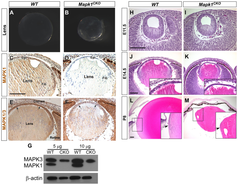
The Mapk1CKO mice were viable and fertile, but their eyes were microphthalmic and their lenses were slightly opaque and smaller than normal (Fig. 1A,B). To confirm MAPK1 deletion in Mapk1CKO lenses, immunostaining was performed at E14.5. Intensive MAPK1 staining was detected in the epithelial cell nuclei and in the fiber cells of the WT lens (Fig. 1C). By contrast, MAPK1 staining was absent in the Mapk1CKO lens (Fig. 1D). Immunohistochemistry against MAPK1/3 revealed uniform immune reactivity in the epithelial layer and in the fiber cells at a lower level in WT lenses (Fig. 1E). In comparison, the signal was significantly reduced in Mapk1CKO lenses; however, weak staining was still detected resulting from the presence of MAPK3 proteins (Fig. 1F). Western blot analysis confirmed that MAPK1 was lost, whereas the MAPK3 level was unchanged in Mapk1CKO lenses (Fig. 1G). Cre expression in the pancreas has been reported in Le-Cre mice (Ashery-Padan et al., 2000), but because the Mapk1CKO mice appeared healthy the MAPK1 levels in this tissue were not examined.
Fig. 1.
Mapk1 conditional deletion in mouse lens. (A,B) Lens gross morphology at P2. The Mapk1CKO lens was smaller than the WT lens and was slightly opaque. (C,D) MAPK1 immunohistochemistry at E14.5. MAPK1 was found in the epithelium (Epi) and fiber cells (Fib) in WT lens. MAPK1 immunoreactivity was absent in Mapk1CKO lens. (E,F) MAPK1/3 immunohistochemistry. MAPK1/3 levels were higher in the epithelial layer (arrows in E) than in the fiber cells in WT lens, and were significantly reduced in Mapk1CKO lens (arrows in F). (G) MAPK1/3 western blot. MAPK1 was absent whereas the MAPK3 level was unchanged in P0 Mapk1CKO lenses after normalization to the internal β;-actin control level. (H-M) Lens histology (Hematoxylin and Eosin). At E11.5, the lens vesicle in Mapk1CKO mice appeared the same as in WT (H,I). At E14.5, Mapk1CKO lens was smaller and the epithelial layer was thinner than for WT lens (J,K). At P8, Mapk1CKO lens was severely hypoplastic and developed vacuoles at the cortical region (M). However, the transitional zone (arrows in L and M insets) can still be identified in Mapk1CKO lens. Scale bars: 100 μm.
The effect of MAPK1 deletion on lens development was analyzed by histology (Fig. 1H-M). In the E11.5 WT embryo, the lens vesicle had formed and cells in the posterior half of the vesicle had begun to elongate to form the primary fiber cells (Fig. 1H). At this stage, the Mapk1CKO lenses were phenotypically indistinguishable from WT lenses (Fig. 1I), suggesting that MAPK1 deletion did not affect lens induction and formation. Although the E14.5 Mapk1CKO lenses retained normal polarity and architecture, they appeared substantially smaller than normal (Fig. 1J,K). The epithelial layer was thinner and with a lower cell density in Mapk1CKO compared with WT lenses (Fig. 1J,K, insets). By postnatal day (P) 8, Mapk1CKO lenses were severely hypoplastic, vacuoles had formed in the cortex (Fig. 1M; supplementary material Fig. S1D,H) and fiber cells were disorganized (supplementary material Fig. S1A,B). However, the equatorial region, in which epithelial cells are induced to differentiate into fiber cells (arrows in Fig. 1L,M insets), could still be identified in Mapk1CKO lenses. The Mapk1CKO mice also exhibited multiple defects in the anterior segments, but early retinal development appeared to be normal (supplementary material Fig. S1C-H). We also confirmed that MAPK3 deficiency did not affect lens or eye development (supplementary material Fig. S1I,J).
To monitor the progressive thinning of the lens epithelium in Mapk1CKO lenses, we compared the total number of epithelial cells with WT lenses (Fig. 2). At E11.5, the WT and Mapk1CKO lens epithelium had about the same number of cells (57±3 for WT and 54±2 for Mapk1CKO, P>0.5). During normal lens development, the epithelial cell number increases along with the expansion of the lens surface area and size. In WT lenses, epithelial cell numbers were 149.9±3.1, 209.7±3.6 and 235.7±2.3 per section at E14.5, E17.5 and P0, respectively. By contrast, epithelial cell numbers in Mapk1CKO lenses at these stages were 98.8±1.9, 93.6±2.3 and 88±1.9, a slight decrease as the lens grew. These results indicated that lens growth was severely inhibited by the loss of MAPK1.
Fig. 2.
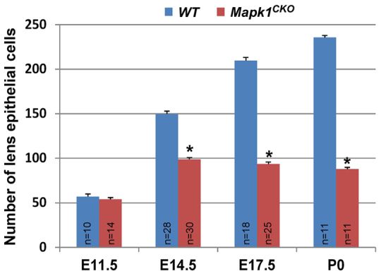
Reduction in epithelial cells in Mapk1CKO lens. The number of epithelial cells was similar in E11.5 Mapk1CKO and WT lenses, but declined as the mutant lenses aged. *P<0.0001 versus WT; error bars indicate s.e.m.
MAPK1 is essential for cell proliferation in the lens germinative zone
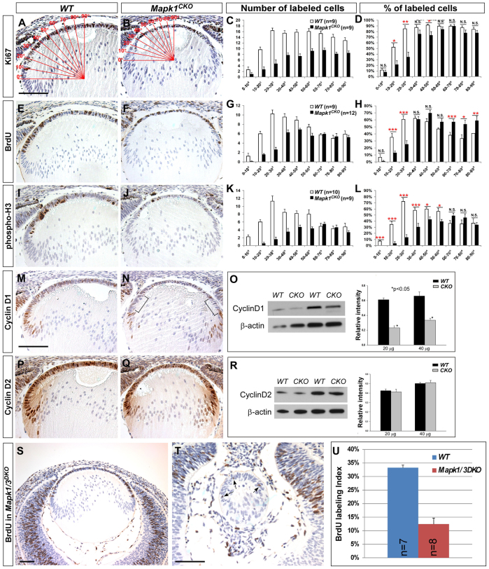
The epithelial defect in Mapk1CKO lenses suggested that MAPK1 might play an important role in cell proliferation, survival, or both during lens development. We first assessed the changes in cell proliferation by examining the expression patterns of several cell cycle markers. Ki67 is a nuclear protein present in proliferating cells (in G1, S, G2 and M phase) but absent from resting (G0) cells (Scholzen and Gerdes, 2000); thus, it is often used as a marker for determining the growth fraction of a given cell population. Our results showed that Ki67 is expressed in the epithelial layer in both WT and Mapk1CKO lenses at E14.5 (Fig. 3A,B). However, the region above the lens equator in Mapk1CKO lenses contained fewer Ki67+ cells than the same region in WT lenses. To quantify the regional distribution of Ki67+ cells, we divided the lens epithelium into 10° sections, with 0° at the lens equator and 90° in the middle of the central epithelium (Fig. 3A,B). At the equator, cells with horizontally located nuclei were considered as epithelial cells. We found that the total number of Ki67+ cells was lower in the Mapk1CKO than in the WT lens epithelium (Fig. 3C). When the Ki67 labeling index (ratio of Ki67+ cells over total epithelial cells) was measured, it was significantly reduced in the region between 10° and 30° in Mapk1CKO lenses, which corresponds to the germinative zone, an area in close contact with the anterior margin of the developing retina (Fig. 3D). Interestingly, beyond the lens germinative zone, the Ki67 labeling index was not significantly altered in Mapk1CKO lenses. Our data suggested that MAPK1 activity is required for cell proliferation in the germinative zone, whereas MAPK3 might be able to compensate for the loss of MAPK1 beyond this region.
Fig. 3.
Cell proliferation analysis at E14.5. (A-D) Ki67 immunohistochemistry. The total number of Ki67+ cells was significantly reduced in the epithelium of Mapk1CKO lens. Ki67 labeling index was decreased only in the germinative zone (10-30°) in Mapk1CKO lens (D). (E-H) BrdU incorporation assay. BrdU index was decreased in the germinative zone (10-30°) but increased in the central zone (60-90°) in Mapk1CKO lens. (I-L) Phospho-H3 immunohistochemistry. A significant reduction in the phospho-H3 index was seen in the Mapk1CKO lens in a broad area (0 to 60°). *P<0.05, **P<0.01, ***P<0.005; N.S., not significant. (M-R) Cyclin D1 (M,N) and cyclin D2 (P,Q) were expressed in lens epithelium and cortical fiber cell nuclei in both WT and Mapk1CKO lenses at E14.5. Compared with WT lens epithelium, Mapk1CKO lens epithelium had less cyclin D1, particularly in the germinative zone (brackets). By contrast, cyclin D2 expression patterns were similar in the two genotypes. (O,R) Western blot analysis. The band intensity for cyclin D1 and D2 was normalized to the internal control (β;-actin). Two sets of samples with different amounts of protein loading are shown. (S-U) Cell proliferation assay in Mapk1/3 double-knockout (DKO) lenses at E14.5. Mapk1/3DKO lens was severely hypoplastic and BrdU+ cells were drastically decreased. Arrows (T) indicate BrdU+ cells. Error bars indicate s.e.m. Scale bars: 100 μm.
To investigate cell cycle changes, we examined BrdU and phosphorylated histone H3 (phospho-H3) expression patterns in WT and Mapk1CKO lenses (Fig. 3E-L). BrdU labels cells in S phase and phospho-H3 is present in cells at G2-M (Gratzner, 1982; Hendzel et al., 1997). Consistent with the Ki67 results in the germinative zone (10-30°), the number of BrdU+ and phospho-H3+ cells and their labeling indices were significantly lower in Mapk1CKO lenses (Fig. 3E-L; supplementary material Fig. S2). Furthermore, the drastic reduction in the ratio of phospho-H3+ over Ki67+ cells suggested that G2-M phase progression was severely inhibited in the germinative zone in Mapk1CKO lenses (supplementary material Fig. S2). In the central epithelium (the region from 60-90°), the labeling index was either slightly higher (for BrdU) or unaffected (for phospho-H3), suggesting that MAPK3 or other signaling activity is sufficient to maintain mitotic activity in this area. Overall, the results supported the conclusion that MAPK1 is essential for cell proliferation and cell cycle progression in the lens germinative zone.
To confirm the redundant roles of MAPK1 and MAPK3 in regulating cell proliferation in central lens epithelium, Mapk1/3 double-deletion (referred to as Mapk1/3DKO) mice were generated and cell proliferation analyzed. It was known that lens development is normal in Mapk3-/- mice (supplementary material Fig. S1). At E14.5, the Mapk1/3DKO lenses were severely hypoplastic and BrdU+ cells were significantly reduced throughout the entire epithelial layer (Fig. 3S-U). By E16.5, BrdU+ cells were absent from Mapk1/3DKO lenses (data not shown). (Further studies on Mapk1/3DKO lenses will be presented in a separate report.)
It is known that mitogenic stimulation activates the D-type cyclins, which are essential for cell cycle entry and G1 phase progression (Cooper, 2000). The mouse lens expresses three isoforms of D-cyclins, with cyclin D2 as the major form (Chen et al., 2000; Fromm and Overbeek, 1996). We compared the expression patterns and levels of cyclin D1 and D2 in WT and Mapk1CKO lenses (Fig. 3M-R). In E14.5 WT lenses, cyclin D1 was expressed in the lens epithelial layer and its expression was upregulated at the lens equator (Fig. 3M). In Mapk1CKO lenses, cyclin D1 was visibly reduced in the epithelial layer, particularly in the germinative zone (indicated by brackets in Fig. 3N). For cyclin D2, the expression pattern appeared similar in the two genotypes (Fig. 3P,Q). Western blot analysis confirmed that the levels of cyclin D1 were decreased, whereas cyclin D2 levels were unchanged in Mapk1CKO lenses (Fig. 3O,R).
Loss of MAPK1 increases apoptosis in lens epithelium
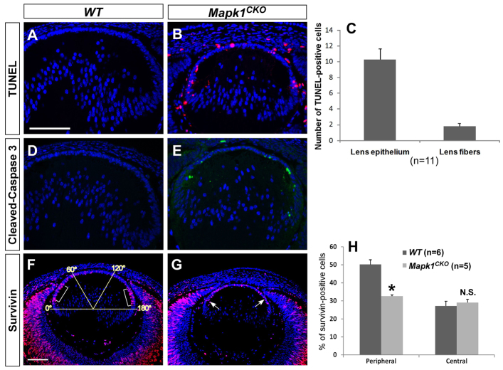
We have shown that MAPK1 deletion severely reduces cell proliferation in the lens germinative zone, whereas the central zone was largely unaffected. Because the number of epithelial cells did not increase with age in Mapk1CKO lenses (Fig. 2), we speculated that cell death could also contribute to the defect. TUNEL assays revealed that TUNEL+ nuclei were not detected in E14.5 WT lenses (Fig. 4A), whereas significant numbers of epithelial cells were undergoing apoptosis in Mapk1CKO lenses (10.3±1.4 TUNEL+ cells per section; Fig. 4B,C). A small number of fiber nuclei were also TUNEL+ (1.8±0.4) in Mapk1CKO lenses. Activation of apoptosis in Mapk1CKO lenses was also demonstrated by cleaved (active) caspase 3 immunofluorescence (Fig. 4D,E). These results together suggest that, in addition to reduced cell proliferation in the germinative zone, apoptosis across the epithelial layer is also a contributing factor to the reduced cell number and stalled growth of Mapk1CKO lenses.
Fig. 4.
Apoptosis analysis at E14.5. (A-C) TUNEL assay. TUNEL+ cells were found in the lens epithelial layer and a few in the fiber compartment in Mapk1CKO lens, whereas none were detected in WT lens. The average number of TUNEL+ cells per section is shown (C). (D,E) Cleaved caspase 3 immunofluorescence. Positive cells were mostly localized in the epithelium of the Mapk1CKO lens. (F-H) Survivin expression. In WT lens epithelium, more survivin+ cells were found in the germinative zone (brackets in F) than in the central zone. Survivin expression was reduced in the germinative zone (arrows in G) but not in the central zone in Mapk1CKO lens. The survivin labeling index (H) was measured by dividing the lens epithelium into three regions, and the number for the peripheral region is the average of the left (0-60°) and right (120-180°) regions. *P<0.005; N.S., not significant. Error bars indicate s.e.m. Scale bars: 100 μm.
Survivin (BIRC5 - Mouse Genome Informatics) is highly expressed during embryonic lens development (Uren et al., 2000; Weber and Menko, 2005). Survivin inhibits apoptosis by blocking the activities of caspase 3, 7 and 9 and also plays an important role in cell proliferation by regulating the G2/M phase of the cell cycle (Chandele et al., 2004; Li et al., 1998; Shin et al., 2001). In vitro studies have shown that the survivin level can be regulated by MAPK activity (Teh et al., 2004; Wang et al., 2010). Survivin was expressed in the epithelial cells of E14.5 WT lenses, and the level was higher in the germinative zone than in the central zone (Fig. 4F,H). Loss of MAPK1 reduced survivin expression only in the germinative zone (Fig. 4G,H), which could be responsible for the increased apoptosis in this region in Mapk1CKO lenses. Other apoptosis-regulatory proteins, such as p53 (TRP53 - Mouse Genome Informatics) (Pan and Griep, 1995) and the anti-apoptotic protein XIAP (XAF1 - Mouse Genome Informatics) (Leonard et al., 2007), were also examined, but their expression patterns were not discernably altered in Mapk1CKO lenses (supplementary material Fig. S3A-D).
MAPK1 is dispensable for cell cycle exit, crystallin expression and fiber cell differentiation
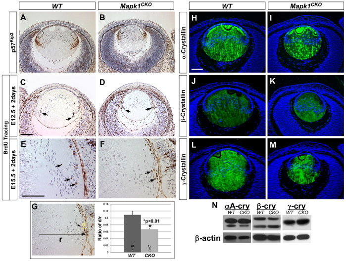
During normal lens development, following cell division in the germinative zone the epithelial cells near the lens equator exit the cell cycle and differentiate into secondary fiber cells (Griep, 2006; Zhang et al., 1998). Fiber differentiation is coupled with upregulation of the cell cycle inhibitor p57Kip2 (CDKN1C - Mouse Genome Informatics) (Fig. 5A). Deletion of atypical protein kinase C λ (aPKCλ; PRKCι - Mouse Genome Informatics) in mouse lens has been shown to reduce cell proliferation in the germinative zone, probably as a result of increased p57Kip2 and premature cell cycle withdrawal in this region (Sugiyama et al., 2009). To determine whether a similar change also occurs in Mapk1CKO lenses, we compared p57Kip2 expression patterns in E14.5 lenses between the two genotypes (Fig. 5A,B). Although cell proliferation was drastically reduced in the germinative zone in Mapk1CKO lenses, unlike aPKCλ-deficient lenses the p57Kip2 expression level was not increased in this region (Fig. 5B). Furthermore, these cells still expressed the epithelial cell marker E-cadherin (data not shown). Therefore, our results suggest that lack of cell proliferation in the germinative zone in Mapk1CKO lenses is not caused by dysregulation of p57Kip2 expression.
Fig. 5.
Fiber cell differentiation. (A,B) p57Kip2 expression was upregulated in the equatorial region in both WT and Mapk1CKO lenses at E14.5. There was no evidence for premature p57Kip2 expression in the germinative zone (brackets) of the Mapk1CKO lens. (C-G) BrdU tracing experiment. Embryos were exposed to BrdU at E12.5 (C,D) or E15.5 (E,F) and the BrdU incorporation pattern in the lens was examined 2 days later. BrdU-labeled nuclei were found in the cortical fiber cells in both WT and Mapk1CKO lenses (arrows), indicating that the proliferating epithelial cells had differentiated into the fiber cells. (G) The distance travelled by the most inwardly migrated BrdU+ nuclei (d) and the lens radius (r) were measured, and the ratio was calculated. BrdU-tagged nuclei moved towards the core more quickly in WT than those in Mapk1CKO lenses. Error bars indicate s.e.m. (H-N) Crystallin expression. In E14.5 WT lens, αA-crystallin was expressed in both lens epithelium and fibers (H), whereas β;- and γ-crystallins were detected in lens fibers (J and L, respectively). Similar expression patterns were found in E14.5 Mapk1CKO lenses (I,K,M). Western blots using P0 lens proteins were quantified after normalizing to β;-actin levels and the result confirmed that crystallin expression levels were unchanged in Mapk1CKO lenses (N). Scale bars: 100 μm.
Cell proliferation in the germinative zone is thought to be a critical step in priming the cells for secondary fiber differentiation (Sue Menko, 2002). To determine whether a cell proliferation defect in the germinative zone would prevent cells from differentiating into fiber cells, we followed lens cell fate using a BrdU incorporation and tracing assay (Kallifatidis et al., 2011). The proliferating cells were marked with BrdU at E12.5 or E15.5 and the fate of these cells was examined at E14.5 or E17.5, respectively (Fig. 5C-F). In WT lenses, the cell nuclei in the lens cortex were strongly labeled with BrdU, indicating that the initially BrdU-tagged epithelial cells had differentiated into fiber cells (Fig. 5C,E, arrows). In Mapk1CKO lenses, BrdU-tagged cells had also differentiated, but had not moved as far into the lens cortex as in WT lenses (Fig. 5F,G). This suggests that epithelial-to-fiber differentiation still occurs despite the defective cell proliferation in the germinative zone.
Lens fiber cell differentiation is characterized by the temporal and spatially restricted expression of crystallin genes (Cvekl and Duncan, 2007). Our data showed that loss of MAPK1 did not affect crystallin gene expression (Fig. 5H-N). The expression patterns of transcription factors such as PAX6, PROX1 and cMAF, which are known to be crucial for crystallin expression, were also not discernibly altered in Mapk1CKO lenses (supplementary material Fig. S3E-J). These data indicated that MAPK1 is not essential for secondary fiber differentiation during lens development.
Increase in phosphorylated MAPK3 in Mapk1CKO lenses
Both MAPK1 and MAPK3 are downstream targets of MAPKK (or MEK) in the growth factor-stimulated receptor tyrosine kinase (RTK)-Ras-MAPK signaling pathway. MAPK1 deletion could potentially alter the phosphorylation level of MAPK3 in the lens. We therefore examined MAPK activity by immunofluorescence and western blotting against phosphorylated MAPK1/3 (pMAPK1/3) (Fig. 6A-F; supplementary material Fig. S4A,B). In WT lenses, pMAPK immunofluorescence was most visible in the equatorial region (Fig. 6A,C, arrows). In Mapk1CKO lenses, pMAPK was still detectable at a level comparable to that in WT lenses, but was more anteriorly localized, probably as a result of the reduced lens size and subsequent anterior shift of the retinal margins (see Fig. 6H). Overall, the high intensity pMAPK staining area in Mapk1CKO lenses was still positioned adjacent to the anterior margin of the retina, a similar spatial relationship to that seen in WT eyes. When pMAPK immunofluorescence was performed without signal enhancement, the pMAPK levels progressively increased in Mapk1CKO lenses from E14.5 to E17.5 (data not shown). By P0, the pMAPK intensity was higher in Mapk1CKO than in WT lenses (supplementary material Fig. S4A,B). The increase in pMAPK3 (but not MAPK3 protein) level in Mapk1CKO lenses was confirmed by western blot analysis (Fig. 6E,F), suggesting that MAPK3 is hyperactive in Mapk1CKO lenses, probably as an adaptive response to compensate for the loss of MAPK1 activity. We also compared the levels of other signaling molecules, including JNK, p38 and Akt (MAPK8, MAPK14 and AKT1 - Mouse Genome Informatics), between the two genotypes. We found that the phosphorylated p38 (p-p38) level was higher in Mapk1CKO lenses (Fig. 6E,F) than in WT lenses. The other two kinases (JNK and Akt) were unchanged (data not shown). Loss of MAPK1 might trigger a stress response and activate p38 in Mapk1CKO lenses.
Fig. 6.
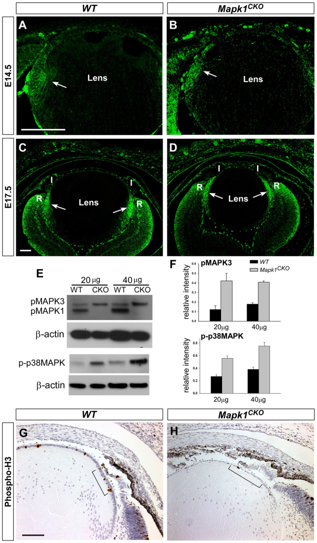
MAPK1/3 and p38 activity levels and phospho-H3 immunostaining. (A-D) pMAPK immunofluorescence. In both WT and Mapk1CKO lenses, pMAPK immunoreactivity was detected in the lens equatorial region (arrows). In E17.5 Mapk1CKO lens, pMAPK appeared to be more anteriorly localized, corresponding to the anterior shift of the retinal margin and the growing iris (I). R, retina. (E,F) Western blot analysis shows that the pMAPK3 level was increased and the pMAPK1 band absent in P0 Mapk1CKO lenses. Additionally, the phosphorylated p38 (p-p38) level was increased in Mapk1CKO lenses. Error bars indicate s.e.m. (G,H) phospho-H3 immunohistochemistry (E17.5). In WT lens, more phospho-H3+ cells were localized in the germinative zone (brackets) than in the central zone. Phospho-H3+ cells were drastically reduced in Mapk1CKO lens. Scale bars: 100 μm.
To determine whether an increase of pMAPK3 in the epithelium can rescue the cell proliferation defect in the germinative zone in Mapk1CKO lenses, phospho-H3 staining was performed on E17.5 and P0 lenses (Fig. 6G,H; supplementary material Fig. S4C,D). Although the overall ocular architecture of the mutant eye had altered as a result of a smaller lens, we could still identify the presumptive lens germinative zone as the area adjacent to the anterior margin of the retina (brackets in Fig. 6G,H). Phospho-H3+ cells in this area were still visibly reduced in Mapk1CKO lenses, suggesting that an increase in pMAPK3 cannot compensate for the loss of MAPK1 function in the germinative zone.
To investigate whether MAPK1 is preferentially activated over MAPK3 in response to growth factor stimulation, we examined pMAPK levels in transgenic mouse lenses overexpressing human platelet-derived growth factor A (PDGF-A) (Reneker and Overbeek, 1996). The lens epithelial cells in PDGFA transgenic mice were hyperproliferative (supplementary material Fig. S4F). MAPK1/3 protein levels were not altered, but pMAPK1/3 levels were higher in PDGFA transgenic lenses than in WT lenses (supplementary material Fig. S4G,H). The increase in pMAPK1 was more substantial than that of pMAPK3, suggesting that MAPK1 is more responsive to PDGF-A stimulation, which led to hyperproliferation in the lens epithelium.
DISCUSSION
The FGFR-Ras-MAPK signaling pathway has been implicated in the control of lens cell proliferation, differentiation and survival during embryonic development (Lovicu and McAvoy, 2001; Xie et al., 2006; Zhao et al., 2008). The mouse lens expresses a higher level of MAPK3 than MAPK1 protein (Fig. 1G), but the pMAPK1 level is higher than that of pMAPK3 (Fig. 6E), suggesting that MAPK1 is preferentially stimulated in the lens. In this study, we used a lens ectodermal driver (Le-Cre) to delete Mapk1 in the lens from the placode stage (E9.0). We demonstrate that the growth of Mapk1CKO lens is severely retarded in contrast to the growth of the MAPK3-deficient lens, which did not display any defects (supplementary material Fig. S1I,J). We showed that MAPK1 is not required for early lens development (before E11.5) and is dispensable for fiber differentiation (Fig. 5), but is essential for cell proliferation in the germinative zone during the lens growth phase. An increase in MAPK3 activity in this region cannot compensate for the loss of MAPK1 (Fig. 6; supplementary material Fig. S4), suggesting that MAPK1 plays a unique role in maintaining the cell proliferation pattern during lens development.
Cell proliferation analysis (Fig. 3; supplementary material Fig. S2) suggests that lens cells in the germinative zone are not actively dividing and they mostly resemble the cells at the quiescent (G0) state. Cells can enter a quiescent stage if completion of one phase of the cycle does not occur successfully. Mechanistically, reduction in the cyclin D1 level could restrict the cells from passing the G1-S phase checkpoint. More importantly, a drastic reduction in the phospho-H3 index in Mapk1CKO lenses (supplementary material Fig. S2) affects G2/M phase progression. Despite the proliferation defect in the lens germinative zone, fiber differentiation markers were not prematurely switched on in this region, suggesting that loss of MAPK1 leads to uncoupling of cell cycle exit and fiber cell differentiation in the equatorial zone of the lens. Thus, the role of MAPK1 in the lens germinative zone is to enhance the cell division rate while preventing cells from premature withdrawal from the cell cycle.
In all vertebrates, the growth of the lens follows a distinct, polarized pattern. Proliferating cells are restricted to the anterior lens epithelium and differentiating fiber cells are confined to the posterior hemisphere (Fig. 7). In the mid 1960s, the lens rotation experiment in chicken eye performed by Coulombre and Coulombre demonstrated that the state of the lens cell, either proliferating or differentiating, depends on its position in the eye (Coulombre and Coulombre, 1963). Within the lens epithelial layer the mitotic activity differs by region, although all of the cells retain the ability to proliferate (Lang, 1997). The highly mitotic region, which is defined as the lens germinative zone, is in close proximity to the anterior neural retina, which later differentiates into the iris and the ciliary body (Fig. 7). Various growth factors have been shown to be expressed in the anterior retina and developing iris/ciliary body (Reneker and Overbeek, 1996; Wilkinson et al., 1989). Both in vitro and in vivo studies have demonstrated that these growth factors can act as mitogens to stimulate cell proliferation (Iyengar et al., 2006; Potts et al., 1994; Reneker and Overbeek, 1996). The endogenous inductive molecules have not yet been identified and they are likely to play redundant roles in stimulating cell proliferation. Based on the data from previous and current studies, we proposed the following model to explain the high mitotic activity in the germinative zone during lens development (Fig. 7). After the lens formation stage (>E12.5), growth factors released from the anterior tip of the retina bind to the nearby lens cells and activate the RTK-Ras-MAPK1 signal transduction pathway to enhance the mitotic activity in this region. The downstream targets known to play important roles in cell cycle progression identified in this study include cyclin D1, phospho-H3 and survivin. Our data imply that MAPK1 has a unique role in the control of the lens cell proliferation pattern and that MAPK1 and MAPK3 are not functionally interchangeable.
Fig. 7.
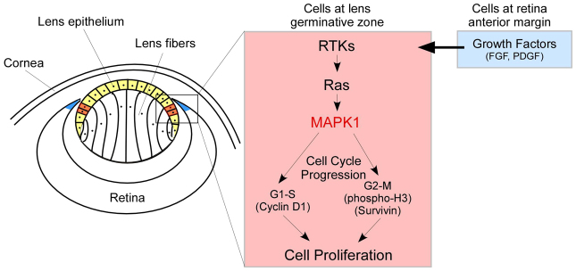
MAPK1 activation is required for cell proliferation in the germinative zone during lens development. MAPK1 plays a unique role in promoting high mitotic activity in the lens germinative zone. Growth factors (such as FGF and PDGF) released from the anterior margin of the retina (blue) stimulate the RTK-Ras-MAPK1 pathway in the adjacent lens cells (red) through a paracrine mechanism. Cell cycle regulatory proteins downstream of MAPK1 identified in this study include cyclin D1, phospho-H3 and survivin. Upregulation of MAPK3 activity in Mapk1CKO lens cannot compensate for the loss of MAPK1. MAPK1 activity in lens epithelium (yellow) is also essential for cell survival during development.
We do not have a clear answer as to why MAPK1 and not MAPK3 functions uniquely in stimulating cell proliferation in the lens germinative zone. One explanation could be that MAPK1 has a higher capacity than MAPK3 to interact with the upstream activator MAPKK and therefore would enhance the signaling output (Vantaggiato et al., 2006). However, this model does not fit well with the observation that an increase in pMAPK3 level failed to restore the lens germinative zone in Mapk1CKO lens (Fig. 6; supplementary material Fig. S4). An alternative explanation could be the different levels of effectiveness of MAPK1 and MAPK3 in activating the downstream targets leading to nuclear signaling. It is known that the nuclear localization of active MAPK is necessary for controlling the gene expression program stimulated by growth factors. MAPK1 and MAPK3 have been shown to differ drastically in their capacity to cross the nuclear envelope, contributing to the observation that MAPK1 is more efficient than MAPK3 in signaling to the nucleus (Marchi et al., 2008). Such a difference could make the transcription-dependent processes, such as G1 phase entry during the cell cycle, more dependent on MAPK1 than on MAPK3 (Baldin et al., 1993). We showed that MAPK1 proteins are mostly localized in the nuclei of the lens epithelial cells (Fig. 1C). We also showed that the cyclin D1 level was dramatically reduced in the germinative zone in Mapk1CKO lens. Thus, our findings appear to support this model. In the lens germinative zone, MAPK1 function is indispensable because high mitotic activity is required. Even when MAPK3 activity was upregulated in P0 Mapk1CKO lens, it did not restore the mitotic pattern (supplementary material Fig. S4). By contrast, in the central region of the lens epithelium, where mitotic activity is relatively low, MAPK3 is sufficient to compensate for the loss of MAPK1 in Mapk1CKO lens.
Our study also indicates that cell proliferation in Mapk1CKO lenses is unaffected before E11.5 (Fig. 1H,I; Fig. 2) and in the central region of the epithelium at a later stage (Fig. 3). To clarify the redundant role of MAPK1 and MAPK3 in cell proliferation during lens development, we recently generated Mapk1/3 double-deletion mice. The preliminary results indicate that cell proliferation before E11.5 is likely to be mediated by MAPK1/3-independent pathways (data not shown). It has been shown that BMP/activin-activated signals are essential for cell proliferation during early lens development (Rajagopal et al., 2009). Additionally, our results indicated that loss of both MAPK1 and MAPK3 in the lens drastically reduced cell proliferation throughout the entire epithelial layer at E14.5 (Fig. 3S-U), suggesting that MAPK1 and MAPK3 play a redundant role in the control of cell proliferation in the central region of the lens epithelium.
In this study, we have shown that apoptosis is significantly increased throughout the entire epithelial layer in the absence of MAPK1 (Fig. 4). Previous studies showed that disruption of cell cycle regulation can trigger apoptosis in the lens (Chen et al., 2000; Pan and Griep, 1995). In our study, cell death was found across the entire lens epithelial layer and was not limited to just the germinative zone, where cell proliferation was severely affected. Therefore, we speculate that apoptosis in the lens epithelium (at least in the central zone) is a direct effect of the absence of MAPK1 activity and not an indirect effect resulting from abnormal cell cycle regulation. We conclude that cell survival in the lens epithelium is dependent on MAPK1 activity and that MAPK3 activity alone is not sufficient.
FGF signaling is known to be essential for lens cell survival. Deletion of Fgfr2 alone, Fgfr1 and Fgfr2 or Fgfr1-3, all induced apoptosis in mouse lens (Garcia et al., 2011; Garcia et al., 2005; Zhao et al., 2008). Loss of FGFRs resulted in reduced or absent MAPK activity in the lens. Our study suggests that the FGFR-mediated survival signal is, at least in part, mediated through MAPK1 activation. How does MAPK1 regulate cell survival? One possibility is by regulating the level of survivin, which is a member of the inhibitor of apoptosis (IAP) family (Fig. 4). The function of survivin is to inhibit caspase 3 activation and thereby negatively regulate apoptosis (Li et al., 1998; Shin et al., 2001). We have demonstrated that MAPK1 deficiency results in a decrease in the survivin level (Fig. 4F,G) and in an increase in cleaved caspase 3 (Fig. 4E) in the peripheral region of the lens epithelium. It has been shown that MAPK activity plays a role in survivin expression and stability (Teh et al., 2004; Wang et al., 2010). However, other MAPK1-dependent survival signals must exist in the central epithelium during lens development that are independent of survivin.
In summary, the data presented in this study provide novel evidence concerning the unique role of MAPK1 in the control of the cell proliferation pattern during mouse lens development. MAPK1 appears to play an essential role in the lens germinative zone, whereas its function in cell proliferation in the central epithelium is likely to be redundant with MAPK3. Our study also shows that the overall MAPK activity in the lens epithelial layer is crucial for cell survival. However, MAPK1 deficiency does not block the process of epithelial-to-fiber differentiation. Future studies will focus on the combined deletion of MAPK1 and MAPK3 in the lens to elucidate the role of total MAPK activity in lens fiber differentiation and lens development.
Supplementary Material
Acknowledgments
We thank Drs Ruth Ashery-Padan and Gilles Pagès for permission to use the Le-Cre and Mapk3-/- mice, respectively; Dr Samuel Zigler for anti-crystallin antibodies; and Drs Venkatesh Govindarajan, Paul Overbeek and Michael Robinson for insightful comments and suggestions.
Footnotes
Funding
This work was supported by the National Institutes of Health [grants EY13146 and EY14795] and unrestricted funding from Research to Prevent Blindness (RPB). Deposited in PMC for release after 12 months.
Competing interests statement
The authors declare no competing financial interests.
Supplementary material
Supplementary material available online at http://dev.biologists.org/lookup/suppl/doi:10.1242/dev.081042/-/DC1
References
- Ashery-Padan R., Marquardt T., Zhou X., Gruss P. (2000). Pax6 activity in the lens primordium is required for lens formation and for correct placement of a single retina in the eye. Genes Dev. 14, 2701–2711 [DOI] [PMC free article] [PubMed] [Google Scholar]
- Baldin V., Lukas J., Marcote M. J., Pagano M., Draetta G. (1993). Cyclin D1 is a nuclear protein required for cell cycle progression in G1. Genes Dev. 7, 812–821 [DOI] [PubMed] [Google Scholar]
- Bost F., Aouadi M., Caron L., Even P., Belmonte N., Prot M., Dani C., Hofman P., Pagès G., Pouysségur J., et al. (2005). The extracellular signal-regulated kinase isoform ERK1 is specifically required for in vitro and in vivo adipogenesis. Diabetes 54, 402–411 [DOI] [PubMed] [Google Scholar]
- Burgess D., Zhang Y., Siefker E., Vaca R., Kuracha M. R., Reneker L., Overbeek P. A., Govindarajan V. (2010). Activated Ras alters lens and corneal development through induction of distinct downstream targets. BMC Dev. Biol. 10, 13 [DOI] [PMC free article] [PubMed] [Google Scholar]
- Chandele A., Prasad V., Jagtap J. C., Shukla R., Shastry P. R. (2004). Upregulation of survivin in G2/M cells and inhibition of caspase 9 activity enhances resistance in staurosporine-induced apoptosis. Neoplasia 6, 29–40 [DOI] [PMC free article] [PubMed] [Google Scholar]
- Chen Q., Hung F. C., Fromm L., Overbeek P. A. (2000). Induction of cell cycle entry and cell death in postmitotic lens fiber cells by overexpression of E2F1 or E2F2. Invest. Ophthalmol. Vis. Sci. 41, 4223–4231 [PubMed] [Google Scholar]
- Chow R. L., Lang R. A. (2001). Early eye development in vertebrates. Annu. Rev. Cell Dev. Biol. 17, 255–296 [DOI] [PubMed] [Google Scholar]
- Cooper S. (2000). The continuum model and G1-control of the mammalian cell cycle. Prog. Cell Cycle Res. 4, 27–39 [DOI] [PubMed] [Google Scholar]
- Coulombre J. L., Coulombre A. J. (1963). Lens development: fiber elongation and lens orientation. Science 142, 1489–1490 [DOI] [PubMed] [Google Scholar]
- Cvekl A., Duncan M. K. (2007). Genetic and epigenetic mechanisms of gene regulation during lens development. Prog. Retin. Eye Res. 26, 555–597 [DOI] [PMC free article] [PubMed] [Google Scholar]
- Fromm L., Overbeek P. A. (1996). Regulation of cyclin and cyclin-dependent kinase gene expression during lens differentiation requires the retinoblastoma protein. Oncogene 12, 69–75 [PubMed] [Google Scholar]
- Fromm L., Shawlot W., Gunning K., Butel J. S., Overbeek P. A. (1994). The retinoblastoma protein-binding region of simian virus 40 large T antigen alters cell cycle regulation in lenses of transgenic mice. Mol. Cell. Biol. 14, 6743–6754 [DOI] [PMC free article] [PubMed] [Google Scholar]
- Garcia C. M., Yu K., Zhao H., Ashery-Padan R., Ornitz D. M., Robinson M. L., Beebe D. C. (2005). Signaling through FGF receptor-2 is required for lens cell survival and for withdrawal from the cell cycle during lens fiber cell differentiation. Dev. Dyn. 233, 516–527 [DOI] [PubMed] [Google Scholar]
- Garcia C. M., Huang J., Madakashira B. P., Liu Y., Rajagopal R., Dattilo L., Robinson M. L., Beebe D. C. (2011). The function of FGF signaling in the lens placode. Dev. Biol. 351, 176–185 [DOI] [PMC free article] [PubMed] [Google Scholar]
- Govindarajan V., Overbeek P. A. (2001). Secreted FGFR3, but not FGFR1, inhibits lens fiber differentiation. Development 128, 1617–1627 [DOI] [PubMed] [Google Scholar]
- Gratzner H. G. (1982). Monoclonal antibody to 5-bromo- and 5-iododeoxyuridine: A new reagent for detection of DNA replication. Science 218, 474–475 [DOI] [PubMed] [Google Scholar]
- Griep A. E. (2006). Cell cycle regulation in the developing lens. Semin. Cell Dev. Biol. 17, 686–697 [DOI] [PMC free article] [PubMed] [Google Scholar]
- Hatano N., Mori Y., Oh-hora M., Kosugi A., Fujikawa T., Nakai N., Niwa H., Miyazaki J., Hamaoka T., Ogata M. (2003). Essential role for ERK2 mitogen-activated protein kinase in placental development. Genes Cells 8, 847–856 [DOI] [PubMed] [Google Scholar]
- Hendzel M. J., Wei Y., Mancini M. A., Van Hooser A., Ranalli T., Brinkley B. R., Bazett-Jones D. P., Allis C. D. (1997). Mitosis-specific phosphorylation of histone H3 initiates primarily within pericentromeric heterochromatin during G2 and spreads in an ordered fashion coincident with mitotic chromosome condensation. Chromosoma 106, 348–360 [DOI] [PubMed] [Google Scholar]
- Iyengar L., Patkunanathan B., Lynch O. T., McAvoy J. W., Rasko J. E., Lovicu F. J. (2006). Aqueous humour- and growth factor-induced lens cell proliferation is dependent on MAPK/ERK1/2 and Akt/PI3-K signalling. Exp. Eye Res. 83, 667–678 [DOI] [PubMed] [Google Scholar]
- Kallifatidis G., Boros J., Shin E. H., McAvoy J. W., Lovicu F. J. (2011). The fate of dividing cells during lens morphogenesis, differentiation and growth. Exp. Eye Res. 92, 502–511 [DOI] [PMC free article] [PubMed] [Google Scholar]
- Lang R. A. (1997). Apoptosis in mammalian eye development: lens morphogenesis, vascular regression and immune privilege. Cell Death Differ. 4, 12–20 [DOI] [PubMed] [Google Scholar]
- Leonard K. C., Petrin D., Coupland S. G., Baker A. N., Leonard B. C., LaCasse E. C., Hauswirth W. W., Korneluk R. G., Tsilfidis C. (2007). XIAP protection of photoreceptors in animal models of retinitis pigmentosa. PLoS ONE 2, e314 [DOI] [PMC free article] [PubMed] [Google Scholar]
- Li F., Ambrosini G., Chu E. Y., Plescia J., Tognin S., Marchisio P. C., Altieri D. C. (1998). Control of apoptosis and mitotic spindle checkpoint by survivin. Nature 396, 580–584 [DOI] [PubMed] [Google Scholar]
- Lovicu F. J., McAvoy J. W. (2001). FGF-induced lens cell proliferation and differentiation is dependent on MAPK (ERK1/2) signalling. Development 128, 5075–5084 [DOI] [PubMed] [Google Scholar]
- Lovicu F. J., McAvoy J. W., de Iongh R. U. (2011). Understanding the role of growth factors in embryonic development: insights from the lens. Philos. Trans. R. Soc. Lond. B Biol. Sci. 366, 1204–1218 [DOI] [PMC free article] [PubMed] [Google Scholar]
- Marchi M., D’Antoni A., Formentini I., Parra R., Brambilla R., Ratto G. M., Costa M. (2008). The N-terminal domain of ERK1 accounts for the functional differences with ERK2. PLoS ONE 3, e3873 [DOI] [PMC free article] [PubMed] [Google Scholar]
- Osborne J. K., Zaganjor E., Cobb M. H. (2012). Signal control through Raf: in sickness and in health. Cell Res. 22, 14–22 [DOI] [PMC free article] [PubMed] [Google Scholar]
- Pagès G., Guérin S., Grall D., Bonino F., Smith A., Anjuere F., Auberger P., Pouysségur J. (1999). Defective thymocyte maturation in p44 MAP kinase (Erk 1) knockout mice. Science 286, 1374–1377 [DOI] [PubMed] [Google Scholar]
- Pan H., Griep A. E. (1995). Temporally distinct patterns of p53-dependent and p53-independent apoptosis during mouse lens development. Genes Dev. 9, 2157–2169 [DOI] [PubMed] [Google Scholar]
- Pan Y., Carbe C., Powers A., Feng G. S., Zhang X. (2010). Sprouty2-modulated Kras signaling rescues Shp2 deficiency during lens and lacrimal gland development. Development 137, 1085–1093 [DOI] [PMC free article] [PubMed] [Google Scholar]
- Potts J. D., Bassnett S., Kornacker S., Beebe D. C. (1994). Expression of platelet-derived growth factor receptors in the developing chicken lens. Invest. Ophthalmol. Vis. Sci. 35, 3413–3421 [PubMed] [Google Scholar]
- Qu X., Hertzler K., Pan Y., Grobe K., Robinson M. L., Zhang X. (2011). Genetic epistasis between heparan sulfate and FGF-Ras signaling controls lens development. Dev. Biol. 355, 12–20 [DOI] [PMC free article] [PubMed] [Google Scholar]
- Rajagopal R., Huang J., Dattilo L. K., Kaartinen V., Mishina Y., Deng C. X., Umans L., Zwijsen A., Roberts A. B., Beebe D. C. (2009). The type I BMP receptors, Bmpr1a and Acvr1, activate multiple signaling pathways to regulate lens formation. Dev. Biol. 335, 305–316 [DOI] [PMC free article] [PubMed] [Google Scholar]
- Reneker L. W., Overbeek P. A. (1996). Lens-specific expression of PDGF-A alters lens growth and development. Dev. Biol. 180, 554–565 [DOI] [PubMed] [Google Scholar]
- Reneker L. W., Xie L., Xu L., Govindarajan V., Overbeek P. A. (2004). Activated Ras induces lens epithelial cell hyperplasia but not premature differentiation. Int. J. Dev. Biol. 48, 879–888 [DOI] [PubMed] [Google Scholar]
- Robinson M. L. (2006). An essential role for FGF receptor signaling in lens development. Semin. Cell Dev. Biol. 17, 726–740 [DOI] [PMC free article] [PubMed] [Google Scholar]
- Saba-El-Leil M. K., Vella F. D., Vernay B., Voisin L., Chen L., Labrecque N., Ang S. L., Meloche S. (2003). An essential function of the mitogen-activated protein kinase Erk2 in mouse trophoblast development. EMBO Rep. 4, 964–968 [DOI] [PMC free article] [PubMed] [Google Scholar]
- Santamaria P. G., Nebreda A. R. (2010). Deconstructing ERK signaling in tumorigenesis. Mol. Cell 38, 3–5 [DOI] [PubMed] [Google Scholar]
- Satoh Y., Endo S., Nakata T., Kobayashi Y., Yamada K., Ikeda T., Takeuchi A., Hiramoto T., Watanabe Y., Kazama T. (2011a). ERK2 contributes to the control of social behaviors in mice. J. Neurosci. 31, 11953–11967 [DOI] [PMC free article] [PubMed] [Google Scholar]
- Satoh Y., Kobayashi Y., Takeuchi A., Pagès G., Pouysségur J., Kazama T. (2011b). Deletion of ERK1 and ERK2 in the CNS causes cortical abnormalities and neonatal lethality: Erk1 deficiency enhances the impairment of neurogenesis in Erk2-deficient mice. J. Neurosci. 31, 1149–1155 [DOI] [PMC free article] [PubMed] [Google Scholar]
- Scholzen T., Gerdes J. (2000). The Ki-67 protein: from the known and the unknown. J. Cell. Physiol. 182, 311–322 [DOI] [PubMed] [Google Scholar]
- Sebolt-Leopold J. S., Herrera R. (2004). Targeting the mitogen-activated protein kinase cascade to treat cancer. Nat. Rev. Cancer 4, 937–947 [DOI] [PubMed] [Google Scholar]
- Shin S., Sung B. J., Cho Y. S., Kim H. J., Ha N. C., Hwang J. I., Chung C. W., Jung Y. K., Oh B. H. (2001). An anti-apoptotic protein human survivin is a direct inhibitor of caspase-3 and -7. Biochemistry 40, 1117–1123 [DOI] [PubMed] [Google Scholar]
- Sue Menko A. (2002). Lens epithelial cell differentiation. Exp. Eye Res. 75, 485–490 [DOI] [PubMed] [Google Scholar]
- Sugiyama Y., Akimoto K., Robinson M. L., Ohno S., Quinlan R. A. (2009). A cell polarity protein aPKClambda is required for eye lens formation and growth. Dev. Biol. 336, 246–256 [DOI] [PMC free article] [PubMed] [Google Scholar]
- Teh S. H., Hill A. K., Foley D. A., McDermott E. W., O’Higgins N. J., Young L. S. (2004). COX inhibitors modulate bFGF-induced cell survival in MCF-7 breast cancer cells. J. Cell. Biochem. 91, 796–807 [DOI] [PubMed] [Google Scholar]
- Uren A. G., Wong L., Pakusch M., Fowler K. J., Burrows F. J., Vaux D. L., Choo K. H. (2000). Survivin and the inner centromere protein INCENP show similar cell-cycle localization and gene knockout phenotype. Curr. Biol. 10, 1319–1328 [DOI] [PubMed] [Google Scholar]
- Vantaggiato C., Formentini I., Bondanza A., Bonini C., Naldini L., Brambilla R. (2006). ERK1 and ERK2 mitogen-activated protein kinases affect Ras-dependent cell signaling differentially. J. Biol. 5, 14 [DOI] [PMC free article] [PubMed] [Google Scholar]
- Wang H., Gambosova K., Cooper Z. A., Holloway M. P., Kassai A., Izquierdo D., Cleveland K., Boney C. M., Altura R. A. (2010). EGF regulates survivin stability through the Raf-1/ERK pathway in insulin-secreting pancreatic β;-cells. BMC Mol. Biol. 11, 66 [DOI] [PMC free article] [PubMed] [Google Scholar]
- Weber G. F., Menko A. S. (2005). The canonical intrinsic mitochondrial death pathway has a non-apoptotic role in signaling lens cell differentiation. J. Biol. Chem. 280, 22135–22145 [DOI] [PubMed] [Google Scholar]
- Wilkinson D. G., Bhatt S., McMahon A. P. (1989). Expression pattern of the FGF-related proto-oncogene int-2 suggests multiple roles in fetal development. Development 105, 131–136 [DOI] [PubMed] [Google Scholar]
- Xie L., Overbeek P. A., Reneker L. W. (2006). Ras signaling is essential for lens cell proliferation and lens growth during development. Dev. Biol. 298, 403–414 [DOI] [PubMed] [Google Scholar]
- Xie L., Chen H., Overbeek P. A., Reneker L. W. (2007). Elevated insulin signaling disrupts the growth and differentiation pattern of the mouse lens. Mol. Vis. 13, 397–407 [PMC free article] [PubMed] [Google Scholar]
- Yao Y., Li W., Wu J., Germann U. A., Su M. S., Kuida K., Boucher D. M. (2003). Extracellular signal-regulated kinase 2 is necessary for mesoderm differentiation. Proc. Natl. Acad. Sci. USA 100, 12759–12764 [DOI] [PMC free article] [PubMed] [Google Scholar]
- Yoon S., Seger R. (2006). The extracellular signal-regulated kinase: multiple substrates regulate diverse cellular functions. Growth Factors 24, 21–44 [DOI] [PubMed] [Google Scholar]
- Zhang P., Wong C., DePinho R. A., Harper J. W., Elledge S. J. (1998). Cooperation between the Cdk inhibitors p27(KIP1) and p57(KIP2) in the control of tissue growth and development. Genes Dev. 12, 3162–3167 [DOI] [PMC free article] [PubMed] [Google Scholar]
- Zhao H., Yang T., Madakashira B. P., Thiels C. A., Bechtle C. A., Garcia C. M., Zhang H., Yu K., Ornitz D. M., Beebe D. C., et al. (2008). Fibroblast growth factor receptor signaling is essential for lens fiber cell differentiation. Dev. Biol. 318, 276–288 [DOI] [PMC free article] [PubMed] [Google Scholar]
Associated Data
This section collects any data citations, data availability statements, or supplementary materials included in this article.


