Abstract
A 76-year old male on warfarin due to atrial fibrillation was admitted with Staphylococcus aureus septicaemia. Echocardiography demonstrated mitral valve endocarditis, and shortly thereafter, he suffered an intracranial haemorrhage as a result of septic embolism. Four weeks later, cardiac magnetic resonance imaging revealed a newly formed pseudoaneurysm. A left ventricular pseudoaneurysm caused by infective endocarditis is very rare, but awareness of this unusual complication may allow surgery to prevent rupture.
Keywords: Endocarditis, Left ventricular pseudoaneurysm, Surgery
CASE PRESENTATION
A 76-year old male was admitted with a 10-day history of fever and neck pain. He had no respiratory, urinary or gastrointestinal symptoms. He had a history of hypertension and atrial fibrillation and was on treatment with metoprolol and warfarin. Two weeks earlier, he underwent surgery for trigger finger and, due to secretion from the surgical wound, he was treated with antibiotics for a week without a drop in his temperature. On admission, his temperature was 38.7°C, pulse was irregular 95 beats per minute, respiratory rate was 17 per minute and blood pressure was 115/65 mmHg. Physical examination and chest X-ray were unremarkable. His white blood cell count was 12.9 × 10⁹/l, C-reactive protein 317 mg/l, platelet count 213 × 109/l and creatinine level 201 µmol/l. The preliminary diagnosis was septicaemia. Blood cultures were secured, and cefotaxime (1 g every 8 h) was started. Spondylitis and an epidural abscess were ruled out by magnetic resonance imaging (MRI). On the third day after admission, with fever persisting, C-reactive protein increasing to 340 mg/l, his blood cultures were positive for Staphylococcus aureus, and a new systolic murmur was noted. Urgent transthoracic echocardiogram showed mild mitral regurgitation and an 8-mm vegetation on the anterior leaflet of the mitral valve. Before transoesophageal echocardiography could be performed, the patient suffered a left-sided haemiparesis. A brain computed tomography scan demonstrated a 5-cm parieto-occipital intracerebral haemorrhage. Four weeks later, while still in hospital recovering from his stroke, repeat echocardiography demonstrated the disappearance of the mitral valve vegetation.
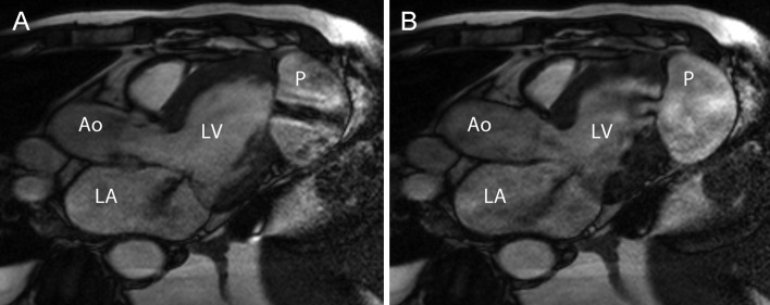
A cardiac MRI (Fig. 1 and Supplementary Video 1) and a transthoracic echocardiography (Supplementary Video 2) demonstrated a left ventricular pseudoaneurysm and showed blood passing in and out through a small left ventricular apical inferolateral wall defect.
Figure 1:
Cardiac MRI clearly shows a jet (black) from blood passing into (A) and back (B) through a small left ventricular inferior wall defect. Three-chamber view, early (A) and late (B) systole. Ao = aorta, LA = left atrium, LV = left ventricle, P = pseudoaneurysm.
Supplementary Video 1:
Cardiac MRI cine loop showing blood flow through a left ventricular apical inferolateral wall defect and a regurgitant jet from mild mitral valve incompetence.
Supplementary Video 2:

Transthoracic echocardiogram showing the pseudoaneurysm and blood passing in and out of the heart through a small left ventricular apical inferolateral wall defect.
DISCUSSION
The left ventricular pseudoaneurysm is a contained rupture of the myocardial wall, whereas a true aneurysm contains all layers of the myocardium and is the result of a thinning of the myocardial wall [1]. The walls of a pseudoaneurysm are composed of visceral pericardium, organized haematoma, thrombus or adhesions and lack any component of the original myocardial wall. It is common for a pseudoaneurysm to have a narrow neck, but a true aneurysm usually has a wide base [2]. When a pseudoaneurysm is formed, there is often a back-and-forth blood flow into a cavity outside of the heart as opposed to true aneurysms where there is a stagnation of blood movement, and spontaneous contrast is observed on echocardiography. It is important to confirm whether the patient has a true aneurysm or a pseudoaneurysm because the latter has a higher risk of rupture and therefore requires urgent surgical treatment [3, 4]. The most common location for a pseudoaneurysm of the heart is in the left ventricle, and the most common aetiology is myocardial infarction [1, 4]. The left ventricular pseudoaneurysm caused by infections is very rare and usually located in the mitral–aortic intervalvular fibrosa [5].
Our patient was initially admitted due to postoperative S. aureus bacteraemia and sepsis, secondary to a surgical site infection. On hospital day 3, echocardiography demonstrated mitral valve endocarditis, and shortly after, the diagnosis was made, he suffered an intracranial haemorrhage as a result of septic embolism to the brain. After approximately 4 weeks of recovery from endocarditis and haemiparesis, follow-up echocardiography revealed a newly formed pseudoaneurysm. Although coronary angiogram was normal at this point, early septic embolism to a coronary artery and subsequent myocardial infarction [2] was the most probable mechanism for pseudoaneurysm formation in this patient. Late gadolinium enhancement MRI [2] of the heart to visualize infarction was not performed due to reduced renal function. He underwent uneventful left ventricular repair and was discharged in good condition.
SUPPLEMENTARY MATERIAL
Supplementary material is available at ICVTS online.
Conflict of interest: none declared.
REFERENCES
- 1.Hulten EA, Blankstein R. Pseudoaneurysms of the heart. Circulation. 2012;125:1920–5. doi: 10.1161/CIRCULATIONAHA.111.043984. doi:10.1161/CIRCULATIONAHA.111.043984. [DOI] [PubMed] [Google Scholar]
- 2.Karamitsos TD, Ferreira V, Banerjee R, Moore NR, Forfar C, Neubauer S. Contained left ventricular rupture after acute myocardial infarction revealed by cardiovascular magnetic resonance imaging. Circulation. 2012;125:2278–80. doi: 10.1161/CIRCULATIONAHA.111.068619. doi:10.1161/CIRCULATIONAHA.111.068619. [DOI] [PubMed] [Google Scholar]
- 3.Atik FA, Navia JL, Vega PR, Gonzalez-Stawinski GV, Alster JM, Gillinov AM, et al. Surgical treatment of postinfarction left ventricular pseudoaneurysm. Ann Thorac Surg. 2007;83:526–31. doi: 10.1016/j.athoracsur.2006.06.080. doi:10.1016/j.athoracsur.2006.06.080. [DOI] [PubMed] [Google Scholar]
- 4.Frances C, Romero A, Grady D. Left ventricular pseudoaneurysm. J Am Coll Cardiol. 1998;32:557–61. doi: 10.1016/s0735-1097(98)00290-3. doi:10.1016/S0735-1097(98)00290-3. [DOI] [PubMed] [Google Scholar]
- 5.Sudhakar S, Sewani A, Agrawal M, Uretsky BF. Pseudoaneurysm of the mitral-aortic intervalvular fibrosa (MAIVF): a comprehensive review. J Am Soc Echocardiogr. 2010;23:1009–18. doi: 10.1016/j.echo.2010.07.015. quiz 112 doi:10.1016/j.echo.2010.07.015. [DOI] [PubMed] [Google Scholar]
Associated Data
This section collects any data citations, data availability statements, or supplementary materials included in this article.


