Editor:
Outflow failure is attributed mostly to mechanical problems such as catheter migration, omental wrapping, constipation, and intraluminal obstruction with fibrin or clot; and it usually occurs shortly after initiation of peritoneal dialysis (PD). Fracture or rupture of the PD catheter is, however, a very unusual cause of drainage failure. We recently experienced a case of early drainage failure because of silicone PD catheter fracture, without previous mechanical or chemical injury, that occurred 1 month after initiation of PD.
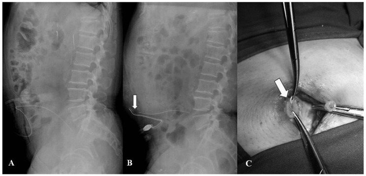
A 62-year-old man visited our PD center because of drainage failure. Because of diabetic end-stage renal disease, a silicone rubber double-cuffed straight Tenckhoff catheter had been successfully inserted without complication a month earlier [Figure 1(A)]. Seven days before presentation, the patient had sensed something abruptly protruding in the abdomen, and subsequently, infused fluid was not being drained. He became swollen and complained of fullness in the abdomen because of drainage failure after repeated exchanges. Hernia was suspected at first, but no palpable mass in the abdomen or edema of the scrotum was observed. The exit site was clear, and no abnormal break was seen in the catheter outside the exit site. During the month after PD initiation, exit-site care was performed with povidone iodine without use of mupirocin ointment, and peritonitis did not occur. Surprisingly, abdominal radiography revealed a clear break of the PD catheter in the subcutaneous area [Figure 1(B)].
Figure 1.
— (A) Plain abdominal radiograph taken immediately after peritoneal dialysis (PD) catheter insertion. The PD catheter was successfully placed in the peritoneal cavity without evidence of damage to the catheter. (B) Spontaneous fracture of the PD catheter on plain abdominal radiography. The radiopaque strip was clearly cut into two (arrow). (C) The fracture site, located just distal to the internal cuff (arrow).
The catheter was removed immediately, and a new catheter was inserted at the same time. The fracture site was found to be located just distal to the internal cuff embedded in the rectus muscle in the operating field [Figure 1(C)].
To date, several reports on this rare complication have been published. Most instances are external breaks associated with mechanical or chemical stress (1-3). Internal breaks are relatively uncommon, but they can be caused by product defects, accidental needle-stick injury, the catheter becoming brittle with aging, and the intrinsic properties of polyurethane (4-8). In particular, it has been widely accepted that polyurethane catheters are highly biocompatible, but vulnerable to various types of physical and chemical stress that lead to polyurethane degradation (7,8). Moreover, Weaver et al. (9) reported that mupirocin containing polyethylene glycol causes structural changes in PD catheters and that the resulting abnormalities increase in severity with longer duration of mupirocin use. It can therefore be speculated that, together, such factors lead to weakening and softening of catheter, eventually resulting in fracture or rupture.
In our patient, however, a polyurethane-based catheter was not used, and mupirocin ointment had never been applied. Catheter aging was unlikely because the drainage failure occurred 1 month after initiation of PD. The patient also denied traumatic accident or strong hand manipulation. We speculate that a defective silicone catheter escaping quality control checks in the production line might lose resistance to mechanical strain. Relevant to our assumption is a prior report by Panuccio et al. (2) of 5 episodes in 4 patients of “epidemic” spontaneous catheter rupture. They suspected a problem with defective silicone catheters during the manufacturing process because no definite cause for the fractures could be found, and most ruptures occurred at similar sites shortly after initiation of PD. Alternatively, damage to the catheter at the time of insertion, such as an accidental needle-stick injury or use of a hemostat to clamp the catheter, might also contribute to an internal break. A thorough inspection and careful manipulation of the catheter before implantation is therefore required to avoid this unusual complication.
CONCLUSIONS
Spontaneous fracture, particularly an internal break, in a PD catheter is extremely rare, but can cause drainage failure and could be considered in the differential diagnosis.
DISCLOSURES
The authors have no financial conflicts of interest to declare.
References
- 1. Guiserix J. Spontaneous rupture of peritoneal catheters. Nephron 1997; 75:100 [DOI] [PubMed] [Google Scholar]
- 2. Panuccio V, Enia G, Crucitti S, Pustorino M, Biondo A, Zoccali C. Another “epidemic” of spontaneous rupture of peritoneal catheters. Nephron 1998; 79:359 [DOI] [PubMed] [Google Scholar]
- 3. Khandelwal M, Bailey S, Izatt S, Chu M, Vas S, Bargman J, et al. Structural changes in silicon rubber peritoneal dialysis catheters in patients using mupirocin at the exit site. Int J Artif Organs 2003; 26:913–17 [DOI] [PubMed] [Google Scholar]
- 4. Closkey GM, Zappacosta AR. CAPD drainage failure due to Tenckhoff catheter fracture: a case report. Perit Dial Int 1992; 12:266–7 [PubMed] [Google Scholar]
- 5. Lee SW, Lee SW, Park GH, Kim SH, Choi YA, Lee CW, et al. Spontaneous fracture of the intraperitoneal portion of a PD catheter. Perit Dial Int 2006; 26:278–80 [PubMed] [Google Scholar]
- 6. Agarwal S, Gandhi M, Kashyap R, Liebman S. Spontaneous rupture of a silicone peritoneal dialysis catheter presenting outflow failure and peritonitis. Perit Dial Int 2011; 31:204–6 [DOI] [PubMed] [Google Scholar]
- 7. Coury AJ, Stokes KB, Cahalan PT, Slaikeu PC. Biostability considerations for implantable polyurethanes. Life Support Syst 1987; 5:25–39 [PubMed] [Google Scholar]
- 8. Crabtree JH. Clinical biodurability of aliphatic polyether based polyurethanes as peritoneal dialysis catheters. ASAIO J 2003; 49:290–4 [DOI] [PubMed] [Google Scholar]
- 9. Weaver ME, Dunbeck DE. Mupirocin (Bactroban) causes permanent structural changes in peritoneal dialysis catheters. Perit Dial Int 1994; 14(Suppl 1):20 [Google Scholar]


