Abstract
Background
Computed tomography is currently the gold standard for the diagnosis of chronic rhinosinusitis. However, this facility is not readily available in many developing countries. Thus, plain sinus radiography is still widely in use in our practice.
Objectives
To assess the diagnostic value of plain radiographs in adult patients with uncomplicated chronic maxillary rhinosinusitis.
Methods
This study was carried out at a tertiary health facility in Northern Nigeria. All adult patients with clinical and radiological diagnosis of chronic maxillary rhinosinusitis were included.
Results
A total of 88 patients were recruited into the study. There were 51 males (58.0%) and 37 (42.0%) females. Their ages ranged from 18 to 60 years; with a mean age of 31.7±9.20 years. Mucosal thickening was the commonest diagnostic plain radiographic feature, and fluid level was the least. Maxillary antra with diagnostic plain radiographic interpretations of fluid level, haziness and opacity had high specificities (100%, 95.2%, and 85.7%) and high positive predictive values (100%, 75%, and 70%) respectively.
Conclusions
Plain radiographs are relevant in the diagnosis of chronic maxillary rhinosinusitis in our locality only when they show features of fluid level: findings of haziness and opacity are of less diagnostic value.
Keywords: Chronic rhinosinusitis, paranasal sinus radiography, maxillary antral lavage, Nigeria
Introduction
Chronic rhinosinusitis is still a common health problem encountered by many physicians: particularly, the Otolaryngologists worldwide1–4. Although this disease is associated with few complications nowadays, the complicated ones are still considered potentially fatal5,6.
Computed Tomography (CT scan) is currently reputed to be the radiographic gold standard for the diagnosis of patients with this disease7–11. Also, it provides a navigational guide of the surgical anatomy of the paranasal sinuses - a prerequisite for Functional Endoscopic Sinus Surgery. 7 Magnetic Rasonance Imaging (MRI) is however of limited value in this condition, being only useful when intracranial complication or allergic fungal rhinosinusitis is suspected.[11]
CT Scan is still not routinely applied in the diagnosis of chronic rhinosinusitis in Nigeria for obvious reasons of high cost,. and also the health risk (high radiation dose)7,9. Moreover, this facility is not readily available in many developing countries including Nigeria.2 Consequently, plain radiography is still in use in our practice for the diagnosis of this disease. Unfortunately, plain radiography lacks reliability even in experienced hands, while also issues of observer error are some of its setback. On the other hand, it has the advantage of easy interpretation, lower radiation dose, and is cheap in comparison with CT scan and MRI. Nevertheless, is the continued use of these plain radiographs in the diagnosis of chronic maxillary rhinosinusitis in our environment justifiable? Previous studies were done decades ago, and to the author's best knowledge, very few were in our setting. 2,12 A study on this will help to update, and also bridge the gap in the current literature in our locality.
Uncomplicated chronic rhinosinusitis is defined as inflammation of the nasal cavity and the paranasal sinuses without any clinically evident extension of inflammation outside these areas for at least 12 weeks. 4,10,13 The treatment of this condition is initially medical; surgery is indicated when medical treatment fails, and when there are anatomical predisposing factors.
This study was intended to access the diagnostic value of plain radiographs in adult patients with uncomplicated chronic maxillary rhinosinusitis. It was limited by the observer error in the interpretation of plain radiographs and maxillary antral effluents.
Methods
This is part of an on-going prospective hospital based study of all consecutive adult patients who presented with clinical features of chronic rhinosinusitis at the Ear, Nose, and Throat (ENT) clinics of Aminu Kano Teaching Hospital, Northern Nigeria. The study was approved by the Institutional Review Board of the hospital and an informed consent was obtained from all eligible patients.
Inclusion criteria
Patients were recruited into the study, if they were 18 years and above, and had a clinical diagnosis of chronic rhinosinusitis. A clinical diagnosis of chronic rhinosinusitis was made, if there were at least any two or more of the following symptoms and signs for a period not less than eight weeks:2] Nasal obstruction: Post-nasal drip, Rhinorrhea which may be mucoid, mucopurulent or purulent, Halithosis/malodorous breath and persistent headache
Exclusion criteria
Patients who suffered complications of chronic rhinosinusitis were excluded from the study. Others excluded were those with:Previous paranasal sinus surgery or trauma and previous antral lavage. For those patients that met the inclusion criteria, a questionnaire was used to obtain their bio-data, relevant medical history, and detailed clinical examinations were carried out on them.
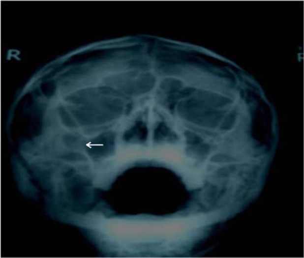
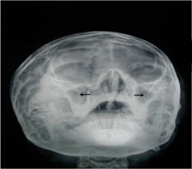
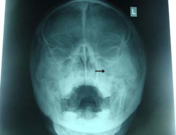
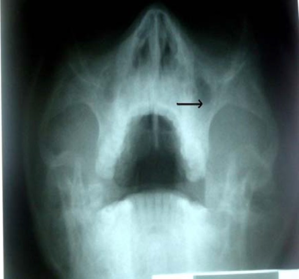
Subsequently, the patients did a standard plain X-ray of the paranasal sinuses - occipitomental, occipitofrontal and lateral views. However, only the occipitomental view (better for analyzing the maxillary sinus) was interpreted in this study. The radiographs were taken at the radiology department of the hospital by an experienced radiographer. The radiographs were interpreted by the author using a viewing box (Kenex-Electro medical Hd, Essex, England). A radiographic diagnosis of chronic maxillary rhinosinusitis was made; if there was any one of the following in the maxillary sinus radiographs: 2 Gross mucosal thickening (>5mm), fluid level, haziness and opacity.
They were considered normal if none of the above was present.
Subsequently, those patients with a radiographic diagnosis of chronic maxillary rhinosinusitis had an antral lavage at the earliest opportunity. The procedure was performed as described by Mackay et el.14
In patients with unilateral disease confirmed radiologically, the two antra were lavaged. The presumed normal sides (with negative radiographic findings) were considered to have undergone a proof puncture. However, if both antra turned out to be radiologically normal, no antral puncture was performed. The lavage was reported as negative if the saline effluent was clear, and positive if it was purulent, mucopurulent, turbid or contained debris.
Data analysis
Data obtained were recorded in a specialized form and analyzed using Statistical Package for Social Sciences (SPSS version 13) computer soft ware program. The plain radiographic interpretations and their corresponding antral lavage results were recorded and analyzed. Test of validity of plain radiograph of the maxillary sinus was determined by calculating the sensitivity, specificity and predictive values using antral lavage findings as a gold standard.
Results
A total of 97 consecutive patients presented with clinical features of chronic rhinosinusitis. Nine patients were excluded because they had recent history of antral lavage - leaving 88 patients for the study. Their general characteristic is represented in table 1.
Table 1.
General characteristic of the study population
| Characteristics | Number | Percentage |
| Sex | ||
| Male | 51 | 58.0 |
| Female | 37 | 42.0 |
| Age (years) | ||
| Range | 18 – 60 | |
| Mean | 31.7 (SD = 9.2) | |
| Duration of symptoms | ||
| Range | 2 months – 30 years | |
| Mean | 5.7 years (SD = 5.8) | |
| Laterality of disease based | ||
| on plain X-ray | ||
| Bilateral | 28 | 59.6 |
| Unilateral | 19 | 40.4 |
Of the 88 patients studied, 68 (77.3%) did plain X-ray of the paranasal sinuses: these resulted in a total of 136 maxillary antra for radiological analysis. Their various diagnostic plain radiographic interpretations are shown in table 2. Twenty patients (22.7%) did not have their plain radiographs because they could not afford the cost.
Table 2.
Diagnostic plain radiographic interpretations for patients with chronic maxillary rhinosinusitis (n=136)
| Plain radiographic feature | Frequency | % |
| Gross mucosal | 55 | 38.2 |
| Fluid level | 6 | 4.2 |
| Haziness | 9 | 6.3 |
| Opacity | 13 | 9.0 |
| Normal (unilateral) | 61 | 42.4 |
| Total | 144* | 100 |
some maxillary antra had more than one feature
Of the 68 patients who had plain X-rays of the paranasal sinuses, 31 (45.6%) had antral lavage. Thirty-seven (54.4%) did not because they either withdrew consent for antral lavage (8 patients) or had normal radiographic interpretations on both sides (29 patients). These antral lavages were done on both sides in 30 patients, while 1 patient had it on one side; the other side was abandoned due to severe pain. Therefore, a total of 61 lavages were performed. Table 3 shows the various diagnostic plain radiographic interpretations and their corresponding antral lavage results.
Table 3.
Diagnostic plain radiographic interpretations and their corresponding antral lavage results
| Plain radiographic feature |
Total | Antral lavage | |
| Positive | Negative | ||
| Gross mucosal | 36 | 24 | 12 |
| thickening | |||
| Fluid level | 5 | 5 | 0 |
| Haziness | 4 | 3 | 1 |
| Opacity | 10 | 7 | 3 |
| Normal (Unilateral) | 13 | 8 | 5 |
| Total | 68 | 47 | 21 |
The results of the sensitivity, specificity and predictive values of the various diagnostic plain radiographic interpretations are shown in table 4.
Table 4.
Diagnostic plain radiographic interpretations and their corresponding Sensitivity, specificity and predictive values
| Plain radiographic |
Sensitivity | Specificity | Positive predictive value |
Nagtive predictive value |
| % | % | % | % | |
| Gross mucosal | 60.0 | 42.9 | 66.7 | 36.0 |
| thickening | ||||
| Fluid level | 12.5 | 100 | 100 | 37 |
| Haziness | 7.5 | 95.2 | 75 | 35.1 |
| Opacity | 17.5 | 85.7 | 70 | 35.3 |
Discussion
Chronic maxillary rhinosinusitis still constitutes a disease burden in most developing countries including Nigeria. In addition, CT scan is generally accepted as the diagnostic gold standard for this disease. 7–10 However, the author could not use this facility in this study because like in most developing countries, it was not readily available and the high cost was a limitation. Hence, the use of maxillary antral lavage findings as a confirmatory test for the presence of chronic maxillary rhinosinusitis. This procedure has been used for diagnostic and therapeutic purposes in cases of chronic maxillary rhinosinusitis (especially those with retained secretions) for at least a century. 3
Although mucosal thickening was found to be the commonest diagnostic plain radiographic feature of chronic maxillary rhinosinusitis in this study, it was the least predictor of this disease. Similarly, Ezeanolue et al, 2 buttressed this in a related study. However, it is note worthy that in their study, gross mucosal thickening was defined as a value ≥4mm, while in this study, >5mm was used. Meanwhile, some authors considered only values 6 – 8mm as diagnostic.7]
This study also found that maxillary antra with plain radiographic interpretations of fluid level, haziness and opacity had high specificities and high positive predictive values. This is particularly true for maxillary antra with fluid levels where 100% positive antral effluents were observed. In other words, plain radiographic diagnostic findings in chronic maxillary rhinosinusitis correlated well with antral lavage findings only where there are fluid levels suggestive of retained secretions. Likewise, other workers had reported similar findings in a related study.2 In other words, they found that fluid level and opacity each had a specificity of 92.3% and positive predictive values of 87.5% and 96.0% respectively. 2 On the contrary, this study found a low to fair degree of sensitivity for all the various diagnostic plain radiographic interpretations (7.5% to 60.0%). In agreement, Patel et al, had highlighted the low sensitivity of plain radiographs in the diagnosis of this disease.15
Furthermore, this study found that normal plain radiographic finding was an unreliable predictor of the absence of chronic maxillary rhinosinusitis. For instance, 8 of the 13 antra with normal features gave positive lavage results. On the other hand, some authors had found normal maxillary plain radiographic feature to reasonably represent the absence of this disease. 2,16]
Conclusion
Plain radiographs are relevant in the diagnosis of chronic maxillary rhinosinusitis in our locality only when they show features of fluid level: findings of haziness and opacity are of less diagnostic value. However, these findings might be biased because of the relatively small size of the study population. A large multi-center prospective study is recommended in order to validate these findings. Preferably, using CT scan as a gold standard to test for validity of plain maxillary radiographs.
Figure 1.

photograph of an occipitomental view of the paranasal sinuses showing mucosal thickening of the right maxillary sinus (see arrow)
Figure 2.

photograph of an occipitomental view of the paranasal sinuses showing bilateral hazy maxillary antra (see arrows)
Figure 3.

photograph of an occipitomental view of the paranasal sinuses showing complete opacification of the left maxillary antrum (see arrow)
Figure 4.

photograph of an occipitomental view of the paranasal sinuses showing fluid level in the left maxillary antrum (see arrow).
References
- 1.Puruckherr M, Byrd R, Roy T, Krishnaswamy G. The diagnosis and management of chronic rhinosinusitis. [web site on the internet]. Cited 2010 Oct 8. Available from: http://www.priory.com/med/rhinitis.htm.
- 2.Ezeanolue BC, Aneke EC, Nwagbo DFE. Correlation of plain radiological diagnostic features with antral lavage results in chronic maxillary sinusitis. West Afr J Med. 2000;19(1):16–18. [PubMed] [Google Scholar]
- 3.Pang YT, Willatt DJ. Do antral washouts have a place in the current management of chronic sinusitis? J Laryngol Otol. 1996;110(10):926–928. doi: 10.1017/s0022215100135376. [DOI] [PubMed] [Google Scholar]
- 4.Fokkens W, Lund V, Mullol J. European position paper on rhinosinusitis and nasal polyps 2007. Rhinol. 2007;20 suppl:1–136. [PubMed] [Google Scholar]
- 5.Singh B, Van Dellen J, Ramjettan S, Maharaj TJ. Sinogenic intracranial complications. J Laryngol Otol. 1995;109(10):945–950. doi: 10.1017/s0022215100131731. [DOI] [PubMed] [Google Scholar]
- 6.Ogunleye AOA, Nwaorgu OGB, Lasisi AO. Complications of sinusitis in Ibadan, Nigeria. West Afr J Med. 2001;20(2):98–101. [PubMed] [Google Scholar]
- 7.Evans KL. Diagnosis and management of sinusitis. BMJ. 1994;309(6966):1415–1422. doi: 10.1136/bmj.309.6966.1415. [DOI] [PMC free article] [PubMed] [Google Scholar]
- 8.Ahmad BM, Tahir AA. Rhinosinusitis in northeastern Nigeria: computerized tomographic scan findings. Niger J Surg Res. 2003;5(3 – 4):110–113. [Google Scholar]
- 9.Aaløkken TM, Hagtvedt T, Dalen I, Kolbenstvedt A. Conventional sinus radiography compared with CT in the diagnosis of acute sinusitis. Dentomaxillofac Radiol. 2003;32(1):60–62. doi: 10.1259/dmfr/65139094. [DOI] [PubMed] [Google Scholar]
- 10.Rosenfeld RM, Andes D, Bhattacharyya N, et al. Clinical practice guideline: adult sinusitis. Otolaryngol Head Neck Surg. 2007;137(Suppl 3):S1–S31. doi: 10.1016/j.otohns.2007.06.726. [DOI] [PubMed] [Google Scholar]
- 11.Ramadan HH, Brown KR. Pediatric sinusitis, medical treatment. [web site on the internet]. Cited 2010 Oct 10. Available from: http://www.emedicine.com/Ent/topic612.htm.
- 12.Kay NJ, Setia RN, Stone J. Relevance of conventional radiography in indicating maxillary antral lavage. Ann Otol Rhinol Laryngol. 1984;93(1 pt 1):37–38. doi: 10.1177/000348948409300109. [DOI] [PubMed] [Google Scholar]
- 13.Benninger MS, Ferguson BJ, Hadley JA, Hamilton DJ, Jacobs M, Kennedy DW, et al. Adult chronic rhinosinusitis: Definations, diagnosis, epidemiology and pathophysiology. Otolaryngol Head Neck Surg. 2003;129:S1–S32. doi: 10.1016/s0194-5998(03)01397-4. [DOI] [PubMed] [Google Scholar]
- 14.Mackay IS, Lund VJ. Surgical management of sinusitis. In: Mackay IS, Bull TR, editors. Scott - Brown's Otolaryngology. 6th ed. London: Butterworth-Heinemann; 1997. pp. 4/12/1–4/12/24. [Google Scholar]
- 15.Patel AM, Vaughan WC. Sinusitis, maxillary, chronic, surgical Treatment. [web site on the internet]. Cited 2010 Oct 12. Available from: http://www.emedicine.com/ent/topic339.htm.
- 16.Vartanian AJ, Joe S. CT Scan, Paranasal Sinuses. [web site on the internet] Cited 2010 Jun 13. Available from: http://www.emedicine.com/ent/topic387.htm.


