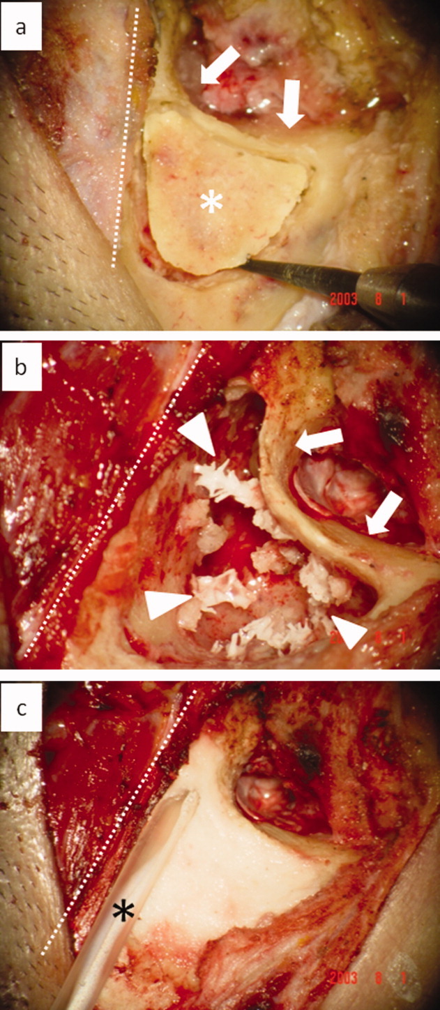
Fig. 1.

Regenerative first-stage operation with mastoid cortex plasty (right ear). white dotted line, temporal line; white asterisk, mastoid cortex bone lid; white arrow, posterior wall of external auditory meatus; white arrow head, artificial pneumatic bones; black asterisk, drainage tube.
a. Before the mastoidectomy is performed, a groove is made on the surface of the post-auricular cortex bone using a minimum-size cutting bar. Bone powder, for making bone putty, is taken from outside the groove. Mastoid cortical bony plate was quarried out from the surface of the temporal bone behind the external auditory canal before mastoidectomy to cover the opened mastoid cavity at the end of the operation.
b. After the cortex lid is chiseled, mastoidectomy is performed in the usual manner. Cholesteatoma, granulation and other lesions are removed while preserving the healthy mucosa as much as possible in this region. Collagen-coated hydroxyapatite fragments (artificial pneumatic bones) are transplanted sparsely into the opened mastoid cavity and fixed in place by fibrin glue.
c. Bone putty mixed with fibrin glue is applied to the edge of the opened mastoid cavity, then the cortex lid is returned to its original position. The cortex lid is then fixed and covered with bone putty. The drainage tube is placed into the mastoid cavity, and the surface of this region is made smooth using the finger cushion. Finally, fibrin glue is sprinkled over this region. [Color figure can be viewed in the online issue, which is available at wileyonlinelibrary.com.]
