Abstract
The Centromere is a unique chromosomal locus where the kinetochore is formed to mediate faithful chromosome partitioning, thus maintaining ploidy during cell division. Centromere identity is inherited via an epigenetic mechanism involving a histone H3 variant, called centromere protein A (CENP-A) which replaces H3 in centromeric chromatin. In spite of extensive efforts in field of centromere biology during the past decade, controversy persists over the structural nature of the CENP-A-containing epigenetic mark, both at nucleosomal and chromatin levels. Here, we review recent findings and hypotheses regarding the structure of CENP-A-containing complexes.
Keywords: Centromeres, Chromosomes, Chromatin, Centromere protein A, Epigenetics
Introduction
Centromere regions are unique in that they direct kinetochore assembly where the spindle microtubules attach and mediate chromosome segregation, thus maintaining ploidy (Cleveland et al. 2003). Defects in the centromeric chromatin may lead to missegregation of chromosomes resulting in aneuploidy, a frequently observed phenomenon in cancer (Tomonaga et al. 2003). Centromeres are highly divergent throughout evolution and even from chromosome to chromosome within a given species (Fukagawa 2004). However, a singular conserved feature of all centromeres is the presence of a centromere-specific histone H3 variant known as centromere protein A (CENP-A) in centromeric nucleosomes (Palmer et al. 1991). CENP-A in humans is a 140 amino acid protein with an N-terminus divergent in sequence from that of the canonical histone H3 (Yoda et al. 2000), a C-terminal histone fold domain (HFD) (Sullivan et al. 1994), and a C-terminal tail required for the recruitment of kinetochore proteins such as CENP-C and -N (Guse et al. 2011). Thus, CENP-A is often regarded to as the first player in kinetochore assembly and centromere identity.
Sequence alignment shows that the HFD-containing C-terminus of CENP-A is 62 % identical to that of canonical H3 (Sullivan et al. 1994). Within this region, deuterium exchange measured by mass spectrometry identified a unique structural element, termed the CENP-A targeting domain (CATD) comprising of the L1 and α2 helices (Black et al. 2007a). Chimeric molecules where the CATD of CENP-A is exchanged with the corresponding region in H3 (and vice versa) revealed that the CATD is a major determinant for targeting of CENP-A to the centromeres (Black et al. 2007b). Interestingly, structural data indicate that CATD also serves as an interface between CENP-A and H4 in the sub-nucleosomal (CENP-A:H4)2 complex (Sekulic et al. 2010) as well as the CENP-A: CENP-A dimer in the putative octameric CENP-A nucleosomes (Bassett et al. 2012; Sekulic et al. 2010). Importantly, the CATD has been shown to interact with the Holliday Junction recognition protein (HJURP) which is an essential chaperon for CENP-A centromeric deposition (Black et al. 2004; Foltz et al. 2009; Hu et al. 2011). Despite detailed structural data discussed above, questions remain as to the precise nature of CENP-A chromatin. Complications in this regard mainly arise from inconsistencies between data obtained from biochemical work using in vitro nucleosome reconstitution approaches, proposing a canonical nucleosome-like octameric entity for CENP-A nucleosomes, and some lines of evidence obtained from studying endogenously purified centromeric chromatin revealing that CENP-A may not be present in a nucleosome form (octamers) and might exist in cells as some sort of tetrameric half-nucleosomes (hemisomes) at the centromere. Here we review our current understanding of the structure of CENP-A containing complexes highlighting the biological outcomes of the octamer vs. tetramer debate.
Soluble CENP-A
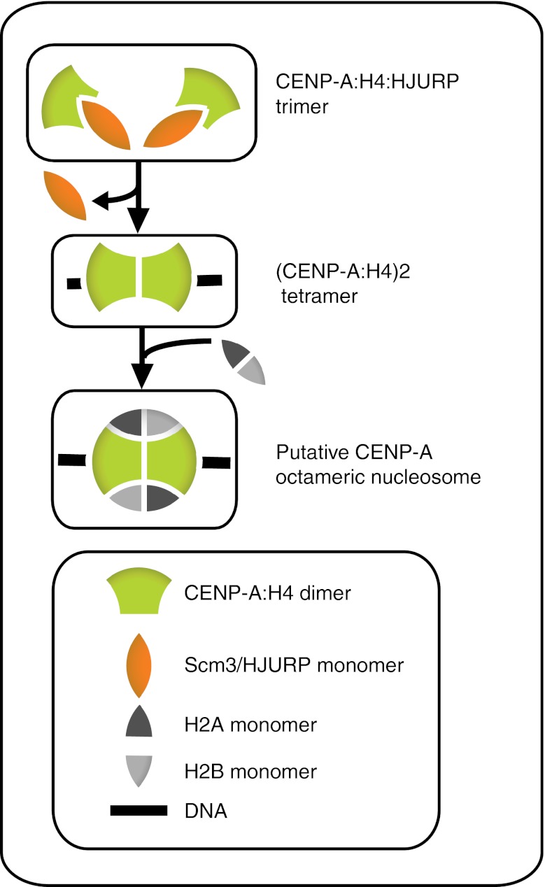
Upon production of CENP-A in G2 (Howman et al. 2000; Shelby et al. 2000), the newly synthesized protein is thought to form a dimer with histone H4 and further in the cell cycle recognized by HJURP (known as suppressor of chromosome missegregation 3, Scm3 in yeast) resulting in an equimolar complex of CENP-A:H4: HJURP (Fig. 1) (Cho and Harrison, 2011; Hu et al. 2011). In yeast, Scm3 binds the CATD of Cse4 (CENP-A homolog), and α2 and α3 of H4 via its Cse4 binding domain (CBD), with key residues conserved in HJURP (Zhou et al. 2011). This interaction of HJURP with CENP-A is required to stabilize CENP-A as depletion of HJURP in human cells results in dramatically decreased CENP-A protein levels (Dunleavy et al. 2009; Foltz et al. 2009; Shuaib et al. 2010). Structural data suggest that this complex (Cse4:H4: Scm3) is not capable of interacting with DNA due to the induction of major conformational alterations in Cse4 and H4 (e.g., displacement of DNA-binding Loop 2 of H4) (Zhou et al. 2011). Moreover, it has been suggested that the presence of Scm3 in the pre-nucleosomal complex prevents the sub-nucleosomal (Cse4:H4)2 tetramer formation (Fig. 1), a step required for the nucleosome assembly (Zhou et al. 2011). Therefore, it is intuitive to assume that Scm3 needs be recognized by another/other component(s) in order to bind the chromatin and that it has to be removed for stable incorporation of CENP-A on to centromeres.
Fig. 1.
Proposed mechanism for the formation of putative CENP-A octameric nucleosomes. Newly synthesized CENP-A along with histone H4 is suggested to bind HJURP to form a pre-nucleosomal trimeric complex. Next, in order to interact with DNA, HJURP has to be released leaving a (CENP-A:H4)2 tetramer. Addition of H2A:H2B dimers then complete the octamer formation
CENP-A nucleosomal structure
The octamer model
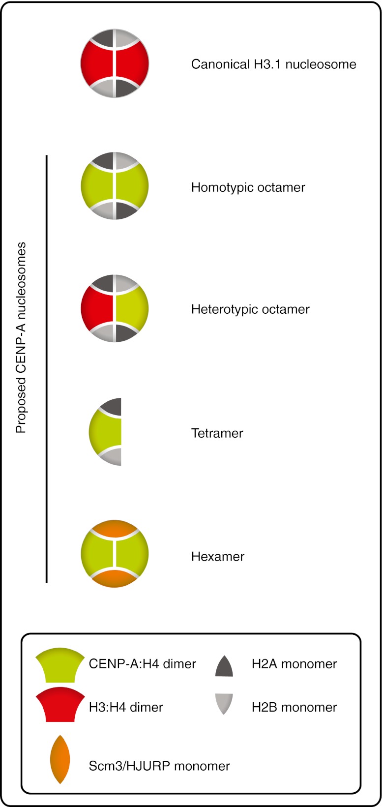
The crystal structure of the human CENP-A-containing nucleosome reconstituted in vitro from bacterially purified histones indicates homotypic octamers containing two copies of each histone molecule (Tachiwana et al. 2011). This study also revealed key features of CENP-A nucleosomes distinguishing them from canonical H3 nucleosomes. For instance, CENP-A contains a shorter αN helix lacking a key Arginine in position 49, which is an essential amino acid for DNA interaction. These findings are consistent with the data obtained independently from stepwise assembly of CENP-A nucleosomes not only confirming the octameric structure of CENP-A nucleosomes but also the loosening of the interaction between DNA superhelical termini and CENP-A (Conde e Silva et al. 2007; Panchenko et al. 2011). CENP-A octamers formed in vitro have also been reported to induce conventional left-handed negative supercoiling to DNA (Barnhart et al. 2011; Conde e Silva et al. 2007; Panchenko et al. 2011; Tachiwana et al. 2011; Yoda et al. 2000). It was recently demonstrated that the mutation of the putative CENP-A: CENP-A dimer interface can abrogate centromeric targeting of CENP-A in Drosophila and mammalian tissue culture cells (Bassett et al. 2012; Zhang et al. 2012). In agreement with an octamer, over-expression of Cse4 (the CENP-A homolog) in budding yeast was reported to result in misincorporation of octamer-sized nucleosomes in chromosome arms (Camahort et al. 2009). These observations, along with the crystal structures available, provide solid evidence supporting the existence of octameric CENP-A nucleosomes at the centromere (Fig. 2).
Fig. 2.
Different models for the composition of CENP-A nucleosomes. These models differ in size, DNA wrapping and number as well as identity of their components. For a more detailed description of the differences between each model, please see the text
The tetramer (hemisome) model
In an effort to determine the native in vivo form of CENP-A chromatin, various purification and analysis techniques have been employed. One of the most extensive efforts has focused on nucleosome cross-linking followed by immunoprecipitation and atomic force microscopy (AFM) to investigate CID-containing nucleosomes (CID for centromere identifier, a Drosophila homologue of CENP-A). Challenging the octameric nucleosome concept, AFM data revealed that the height of the CID-containing interphase chromatin is half the height of canonical H3 nucleosomes (approximately 1 vs. 2 nm) (Dalal et al. 2007). Moreover, in the beads-on-a-string structure of CID chromatin, the linker DNA is reported to be 2–3 times longer than that of conventional nucleosomes (Dalal et al. 2007). Surprisingly, the electrophoretic behavior of the purified CID-nucleosomal core particles corresponds to the presence of only one copy of each histone. This composition (CID:H4:H2A:H2B) is referred to as a tetramer, half-nucleosome or hemisome (Fig. 2). Work done in human cells resulted in similar observations regarding the equimolar presence of core histones with particle heights and volumes fitting well with the half-nucleosome model as compared to H3 nucleosomes (Dimitriadis et al. 2010). Immuno-electron microscopy (Immuno-EM) data also suggest that the quantity of histones within each CENP-A nucleosomal particle matches with the half-nucleosome structure (Dimitriadis et al. 2010).
Recently, it was reported that the budding yeast centromeres are composed of a single Cse4-containing nucleosome wrapping about 80 bp of DNA, half the length of a canonical nucleosome, once in a right-handed manner (Henikoff and Henikoff 2012; Krassovsky et al. 2012). The right-handedness of DNA wrapping in centromeric nucleosomes has been reported by other studies as well (Furuyama and Henikoff 2009; Huang et al. 2011). ChIP data also demonstrated the occupancy of H2A at these sites all together consistent with the existence of a Cse4 hemisome at the centromere (Krassovsky et al. 2012). In support of this, the same study that found Cse4 nucleosomes (octamers) in chromosome arms reported tetramers in centromeres (Camahort et al. 2009). It should be noted that in vivo calibrated fluorescence intensity measurements of GFP: Cse4 are not consistent with a single copy of Cse4 at each centromere (Coffman et al. 2011; Lawrimore et al. 2011). Thus, it is entirely possible that the conditions used for purification induce a hemisome-like artifact and that does not exist or is not stable in vivo.
Intriguingly, the observations presented above about centromeric nucleosomes, are reminiscent of an early model suggesting that octameric nucleosomes are in fact constituted from symmetrical half-nucleosome pairs capable of independent existence (Weintraub et al. 1976). This was supported by work on SV40 minichromosome which primarily consists of about 20–25 nucleosomes as shown by EM. Incubation of purified minichromosomes at low ionic strengths was however reported to induce the doubling of the number of beads-on-a-string, reduction of the dimensions of resulting particles and more interestingly longer inter-particle distances, all suggesting the splitting of octameric nucleosomes into half-nucleosomes (Oudet et al. 1978). Similar observations were made with cellular chromatin (Oudet et al. 1978). However, the occurrence of this conversion under cellular conditions remains to be shown to date.
One the other hand, the observation that nucleosomes can be found in various conformational states (Lavelle and Prunell 2007), examples of which include Archaeal nucleosomes consisting solely of (H3:H4)2 tetramers (Reeve et al. 1997); eukaryotic reversomes generated upon depletion of H2A:H2B dimers with right-handed DNA wrapping (Lavelle and Prunell, 2007) and the more recent proposed heterotetramer formation of CENPs-T:W:S:X (Nishino et al. 2012) capable of supercoiling DNA similar to nucleosomes, supports the possibility of tetrameric half-nucleosomes (hemisomes) residing in certain regions of the genome such as the centromere.
Other proposed forms of CENP-A nucleosomes
Using a modified sequential immunoprecipitation technique in budding yeast, H3 was recently reported to co-occupy the centromeric DNA along with Cse4 and other core histones in a cell cycle independent manner (Lochmann and Ivanov 2012) suggesting the potential existence of (Cse4:H4)(H3:H4)(H2A:H2B)2 heterotypic octamers (Fig. 2). However, it is not clear if stable association of H3 with Cse4 containing nucleosome is in the form of a heterotypic octamer or non-nucleosomal associations.
Additionally, a (Cse4:H4)2(Scm3)2 hexameric organization has also been proposed for the centromeric chromatin in budding yeast (Fig. 2) (Mizuguchi et al. 2007). However, a number of key observations soon detracted support for stable occurrence of this structure in centromeric chromatin. These include the previously mentioned structural barriers occluding Cse4 and H4 interaction with DNA (Zhou et al. 2011) and the fact that over-expression of Cse4 in an Scm3Δbackground, can rescue the Scm3 null phenotype (Camahort et al. 2009) suggesting that Scm3 is dispensable for centromere organization.
The tetramer to octamer transition model: towards a dispute settlement?
The controversial observations regarding the nature of CENP-A nucleosomes possibly stem from different chromatin preparation techniques, the stabilization of transient intermediates or the co-existence of more than one CENP-A nucleosome type under certain conditions. No matter the technical difference, a potential structural dynamics model for CENP-A containing nucleosomes through the cell cycle would be an important step forward in understanding centromere biology.
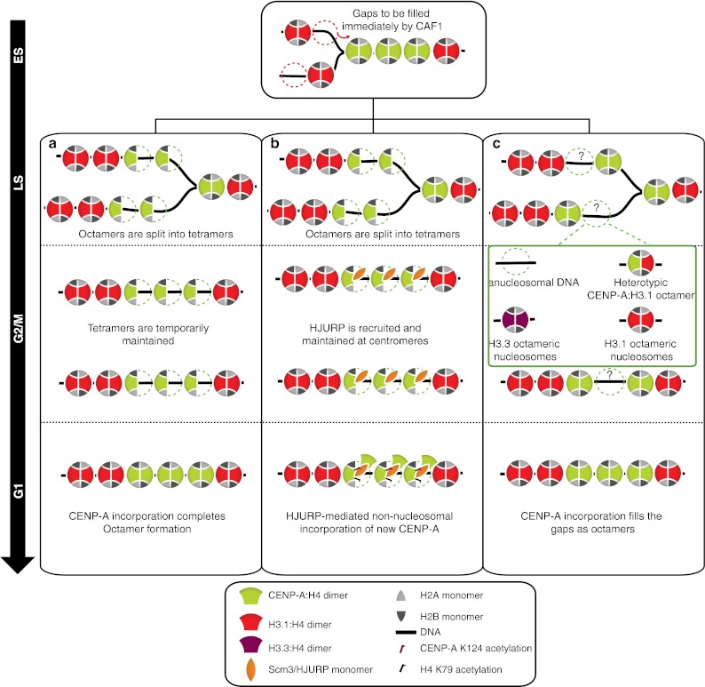
In this regard, an octamer to tetramer conversion model had been previously proposed based on which octameric CENP-A nucleosomes are split into tetrameric half-nucleosomes upon the passage of the replication fork in S phase allowing the equal inheritance of the epigenetic mark to the daughter strands (Fig. 3a) (Allshire and Karpen 2008; Probst et al. 2009). The resultant tetramers were proposed to be maintained throughout G2/M but converted into octamers in G1 following incorporation of new CENP-A by HJURP. This model, while providing a possible mechanism for the preservation of centromeric identity, had never been experimentally validated.
Fig. 3.

Proposed structural dynamics of centromeric nucleosomes throughout the cell cycle. a An octamer to tetramer transition model in which passage of the replication fork splits the pre-existing octameric CENP-A nucleosomes into tetramers allowing equal inheritance of the epigenetic mark to daughter strands. In this model, HJURP exclusively is found at the centromere in G1 and mediates the reconstruction of octamers by incorporation of new CENP-A. b The tetramer to octamer transition model. CENP-A:H4: HJURP complex is recruited to the centromeric chromatin in G1 and is assumed to associate to the pre-existing CENP-A tetramers extra-nucleosomally. Posttranslational modifications (PTMs) of tetrameric CENP-A nucleosomes along with the presence of HJURP impede the stable incorporation of new CENP-A:H4 dimers into nucleosomes. In late G1/early S, action certain proteins chaperones and remodeling complexes is proposed to facilitate the tetramer to octamer transition by affecting the PMTs of histones and chromatin structure resulting in tetramer to octamer transition. Likewise to the previous model, passage of the replication fork splits the octamers to the tetramers. The tetramers are further stabilized by the reassociation of HJURP in G2 and presumed to be maintained in mitosis to form the kinetochore plate. c. In an alternative model, where CENP-A exists in octameric nucleosomes throughout the cell cycle, gaps generated as a result of the passage of the replication fork will be either maintained or could be transiently filled with multiple possible placeholder structures. In this model, HJURP is also found at the centromere exclusively in G1 to participate in new CENP-A assembly
Interestingly, two recent studies co-published in Cell (Bui et al. 2012; Shivaraju et al. 2012) provide evidence for a novel cell cycle-coupled structural transition of CENP-A nucleosomes in human cells and budding yeast (Fig. 3b). AFM-based analysis of immuno-precipitated CENP-A nucleosomes from cell cycle staged human cells revealed that centromeric nucleosomes changed in size depending on cell cycle timing. Tetrameric dimensions of CENP-A nucleosomes in G1 were reported to undergo a transition to octameric dimensions during S phase and revert back to tetrameric state in G2 and maintained through mitosis (Bui et al. 2012). Intriguingly, the authors also report cyclic association, dissociation, and reassociation of HJURP to the centromeric chromatin in G1, S, and G2 phases, respectively (Fig. 3b). Purification of DNA-bound CENP-A:H4 from G1/S arrested cells followed by mass-spectroscopic analysis identified two previously unknown covalent modifications: acetylation of CENP-A K124 and H4K79. These modifications and the presence of HJURP were proposed to prevent stable octamer formation in G1/S. However, as the cell enters the S phase, the authors speculated that opening of centromeric chromatin would be concomitant with the resolution of these modifications. This biochemical change could be coupled to the release of HJURP physically allowing the action of chromatin remodelers to trigger the generation octamers. The dynamics of HJURP at the centromere and the reformation of tetramers in G2 might indicate the role of HJURP in this reversal transition following DNA replication. However, HJURP in human cells has been previously shown to localize to centromeres exclusively during CENP-A loading in G1 (Dunleavy et al. 2009; Foltz et al. 2009) and the G2 reappearance of HJURP at the centromeres has never been reported by other groups. Given the fact that nascent CENP-A is chaperoned by HJURP after synthesis in G2, cytoplasmic contamination of chromatin lysate could be a potential source for the detection of HJURP in G2/M CENP-A pull down. On the other hand, the authors report that HJURP is absent from S phase CENP-A pull down. If the possibility of cytoplasmic contamination is true, this would nicely explain the absence of HJURP in S phase CENP-A pull down since HJURP has already completed deposition of its cargo and thus even if the chromatin prep does contain cytoplasmic contamination, HJURP will not copurify with CENP-A any longer.
Work in Saccharomyces cerevisiae and Candida albicans using fluorescence correlation spectroscopy (FCS) also suggest the presence of a single copy of Cse4 at each centromere (Shivaraju et al. 2012). This seems to be the case for G1, S, G2, and metaphase; however, during anaphase B the authors note a “doubling” of Cse4 and speculate this is due to a tetramer to octamer transition. FCS can very accurately measure protein complexes in living cells by recording peak intensities of diffusing molecules and comparing these over varying time scales (Bulseco and Wolf 2007) (usually up to 2 min), detecting auto-correlations. A major caveat with this technique is that while extremely accurate for freely diffusing complexes, it is less accurate for slow diffusing structures (Krichevsky and Bonnet 2002) (in this case mitotic centromeres bound to microtubules). This is in part due to the fact that FCS is based on peak intensity of a diffraction limited spot. Thus, slowly diffusing or slightly dispersed (greater than the measurement spot) structures such as centromeres will not all be measured in the time scale required. Interestingly, centromeres are much less dispersed in anaphase (compared to metaphase) (Pearson et al. 2001), which could allow for more accurate measurements and thus explain the difference reported. Nonetheless, using FRET and sequential ChIPs the authors show that Cse4: Cse4 interaction does indeed take place increasingly in anaphase B (Shivaraju et al. 2012). The tetramer to octamer transition is also concomitant with the transient disappearance of Scm3 from centromeres in a short time window corresponding to anaphase B. The mutually exclusive relationship between presence of Scm3/HJURP and the Cse4/CENP-A dimers could be attributed to the unique HFD of CENP-A harboring a shared interface for interaction with Scm3/HJURP or another molecule of CENP-A.
Even though the generation of differential CENP-A nucleosome types and the transition mechanisms remain largely unknown, but it is presumable that such a dynamic behavior may require a tight regulation for timing and the concerted action of chromatin remodeling factors. It would be interesting to investigate the occurrence of K124- and K79-like modifications in yeast Cse4 and H4. In addition, the previous detection of CID-nucleosomes corresponding in dimensions to tetramers in interphase but octamers in mitotic Drosophila cells (Dalal et al. 2007) might reflect a similar cell-cycle-regulated transition formerly unexplored.
Implications of the structure of CENP-A nucleosomes: why does it matter after all?
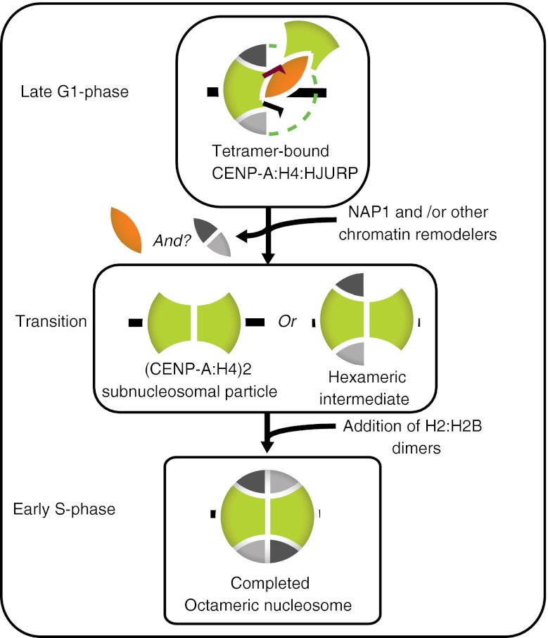
Emerging evidence suggests that the assembly of CENP-A and thus propagation of the epigenetic mark occurs through three major steps: licensing by KNL-2/M18BP1 (Fujita et al. 2007; Maddox et al. 2007) and Mis18 (Hayashi et al. 2004), incorporation via HJURP (Bernad et al. 2011; Dunleavy et al. 2009; Foltz et al. 2009) and maintenance by MgcRacGAP (Lagana et al. 2010). During S phase, as the replication fork forges ahead, the pre-existing population of CENP-A nucleosomes is halved and thereby inherited to daughter strands to preserve centromeric identity (Allshire and Karpen, 2008). The dilution of CENP-A nucleosomes and the replication-independent incorporation of CENP-A raise the possibility of the formation of various placeholder structures in S phase (Dunleavy et al. 2011) (Fig. 3c). In case of human cells, according to the recent data, the CENP-A nucleosomal population is proposed to undergo the tetramer to octamer transition in front of the replication fork approaching the centromeric DNA (Bui et al. 2012). Figure 4 depicts possible steps in this transition.
Fig. 4.
Possible steps of the tetramer to octamer transition. Upon transition of the cell to S phase, HJURP is released from the centromeric chromatin and certain posttranslational modifications and of CENP-A tetramers are resolved by yet-to-be-identified factors. In the first scenario, H2A:H2B dimers are also temporarily removed allowing the tetrameric (CENP-A:H4)2 complex to form and interact with DNA. This reaction is speculated to be mediated by NAP1, which is a chromatin remodeler. Addition of a pair of H2A:H2B dimers will complete the octamer formation. In the second scenario, however, incorporation of new CENP-A:H4 as well as H2B:H2B dimers does not require disassembly of the pre-existing CENP-A tetramers
It is not clear what exactly signals the speculated reversion of octamers into the tetramers at the end of S phase. Passage of the fork may, via unknown mechanisms or interactions, trigger not only the splitting of the pre-existing CENP-A nucleosomes, but also the reversion of octamers into tetramers. This may in turn coincide with the reappearance of HJURP at the end of S phase in human cells. In contrast to canonical nucleosomes, incorporation of newly synthesized CENP-A nucleosomes does not accompany DNA replication (Jansen et al. 2007). CENP-A assembly in the mammalian system requires exit from mitosis (Jansen et al. 2007) and takes place during late M/G1 phase of the cell cycle in the mammalian and embryonic Drosophila systems (Schuh et al. 2007; Mellone et al. 2011). However, the mechanism of specific recognition of the centromere by CENP-A assembly proteins remains largely unknown. It is assumable that these proteins might recognize a specialized chromatin structure, certain contact sites on CENP-A-containing nucleosomes or a non-conventional nucleosome form exclusively found at the centromeric chromatin. Given the proposed atypical CENP-A nucleosomes, regardless of the model, an entertaining speculation would be that the heteroclite structure of CENP-A nucleosomes might provide the green light for CENP-A assembly machinery to repopulate the centromere in preparation for the subsequent mitosis. In addition, these atypical structures may serve as recognition sites for kinetochore assembly during mitosis.
Conclusion
This review summarizes major features of CENP-A-containing complexes on sub-nucleosomal, nucleosomal and chromatin levels. The debate over the true molecular nature of the CENP-A epigenetic mark remains to be resolved as many questions are still unanswered. For example, what could be the biological significance of the tetramer to octamer transition in S phase for mammalian cells or anaphase B in case of yeast? What are the factors and mechanisms involved? In the coming exciting years of research, high-resolution imaging and biochemical approaches hold promise to pave the way for answering these questions.
Acknowledgments
We are grateful to all members of Paul and Amy Maddox labs. Special thanks to Joel Ryan for his valuable assistance with the figures. P.S.M. is the Canada Research Chair in Cell Division and Chromosomal Organization and supported by research grants from the CIHR (MOP-106548) and CCSRI (700824).
Abbreviations
- CENP-A
Centromere protein A
- Cse4
Chromosome segregation 4
- CID
Centromere identifier
- CATD
CENP-A targeting domain
- HFP
Histone fold domain
- HJURP
Holliday Junction recognition protein
- Scm3
Suppressor of chromosome missegregation 3
- CBD
Cse4 binding domain
- AFM
Atomic force microscopy
- EM
Electron microscopy
- FCS
Fluorescence correlation spectroscopy
- ChIP
Chromatin immunoprecipitation
- PTM
Posttranslational modification
References
- Allshire RC, Karpen GH. Epigenetic regulation of centromeric chromatin: old dogs, new tricks? Nat Rev Genet. 2008;9:923–937. doi: 10.1038/nrg2466. [DOI] [PMC free article] [PubMed] [Google Scholar]
- Barnhart MC, Kuich PH, Stellfox ME, Ward JA, Bassett EA, Black BE, Foltz DR. HJURP is a CENP-A chromatin assembly factor sufficient to form a functional de novo kinetochore. J Cell Biol. 2011;194:229–243. doi: 10.1083/jcb.201012017. [DOI] [PMC free article] [PubMed] [Google Scholar]
- Bassett EA, DeNizio J, Barnhart-Dailey MC, Panchenko T, Sekulic N, Rogers DJ, Foltz DR, Black BE. HJURP uses distinct CENP-A surfaces to recognize and to stabilize CENP-A/histone H4 for centromere assembly. Dev Cell. 2012;22:749–762. doi: 10.1016/j.devcel.2012.02.001. [DOI] [PMC free article] [PubMed] [Google Scholar]
- Bernad R, Sanchez P, Rivera T, Rodriguez-Corsino M, Boyarchuk E, Vassias I, Ray-Gallet D, Arnaoutov A, Dasso M, Almouzni G, et al. Xenopus HJURP and condensin II are required for CENP-A assembly. J Cell Biol. 2011;192:569–582. doi: 10.1083/jcb.201005136. [DOI] [PMC free article] [PubMed] [Google Scholar]
- Black BE, Brock MA, Bedard S, Woods VL, Jr, Cleveland DW. An epigenetic mark generated by the incorporation of CENP-A into centromeric nucleosomes. Proc Natl Acad Sci U S A. 2007;104:5008–5013. doi: 10.1073/pnas.0700390104. [DOI] [PMC free article] [PubMed] [Google Scholar]
- Black BE, Foltz DR, Chakravarthy S, Luger K, Woods VL, Jr, Cleveland DW. Structural determinants for generating centromeric chromatin. Nature. 2004;430:578–582. doi: 10.1038/nature02766. [DOI] [PubMed] [Google Scholar]
- Black BE, Jansen LE, Maddox PS, Foltz DR, Desai AB, Shah JV, Cleveland DW. Centromere identity maintained by nucleosomes assembled with histone H3 containing the CENP-A targeting domain. Mol Cell. 2007;25:309–322. doi: 10.1016/j.molcel.2006.12.018. [DOI] [PubMed] [Google Scholar]
- Bui M, Dimitriadis EK, Hoischen C, An E, Quenet D, Giebe S, Nita-Lazar A, Diekmann S, Dalal Y. Cell-cycle-dependent structural transitions in the human CENP-A nucleosome in vivo. Cell. 2012;150:317–326. doi: 10.1016/j.cell.2012.05.035. [DOI] [PMC free article] [PubMed] [Google Scholar]
- Bulseco DA, Wolf DE. Fluorescence correlation spectroscopy: molecular complexing in solution and in living cells. Methods Cell Biol. 2007;81:525–559. doi: 10.1016/S0091-679X(06)81025-3. [DOI] [PubMed] [Google Scholar]
- Camahort R, Shivaraju M, Mattingly M, Li B, Nakanishi S, Zhu D, Shilatifard A, Workman JL, Gerton JL. Cse4 is part of an octameric nucleosome in budding yeast. Mol Cell. 2009;35:794–805. doi: 10.1016/j.molcel.2009.07.022. [DOI] [PMC free article] [PubMed] [Google Scholar]
- Cho US, Harrison SC. Recognition of the centromere-specific histone Cse4 by the chaperone Scm3. Proc Natl Acad Sci U S A. 2011;108:9367–9371. doi: 10.1073/pnas.1106389108. [DOI] [PMC free article] [PubMed] [Google Scholar]
- Cleveland DW, Mao Y, Sullivan KF. Centromeres and kinetochores: from epigenetics to mitotic checkpoint signaling. Cell. 2003;112:407–421. doi: 10.1016/S0092-8674(03)00115-6. [DOI] [PubMed] [Google Scholar]
- Coffman VC, Wu P, Parthun MR, Wu JQ. CENP-A exceeds microtubule attachment sites in centromere clusters of both budding and fission yeast. J Cell Biol. 2011;195:563–572. doi: 10.1083/jcb.201106078. [DOI] [PMC free article] [PubMed] [Google Scholar]
- Conde e Silva N, Black BE, Sivolob A, Filipski J, Cleveland DW, Prunell A. CENP-A-containing nucleosomes: easier disassembly versus exclusive centromeric localization. J Mol Biol. 2007;370:555–573. doi: 10.1016/j.jmb.2007.04.064. [DOI] [PubMed] [Google Scholar]
- Dalal Y, Wang H, Lindsay S, Henikoff S. Tetrameric structure of centromeric nucleosomes in interphase Drosophila cells. PLoS Biol. 2007;5:e218. doi: 10.1371/journal.pbio.0050218. [DOI] [PMC free article] [PubMed] [Google Scholar]
- Dimitriadis EK, Weber C, Gill RK, Diekmann S, Dalal Y. Tetrameric organization of vertebrate centromeric nucleosomes. Proc Natl Acad Sci U S A. 2010;107:20317–20322. doi: 10.1073/pnas.1009563107. [DOI] [PMC free article] [PubMed] [Google Scholar]
- Dunleavy EM, Almouzni G, Karpen GH. H3.3 is deposited at centromeres in S phase as a placeholder for newly assembled CENP-A in G(1) phase. Nucleus. 2011;2:146–157. doi: 10.4161/nucl.2.2.15211. [DOI] [PMC free article] [PubMed] [Google Scholar]
- Dunleavy EM, Roche D, Tagami H, Lacoste N, Ray-Gallet D, Nakamura Y, Daigo Y, Nakatani Y, Almouzni-Pettinotti G. HJURP is a cell-cycle-dependent maintenance and deposition factor of CENP-A at centromeres. Cell. 2009;137:485–497. doi: 10.1016/j.cell.2009.02.040. [DOI] [PubMed] [Google Scholar]
- Foltz DR, Jansen LE, Bailey AO, Yates JR, 3rd, Bassett EA, Wood S, Black BE, Cleveland DW. Centromere-specific assembly of CENP-a nucleosomes is mediated by HJURP. Cell. 2009;137:472–484. doi: 10.1016/j.cell.2009.02.039. [DOI] [PMC free article] [PubMed] [Google Scholar]
- Fujita Y, Hayashi T, Kiyomitsu T, Toyoda Y, Kokubu A, Obuse C, Yanagida M. Priming of centromere for CENP-A recruitment by human hMis18alpha, hMis18beta, and M18BP1. Dev Cell. 2007;12:17–30. doi: 10.1016/j.devcel.2006.11.002. [DOI] [PubMed] [Google Scholar]
- Fukagawa T. Centromere DNA, proteins and kinetochore assembly in vertebrate cells. Chromosome Research: an International Journal on the Molecular, Supramolecular and Evolutionary Aspects of Chromosome Biology. 2004;12:557–567. doi: 10.1023/B:CHRO.0000036590.96208.83. [DOI] [PubMed] [Google Scholar]
- Furuyama T, Henikoff S. Centromeric nucleosomes induce positive DNA supercoils. Cell. 2009;138:104–113. doi: 10.1016/j.cell.2009.04.049. [DOI] [PMC free article] [PubMed] [Google Scholar]
- Guse A, Carroll CW, Moree B, Fuller CJ, Straight AF. In vitro centromere and kinetochore assembly on defined chromatin templates. Nature. 2011;477:354–358. doi: 10.1038/nature10379. [DOI] [PMC free article] [PubMed] [Google Scholar]
- Hayashi T, Fujita Y, Iwasaki O, Adachi Y, Takahashi K, Yanagida M. Mis16 and Mis18 are required for CENP-A loading and histone deacetylation at centromeres. Cell. 2004;118:715–729. doi: 10.1016/j.cell.2004.09.002. [DOI] [PubMed] [Google Scholar]
- Henikoff S, Henikoff JG. “Point” centromeres of Saccharomyces harbor single centromere-specific nucleosomes. Genetics. 2012;190:1575–1577. doi: 10.1534/genetics.111.137711. [DOI] [PMC free article] [PubMed] [Google Scholar]
- Howman EV, Fowler KJ, Newson AJ, Redward S, MacDonald AC, Kalitsis P, Choo KH. Early disruption of centromeric chromatin organization in centromere protein A (CENP-A) null mice. Proc Natl Acad Sci U S A. 2000;97:1148–1153. doi: 10.1073/pnas.97.3.1148. [DOI] [PMC free article] [PubMed] [Google Scholar]
- Hu H, Liu Y, Wang M, Fang J, Huang H, Yang N, Li Y, Wang J, Yao X, Shi Y, et al. Structure of a CENP-A-histone H4 heterodimer in complex with chaperone HJURP. Genes Dev. 2011;25:901–906. doi: 10.1101/gad.2045111. [DOI] [PMC free article] [PubMed] [Google Scholar]
- Huang CC, Chang KM, Cui H, Jayaram M. Histone H3-variant Cse4-induced positive DNA supercoiling in the yeast plasmid has implications for a plasmid origin of a chromosome centromere. Proc Natl Acad Sci U S A. 2011;108:13671–13676. doi: 10.1073/pnas.1101944108. [DOI] [PMC free article] [PubMed] [Google Scholar]
- Jansen LE, Black BE, Foltz DR, Cleveland DW. Propagation of centromeric chromatin requires exit from mitosis. J Cell Biol. 2007;176:795–805. doi: 10.1083/jcb.200701066. [DOI] [PMC free article] [PubMed] [Google Scholar]
- Krassovsky K, Henikoff JG, Henikoff S. Tripartite organization of centromeric chromatin in budding yeast. Proc Natl Acad Sci U S A. 2012;109:243–248. doi: 10.1073/pnas.1118898109. [DOI] [PMC free article] [PubMed] [Google Scholar]
- Krichevsky O, Bonnet G. Fluorescence correlation spectroscopy: the technique and its applications. Rep Prog Phys. 2002;65:251–297. doi: 10.1088/0034-4885/65/2/203. [DOI] [Google Scholar]
- Lagana A, Dorn JF, De Rop V, Ladouceur AM, Maddox AS, Maddox PS. A small GTPase molecular switch regulates epigenetic centromere maintenance by stabilizing newly incorporated CENP-A. Nat Cell Biol. 2010;12:1186–1193. doi: 10.1038/ncb2129. [DOI] [PubMed] [Google Scholar]
- Lavelle C, Prunell A. Chromatin polymorphism and the nucleosome superfamily: a genealogy. Cell Cycle. 2007;6:2113–2119. doi: 10.4161/cc.6.17.4631. [DOI] [PubMed] [Google Scholar]
- Lawrimore J, Bloom KS, Salmon ED. Point centromeres contain more than a single centromere-specific Cse4 (CENP-A) nucleosome. J Cell Biol. 2011;195:573–582. doi: 10.1083/jcb.201106036. [DOI] [PMC free article] [PubMed] [Google Scholar]
- Lochmann B, Ivanov D. Histone H3 localizes to the centromeric DNA in budding yeast. PLoS Genet. 2012;8:e1002739. doi: 10.1371/journal.pgen.1002739. [DOI] [PMC free article] [PubMed] [Google Scholar]
- Maddox PS, Hyndman F, Monen J, Oegema K, Desai A. Functional genomics identifies a Myb domain-containing protein family required for assembly of CENP-A chromatin. J Cell Biol. 2007;176:757–763. doi: 10.1083/jcb.200701065. [DOI] [PMC free article] [PubMed] [Google Scholar]
- Mellone BG, Grive KJ, Shteyn V, Bowers SR, Oderberg I, Karpen GH. Assembly of Drosophila centromeric chromatin proteins during mitosis. PLoS Genet. 2011;7:e1002068. doi: 10.1371/journal.pgen.1002068. [DOI] [PMC free article] [PubMed] [Google Scholar]
- Mizuguchi G, Xiao H, Wisniewski J, Smith MM, Wu C (2007) Nonhistone scm3 and histones cenH3-H4 assemble the core of centromere-specific nucleosomes. Cell 129(6):1153–1164 [DOI] [PubMed]
- Nishino T, Takeuchi K, Gascoigne KE, Suzuki A, Hori T, Oyama T, Morikawa K, Cheeseman IM, Fukagawa T. CENP-T-W-S-X forms a unique centromeric chromatin structure with a histone-like fold. Cell. 2012;148:487–501. doi: 10.1016/j.cell.2011.11.061. [DOI] [PMC free article] [PubMed] [Google Scholar]
- Oudet P, Germond JE, Bellard M, Spadafora C, Chambon P. Nucleosome structure. Philos Trans R Soc Lond B Biol Sci. 1978;283:241–258. doi: 10.1098/rstb.1978.0021. [DOI] [PubMed] [Google Scholar]
- Palmer DK, O’Day K, Trong HL, Charbonneau H, Margolis RL. Purification of the centromere-specific protein CENP-A and demonstration that it is a distinctive histone. Proc Natl Acad Sci U S A. 1991;88:3734–3738. doi: 10.1073/pnas.88.9.3734. [DOI] [PMC free article] [PubMed] [Google Scholar]
- Panchenko T, Sorensen TC, Woodcock CL, Kan ZY, Wood S, Resch MG, Luger K, Englander SW, Hansen JC, Black BE. Replacement of histone H3 with CENP-A directs global nucleosome array condensation and loosening of nucleosome superhelical termini. Proc Natl Acad Sci U S A. 2011;108:16588–16593. doi: 10.1073/pnas.1113621108. [DOI] [PMC free article] [PubMed] [Google Scholar]
- Pearson CG, Maddox PS, Salmon ED, Bloom K. Budding yeast chromosome structure and dynamics during mitosis. J Cell Biol. 2001;152:1255–1266. doi: 10.1083/jcb.152.6.1255. [DOI] [PMC free article] [PubMed] [Google Scholar]
- Probst AV, Dunleavy E, Almouzni G. Epigenetic inheritance during the cell cycle. Nat Rev Mol Cell Biol. 2009;10:192–206. doi: 10.1038/nrm2640. [DOI] [PubMed] [Google Scholar]
- Reeve JN, Sandman K, Daniels CJ. Archaeal histones, nucleosomes, and transcription initiation. Cell. 1997;89:999–1002. doi: 10.1016/S0092-8674(00)80286-X. [DOI] [PubMed] [Google Scholar]
- Schuh M, Lehner CF, Heidmann S. Incorporation of Drosophila CID/CENP-A and CENP-C into centromeres during early embryonic anaphase. Current Biology: CB. 2007;17:237–243. doi: 10.1016/j.cub.2006.11.051. [DOI] [PubMed] [Google Scholar]
- Sekulic N, Bassett EA, Rogers DJ, Black BE. The structure of (CENP-A-H4)(2) reveals physical features that mark centromeres. Nature. 2010;467:347–351. doi: 10.1038/nature09323. [DOI] [PMC free article] [PubMed] [Google Scholar]
- Shelby RD, Monier K, Sullivan KF. Chromatin assembly at kinetochores is uncoupled from DNA replication. J Cell Biol. 2000;151:1113–1118. doi: 10.1083/jcb.151.5.1113. [DOI] [PMC free article] [PubMed] [Google Scholar]
- Shivaraju M, Unruh JR, Slaughter BD, Mattingly M, Berman J, Gerton JL. Cell-cycle-coupled structural oscillation of centromeric nucleosomes in yeast. Cell. 2012;150:304–316. doi: 10.1016/j.cell.2012.05.034. [DOI] [PMC free article] [PubMed] [Google Scholar]
- Shuaib M, Ouararhni K, Dimitrov S, Hamiche A. HJURP binds CENP-A via a highly conserved N-terminal domain and mediates its deposition at centromeres. Proc Natl Acad Sci U S A. 2010;107:1349–1354. doi: 10.1073/pnas.0913709107. [DOI] [PMC free article] [PubMed] [Google Scholar]
- Sullivan KF, Hechenberger M, Masri K. Human CENP-A contains a histone H3 related histone fold domain that is required for targeting to the centromere. J Cell Biol. 1994;127:581–592. doi: 10.1083/jcb.127.3.581. [DOI] [PMC free article] [PubMed] [Google Scholar]
- Tachiwana H, Kagawa W, Shiga T, Osakabe A, Miya Y, Saito K, Hayashi-Takanaka Y, Oda T, Sato M, Park SY, et al. Crystal structure of the human centromeric nucleosome containing CENP-A. Nature. 2011;476:232–235. doi: 10.1038/nature10258. [DOI] [PubMed] [Google Scholar]
- Tomonaga T, Matsushita K, Yamaguchi S, Oohashi T, Shimada H, Ochiai T, Yoda K, Nomura F. Overexpression and mistargeting of centromere protein-A in human primary colorectal cancer. Cancer Res. 2003;63:3511–3516. [PubMed] [Google Scholar]
- Weintraub H, Worcel A, Alberts B. A model for chromatin based upon two symmetrically paired half-nucleosomes. Cell. 1976;9:409–417. doi: 10.1016/0092-8674(76)90085-4. [DOI] [PubMed] [Google Scholar]
- Yoda K, Ando S, Morishita S, Houmura K, Hashimoto K, Takeyasu K, Okazaki T. Human centromere protein A (CENP-A) can replace histone H3 in nucleosome reconstitution in vitro. Proc Natl Acad Sci U S A. 2000;97:7266–7271. doi: 10.1073/pnas.130189697. [DOI] [PMC free article] [PubMed] [Google Scholar]
- Zhang W, Colmenares SU, Karpen GH. Assembly of Drosophila centromeric nucleosomes requires CID dimerization. Mol Cell. 2012;45:263–269. doi: 10.1016/j.molcel.2011.12.010. [DOI] [PMC free article] [PubMed] [Google Scholar]
- Zhou Z, Feng H, Zhou BR, Ghirlando R, Hu K, Zwolak A, Miller Jenkins LM, Xiao H, Tjandra N, Wu C, et al. Structural basis for recognition of centromere histone variant CenH3 by the chaperone Scm3. Nature. 2011;472:234–237. doi: 10.1038/nature09854. [DOI] [PMC free article] [PubMed] [Google Scholar]


