

Abstract
Objective:
Recent evidence suggests that women with gestational hypertension (GH) have a high rate of sleep disordered breathing (SDB), and treatment for even marginal SDB may improve blood pressure control in women with GH. We assessed whether the application SDB treatment could improve blood pressure in women with GH.
Methods:
This was a single-center randomized study. Subjects underwent an unattended home-based diagnostic sleep study. The study was then repeated with subjects wearing one of two randomly assigned treatments: auto-titrating continuous positive airway pressure (auto-CPAP) or mandibular advancement device (MAD) + nasal strip. First morning blood pressure and blood for standard GH measures plus inflammatory markers were taken after each study. Subjects completed a series of questionnaires addressing sleep quality and tolerance of assigned therapy.
Results:
Twenty-four women completed the protocol—13 in the MAD group and 11 in auto-CPAP. The overall rate of SDB was 38%. Auto-CPAP was more effective at treating SDB than MAD + nasal strip, although the women randomized to MAD + nasal strip reported the greater comfort with therapy. First morning blood pressure was not consistently improved with either therapy. When subjects were stratified according to those whose blood pressure increased or decreased with therapy, an association was suggested between blood pressure improvement and reduced levels of tumour necrosis factor-α.
Conclusion:
We demonstrated that 38% of women with GH had concurrent SDB. We did not find an improvement in blood pressure or inflammatory markers with a single night of either the auto-CPAP or MAD + nasal strip interventions. However important lessons from this study may guide future investigations in this area.
Citation:
Reid J; Taylor-Gjevre R; Gjevre J; Skomro R; Fenton M; Olatunbosun F; Gordon JR; Cotton D. Can gestational hypertension be modified by treating nocturnal airflow limitation? J Clin Sleep Med 2013;9(4):311-317.
Keywords: Hypertension, preeclampsia, sleep apnea, sleep disordered breathing, treatment
Gestational hypertension (GH) complicates nearly 10% of all pregnancies.1 If a patient with GH has evidence of proteinuria (≥ 300 mg/day) or signs of other organ damage, then she is said to have preeclampsia.2 The underlying cause of GH is unknown, but recent evidence suggests that sleep disordered breathing (SDB) may be a contributing factor in the development or exacerbation of GH.3–8 Certainly, GH and SDB share common risk factors and SDB is a recognized cause of hyper-tension in non-pregnant adults.9
Continuous positive airway pressure (CPAP) treatment is considered a safe treatment in pregnant women with SDB, and small studies have suggested its use may improve blood pressure in women with or at risk for GH. Edwards et al. demonstrated improvements in blood pressure with only a single night of auto-adjusting CPAP (auto-CPAP) in women with severe preeclampsia, as well as a reduction in uric acid levels—a metabolic marker of preeclampsia.6 These results were particularly interesting because although all subjects demonstrated subtle decrements to inspira-tory airflow, none had an AHI ≥ 5, suggesting that perhaps even a mild degree of airway obstruction during sleep interacts negatively with the heightened inflammatory state of GH. If these results could be replicated in more “real world” conditions, it might obviate the need for diagnostic testing and thereby simplify and expedite the treatment of patients for whom time is of the essence.
BRIEF SUMMARY
Current knowledge/Study Rationale: To assess the frequency of SDB in women with GH using a portable monitoring system. Also, to assess the effect of two different SDB treatment strategies on first morning blood pressure and markers of inflammation in these women.
Study Impact: Our findings support the hypothesis that at least mild SDB in common in unselected women with GH. While we failed to achieve our pirmary endpoint, important lessons from this study will be innstuctive for further research in this area.
To date the published literature has focussed solely on CPAP for treatment of SDB in GH.5,6,10 Although mandibular advancement devices (MAD) are an alternative to CPAP therapy for mild to moderate SDB11,12 and are often preferred to CPAP by young patients, MADs have not been evaluated in the context of GH. One reason may be that MADs usually take weeks to months to be properly adjusted. However, temporary MADs can be fitted at home in ten minutes without the assistance of a dental professional. The SomnoGuard AP is one such device that has been shown to improve mild to moderate SDB.13,14 Additionally, nasal strips are widely used to diminish snoring,15,16 but they alone are not effective at treating SDB. As nasal congestion is an extremely common symptom in women with GH,4,7 we postulated that the addition of a nasal strip may prove to be a simple, safe, and inexpensive augmentation to MAD in this population.
There are often only a few weeks or less from the time of GH diagnosis until delivery and if GH progresses to preeclampsia then the expectant mother is routinely admitted to hospital and may be urgently delivered. Patients with GH require the prompt initiation of effective therapies, from which obstetricians expect results within days and sometimes even hours. These circumstances render laboratory-based polysomnography (PSG) impractical, but home-based testing strategies such as nocturnal oximetry and conventional level 3 monitoring are insufficiently sensitive to diagnose the mild degree of SDB usually seen in women with GH—likely explaining the disparate reports in the literature.6,7,17,18
We postulated that by using a portable sleep monitor with EEG capability we would detect a high rate of mild SDB in women with GH and furthermore that most, if not all, subjects would demonstrate inspiratory airflow limitation. We also assessed whether the combination of a MAD + nasal strip would be as effective as auto-CPAP in improving SDB and inspiratory airflow. Finally and most importantly, to assess whether the findings of Edwards et al.6 could be translated to “real world” conditions, we assessed whether either assigned therapy was associated with an improvement in first morning blood pressure, regardless of RDI. This was our primary endpoint. Additionally, as an exploratory assessment, we measured first morning metabolic/inflammatory marker responses to treatment in all our subjects. Although it would have been ideal to evaluate the treatment effect over several weeks, the unpredictable course of late-term complicated pregnancies along with frequent medication changes made this impractical. Additionally, as the clinical arena demands therapies to effectively lower blood pressure within days we felt it appropriate to test our interventions against this standard and therefore focussed our assessment to a single night intervention.
METHODS
This was a single-center randomized pilot study comparing 2 SDB treatment modalities (auto-CPAP versus MAD + nasal strip) in women with GH (with or without proteinuria). The study comprised 2 parts: part one was a home-based diagnostic evaluation for SDB; in part 2, subjects repeated the home-based sleep study while wearing one of the 2 randomly assigned treatments. After each study, first morning blood pressure was measured by a registered nurse and blood was drawn for inflammatory markers. Subjects also completed a series of questionnaires which assessed sleep quality and tolerance of assigned therapy. The study was approved by the University of Saskatchewan Research Ethics Board and registered with the National Clinical Trials Registry (NCT 00757718).
Participants
Women ≥ 18 years of age with singleton pregnancies and the diagnosis of GH (with or without proteinuria) were recruited from the Fetal Assessment Unit and Antepartum ward of Royal University Hospital, Saskatoon, Saskatchewan, Canada. GH was defined according to standard criteria.2 All potentially eligible women identified were screened for eligibility by chart review and if possible, by interview.
Procedures
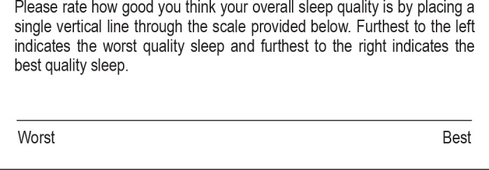
Baseline data were collected for age, height, weight, weight gain in pregnancy, snoring, smoking status, and obstetrical and general medical history. Subjects completed a series of short questionnaires which addressed both sleepiness and common signs or symptoms of SDB. These included the Epworth Sleepiness Score (ESS),19 the Pittsburgh Sleep Quality Index (PSQI),20 the Berlin Questionnaire,21 and a visual analogue scale ([VAS] with 0 for worst and 100 for best) of sleep quality (Appendix 1). A full-night unattended portable Sleep Study using the Embletta X100 (Embla, Denver, CO, USA) was then performed either at the subjects' home or hospital bed. Monitoring channels included 2-lead electroencephalogram, 1-lead electrooculogram, submental electromyogram, pulse oximetry, nasal airflow pressure sensor and oronasal thermistor airflow, and snore vibration sensor chest and abdominal movements by Xact Trace inductive plethysmography. Following the study, first morning blood pressure was taken by a registered nurse with experience in high-risk obstetrics. Fasting blood was drawn the morning after the study and tested the same day for CBC, liver enzymes, and highly sensitive C-reactive protein (CRP). A final tube of blood was prepared and stored at −80° centigrade until study completion, at which time all samples were tested as a single batch for interleukin (IL)-2, IL-6, IL-8, and tumour necrosis factor alpha (TNF-α).
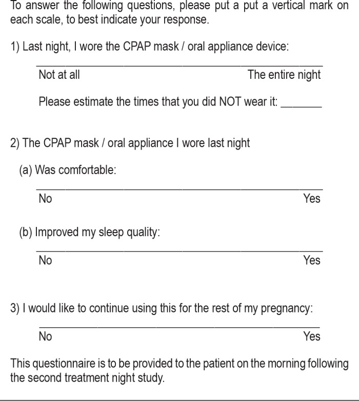
Subjects then completed a second sleep study while wearing the randomly assigned treatment: auto-CPAP (Respironics [Koninklijke Philips Electronics N.V. Amsterdam The Netherlands] REMstar Auto M series with range 5-20 cm H2O and C-Flex set at 2) with heated humidification or a combination of SomnoGuard AP (Tomed Dr. Toussaint GmbH, Bensheim Germany) MAD and Breathe Right nasal strip (GSK, Brentford, Middlesex, UK). The treatment night was conducted as soon as possible after the diagnostic study, usually the subsequent night, and almost always within 72 hours. Treatment allocation was determined by envelope randomization performed at the time of study enrolment and all subjects received a treatment night regardless of the results of the first night diagnostic study. Auto-CPAP subjects were fitted for a mask and instructed on use of the CPAP by a registered sleep technician. All patients assigned to the MAD group had the device fitted by a single investigator (JKR) according to manufacturer's instructions, with the final position of the lower jaw determined by the optimal balance between forward mandibular advancement (mean of 5 mm from resting position, which was 68% of maximum) and subject comfort. The appropriate size of nasal strip (Sm/Med or Large) was assessed by the same investigator, and subjects were instructed on the proper application of both devices. Blood pressure and fasting blood work was repeated in same manner as for the diagnostic study. On the morning following the treatment study, subjects completed a short questionnaire with visual analogue scales (0 for worst and 100 for best, Appendix 2), which evaluated the quality of their sleep with the assigned device, whether it improved their sleep, and whether they would like to continue using this treatment for the remainder of the pregnancy. Our clinical experience has been that many patients initially agree to CPAP therapy but do not follow through with this once at home. We therefore chose visual analogue scales rather than categorical questions because we wanted to appreciate the degree of positive and negative reactions subjects had to their assigned therapies. Although the study evaluation was complete after the single treatment night, women in the Auto-CPAP group were allowed to keep the CPAP unit until the end of pregnancy if they wished. Women in the MAD group kept the MAD for use as they desired and were provide with a supply of nasal strips. Additionally, all subjects were instructed that if they wished to try the therapy not assigned to them, we would provide that for the duration of the pregnancy.
Studies were scored according the American Academy of Sleep Medicine standard criteria22 by a single registered sleep technician who was blinded to clinical data. Respiratory events were categorized as apneas (a decrease in airflow ≥ 90% from baseline for ≥ 10 sec); hypopneas (decrease in airflow ≥ 30% for ≥ 10 sec and followed by a desaturation ≥ 4% from the pre-event baseline); and respiratory event-related arousals (RERAs), defined as a sequence of breaths lasting ≥ 10 sec, associated with flattening of the nasal pressure waveform but not meeting criteria for an apnea or hypopnea, and leading to an arousal from sleep.22 The total number of apneas, hypopneas and RERAs divided by the hours of sleep was expressed as the respiratory disturbance index (RDI).
Cytokine Assays
IL-1, IL-6, IL-8, and TNF-α levels were determined using standard capture ELISA assays, with matched capture and biotinylated specific detection antibodies and recombinant protein standards.23 All samples were run in duplicate, and all assays were sensitive to ≈10 pg/mL of recombinant cytokine
Statistical Analysis
The primary outcome measure was change in morning blood pressure with therapy. Secondary outcomes included changes in RDI and inflammatory cytokine levels with therapy, as well as treatment compliance and subjective perception of improvement in sleep quality. SPSS v.17.0 was employed for data entry and analysis. Descriptive and frequency data was reported. Independent 2-tailed t-tests were used for comparison of continuous data. Comparison of continuous data measures between time points within individuals was performed by paired 2-tailed t-testing. Chi-square testing was employed for categorical data; Fisher exact test was used when cell size was < 5.
The sample size for this study was based on 2-group comparison of the binary outcome of DBP above or below 90 mm Hg. We employed a 2-sided α value of 0.05, a β of 0.2, estimated PA for the CPAP treatment group of 0.85, and PB for the MAD treatment group of 0.30. Utilizing standard sample size calculation formulae provided a requirement for 24 patients in total.24
RESULTS
Study enrolment ran from December 2008 to July 2009, during which time there were 2,712 deliveries, including 101 patients diagnosed with GH (with or without proteinuria). Three-quarters of potentially eligible subjects were identified and approached for study enrollment, with 26 consenting to participate. Twenty-four subjects completed both nights of the study with assigned therapy (11 auto-CPAP and 13 MAD + nasal strip subjects), and these 24 were used for analysis. Seven of the 24 subjects had proteinuria (3 in auto-CPAP and 4 in MAD + nasal strip). Baseline characteristics are presented in Table 1. Despite no subjects having had a prior diagnosis or suspicion of SDB, nasal congestion (19 subjects) and snoring (21 subjects) were commonly reported, and 10 subjects reported nocturnal gasping/choking. There were no significant differences between groups for smoking, nasal congestion, witnessed apneas, snoring, nocturnal gasping or choking, use of antihypertensive medications, gestational diabetes, current or previous preeclampsia. Subjects randomized to MAD + nasal strip did report slightly better sleep at baseline by VAS than the CPAP women (p = 0.034). The treatment study was conducted on the night subsequent to the diagnostic study for 18 of the 24 subjects, and all but one (a 7-day delay in one CPAP subject) were studied within 72 hours. Thirteen subjects had both studies conducted at home, and 8 had both done in the hospital. Three subjects had their diagnostic study in hospital and treatment study at home.
Table 1.
Comparison of baseline characteristics (n = 24)

On the diagnostic study, most subjects demonstrated substantial inspiratory flow limitation, and 9 subjects had at least mild (RDI ≥ 5) SDB. With a mean pressure of 6.8 ± 1.3 cm H2O, auto-CPAP eliminated inspiratory flow limitation and lowered the RDI (10.73 ± 17.07 to 1.38 ± 2.12; p = 0.075; 95% CI: -1.12, 19.81). Downloaded data from the CPAP units showed average usage of nearly 6.5 h for treatment nights (Table 3). This corresponded to 78% of the subjects' estimated total sleep time and correlated surprisingly closely to their subjective estimate of CPAP usage, which was 84% of the night. MAD subjects estimated that they wore the therapy for 75% of the night, but we had no objective measure for verification. The therapeutic effect of the MAD + nasal strip was inconsistent, with 3 subjects actually having an increased RDI and flow limitation with therapy. We did not find any significant, consistent treatment response within individuals for blood pressure (our primary objective), cytokine levels, or sleep parameters over the 2 study nights in either the auto-CPAP or the MAD + nasal strip treatment groups. Full comparison of the diagnostic and treatment night parameters are presented in Table 2.
Table 3.
Comparison of responders vs non-responders, based on diastolic blood pressure (DBP)

Table 2.
Comparison of outcome parameters for diagnostic and treatment studies

As obstetricians consider a diastolic blood pressure (DBP) ≥ 90 mm Hg as the clinically relevant threshold for GH treatment,2 we further compared patients within and between treatment groups based on this dichotomy. In the morning after the diagnostic study, 45.5% (5/11) of CPAP subjects and 53.8% (7/13) MAD subjects had a DBP < 90 mm Hg, compared to post-treatment night percentages of 54.5% (6/11) and 61.5% (8/13), respectively. For CPAP subjects, 18% (2/11) converted to a DBP < 90 mm Hg with treatment, and 9% (1/11) progressed in the opposite direction to a DBP ≥ 90 mm Hg. For the MAD group, 1/13 (7.7%) converted to a DBP < 90 mm Hg, and none converted in the opposite direction. These proportional changes in category between treatment groups were not statistically significant (p = 0.371). Further evaluation of subjects according to DBP response (Table 3) revealed that the TNF-α level significantly decreased in women with blood pressure improvement (Table 3, Figure 1). Similar results were not found for the other cytokine measures.
Figure 1. Comparison of the change in TNF-α levels, based on diastolic blood pressure response.

DISCUSSION
By using a relatively simple home-based portable sleep monitor with limited EEG capability, we found 38% of participants with GH to have coexistent SDB, and most of those with RDI < 5 demonstrated prolonged periods of inspiratory flow limitation during sleep. This is in agreement with other studies that have utilized more extensive monitoring techniques3,6,7 and further supports the concept of SDB in GH as being primarily due to partial upper airway obstruction with little or no arterial oxygen desaturation. While the pathophysiologic importance of this subtle breathing disturbance in GH needs further elucidation, we feel that future research in this evolving area should not rely on oximetry-based monitoring strategies as most abnormalities will likely be missed.17
Regarding the SDB treatment arms, it is not surprising that the MAD was less effective at lowering the RDI than auto-CPAP. MADs are generally less efficacious therapy than CPAP with results that depend on a constellation of individual characteristics; and even when these are considered, a positive response to therapy can still be difficult to predict.25 Furthermore, in our study the MAD could not be automatically adjusted in response to upper airway resistance as the auto-CPAP could. An important reason for our choice of the SomnoGuard AP was its two-piece design that incorporated a small aperture to allow for oral breathing. However, some of our subjects had profound nasal congestion that was not improved by the nasal strip, and it is possible that for these women the MAD actually increased airflow obstruction at the mouth opening.
Ideally, we would have utilized several nights to optimize compliance and adjust therapy as necessary, but the unpredictable clinical course of GH risked us losing a number of subjects to early delivery with this strategy. Furthermore, the physiologic status and medications of GH patients are subject to rapid changes and time delays between the diagnostic night and therapeutic night studies could have complicated the evaluation of treatment. We had therefore planned to have the treatment night as soon as possible after the diagnostic study. We were interested to see that the MAD subjects indicated a greater desire to continue with therapy (Table 2) as well as trend to greater comfort compared to CPAP. This suggests to us that the MAD + nasal strip may still be a therapy worth exploring as an intervention, but earlier in gestation when sufficient time would be available for appropriate patient selection and device adjustment.
With regard to our primary endpoint of blood pressure improvement with therapy, we failed to demonstrate a significant improvement for either treatment arm. It could be reasonably argued that this was an expected consequence of applying treatment to many subjects who did not satisfy the standard diagnostic criteria for SDB. However, we chose our approach to test the hypothesis that most, if not all, women with GH have at least inspiratory airflow limitation during sleep and that this carries with it hemodynamic and inflammatory consequences.6 We do not believe our negative results justify discarding this hypothesis as null because there remain several other potential reasons for our failure to reproduce the results of Edwards. One such reason is that we included all patients with GH, not only those with severe preeclampsia, which may account for the inconsistent changes in hemodynamic parameters observed. We chose this broad inclusion criteria because our previous work had established that SDB frequency is similar in GH women with or without preeclampsia,7 and also because over a third of women with the initial diagnosis of GH will eventually progress to preeclampsia.2,26 However, for a small study such as ours with only a single night intervention, it may have been better in retrospect to have selected subjects with the greatest likelihood of benefit from therapy (i.e., preeclamptics). Accordingly, we find it interesting that those subjects whose DBP did improve with therapy showed a nonsignificant trend toward having a higher DBP at baseline (p = 0.179, Table 3).
Our study has a number of limitations, first being the aforementioned small sample size. Nonetheless, ours is the largest trial we are aware of in this novel area. We do believe that a large multicenter trial is in order to definitively evaluate whether treatment of SDB can improve the management of GH. Secondly, it may be that only one night of treatment in late third trimester is not enough to influence the inflammatory and hemodynamic perturbations of GH; and intervening earlier in gestation, as considered by Guilleminault et al.10 may hold more promise. Such a strategy could allow sufficient time to optimize treatment and provide longitudinal follow-up of nightly therapy. Since women who develop GH earlier in gestation are more likely to be the ones who progress to preeclampsia,26 intervening earlier in gestation (i.e., < 30 weeks) may target those women who are most likely to benefit from therapy.
Furthermore, although the home-based design of our study allowed for generalization to “real-world” conditions, it also presented some limitations. We did not have the ability to make adjustments to the assigned therapy “on the fly” as we could in a laboratory-based study—potentially limiting both the effectiveness of therapy and subject compliance. Additionally, we could not measure therapeutic compliance for the MAD subjects. We do note that for the CPAP group the subjective estimate of usage closely matched actual usage (data downloaded from the machines) and speculate this may also have been true for the MAD group. Just as importantly, our blood pressure measurement technique differed substantially from Edwards et al., who utilized a Finapres (FMS, Amsterdam, The Netherlands) to continuously monitor pressure by the noninvasive volume-clamp method. We have used similar technology in our lab, but found it to be too cumbersome and unreliable to implement with portable testing. Instead we recorded early morning BP measured by an obstetrical nurse while subjects were awake. We feel this approach was appropriate because these are the measurements on which clinical decisions are made. However, such “real-world” measurements likely introduced additional random error for which our study was underpowered to accommodate.
Our biochemical analyses included standard measures for GH (platelets, uric acid) as well as those not evaluated clinically but which have been shown to be increased in GH and/or disturbances of sleep.27,28 In particular, TNF-α is an inflamma-tory biomarker which has been associated with sleep loss29 and improves with CPAP therapy in non-pregnant OSA patients.30 We therefore find it intriguing that the TNF-α level decreased in those subjects who did experience a blood pressure improvement with treatment. More study will be required to determine the clinical relevance of TNF-α in GH women, if it is impacted by the coexistence of SDB, and if so, if SDB treatment can consistently influence that relationship. We hope that our findings will encourage further research into these interactions.
CONCLUSION
Through portable testing with EEG capability, we demonstrated a high rate of SDB in women with GH and auto-titrating CPAP provided effective SDB treatment. While the combination of MAD + nasal strip provided inconsistent results, future studies may delineate clinical characteristics that allow the therapy to be better customized to individuals. Although we were unable to demonstrate a consistent relationship between SDB treatment and improvement in blood pressure or inflammatory markers, we believe the information from this pilot study will guide future research in this evolving field.
DISCLOSURE STATEMENT
This was not an industry supported study. The authors have indicated no financial conflicts of interest.
ACKNOWLEDGMENTS
All research was conducted at the Royal University Hospital Sleep Disorders Centre, University of Saskatchewan. The authors thank Maryla Stiles, Xiaobei Zhang, Jennifer Town, Lori Reid, and the staff in the Royal University Hospital Sleep Lab and Antepartum Ward for their assistance. The authors also acknowledge the Saskatoon Health Region for their support. This project was jointly funded by the Royal University Hospital Foundation and The Lung Association of Saskatchewan. The funding agencies had no role in the acquisition, storage or interpretation of the data. Dr. Reid had full access to all the data in the study and takes full responsibility for the integrity of the data and accuracy of the analysis.
APPENDICES
Appendix 1. Visual analogue scale of sleep quality.
Appendix 2. Feedback form for treatment night.
REFERENCES
- 1.Granger JP, Alexander BT, Bennett WA, Khalil RA. Pathophysiology of pregnancy-induced hypertension. Am J Hypertens. 2001;14:178S–85S. doi: 10.1016/s0895-7061(01)02086-6. [DOI] [PubMed] [Google Scholar]
- 2.Martel MJ. Diagnosis, evaluation, and management of the hypertensive disorders of pregnancy. J Obstet Gynaecol Can. 2008;30:562–3. doi: 10.1016/s1701-2163(16)32887-0. author reply 563. [DOI] [PubMed] [Google Scholar]
- 3.Champagne K, Schwartzman K, Opatrny L, et al. Obstructive sleep apnoea and its association with gestational hypertension. Eur Respir J. 2009;33:559–65. doi: 10.1183/09031936.00122607. [DOI] [PubMed] [Google Scholar]
- 4.Franklin KA, Holmgren PA, Jonsson F, Poromaa N, Stenlund H, Svanborg E. Snoring, pregnancy-induced hypertension, and growth retardation of the fetus. Chest. 2000;117:137–41. doi: 10.1378/chest.117.1.137. [DOI] [PubMed] [Google Scholar]
- 5.Guilleminault C, Kreutzer M, Chang JL. Pregnancy, sleep disordered breathing and treatment with nasal continuous positive airway pressure. Sleep Med. 2004;5:43–51. doi: 10.1016/j.sleep.2003.07.001. [DOI] [PubMed] [Google Scholar]
- 6.Edwards N, Blyton DM, Kirjavainen T, Kesby GJ, Sullivan CE. Nasal continuous positive airway pressure reduces sleep-induced blood pressure increments in preeclampsia. Am J Respir Crit Care Med. 2000;162:252–7. doi: 10.1164/ajrccm.162.1.9905006. [DOI] [PubMed] [Google Scholar]
- 7.Reid J, Skomro R, Cotton D, et al. Pregnant women with gestational hyper-tension may have a high frequency of sleep disordered breathing. Sleep. 2011;34:1033–8. doi: 10.5665/SLEEP.1156. [DOI] [PMC free article] [PubMed] [Google Scholar]
- 8.Yinon D, Lowenstein L, Suraya S, et al. Pre-eclampsia is associated with sleep-disordered breathing and endothelial dysfunction. Eur Respir J. 2006;27:328–33. doi: 10.1183/09031936.06.00010905. [DOI] [PMC free article] [PubMed] [Google Scholar]
- 9.Canadian Hypertension Education Program. The 2008 Canadian Hypertension Education Program recommendations: the scientific summary—an annual update. Can J Cardiol. 2008;24:447–52. doi: 10.1016/s0828-282x(08)70618-4. [DOI] [PMC free article] [PubMed] [Google Scholar]
- 10.Guilleminault C, Palombini L, Poyares D, Takaoka S, Huynh NT, El-Sayed Y. Pre-eclampsia and nasal CPAP: part 1. Early intervention with nasal CPAP in pregnant women with risk-factors for pre-eclampsia: preliminary findings. Sleep Med. 2007;9:9–14. doi: 10.1016/j.sleep.2007.04.020. [DOI] [PubMed] [Google Scholar]
- 11.Kushida CA, Morgenthaler TI, Littner MR, et al. Practice parameters for the treatment of snoring and obstructive sleep apnea with oral appliances: an update for 2005. Sleep. 2006;29:240–3. doi: 10.1093/sleep/29.2.240. [DOI] [PubMed] [Google Scholar]
- 12.Holley AB, Lettieri CJ, Shah AA. Efficacy of an adjustable oral appliance and comparison with continuous positive airway pressure for the treatment of obstructive sleep apnea syndrome. Chest. 2011;140:1511–6. doi: 10.1378/chest.10-2851. [DOI] [PubMed] [Google Scholar]
- 13.Maurer JT, Huber K, Verse T, Hormann K, Stuck B. A mandibular advancement device for the ENT office to treat obstructive sleep apnea. Otolaryngol Head Neck Surg. 2007;136:231–5. doi: 10.1016/j.otohns.2006.08.008. [DOI] [PubMed] [Google Scholar]
- 14.Vanderveken OM, Boudewyns AN, Braem MJ, et al. Pilot study of a novel mandibular advancement device for the control of snoring. Acta Otolaryngol. 2004;124:628–33. doi: 10.1080/00016480310015984. [DOI] [PubMed] [Google Scholar]
- 15.Ulfberg J, Fenton G. Effect of Breathe Right nasal strip on snoring. Rhinology. 1997;35:50–2. [PubMed] [Google Scholar]
- 16.Todorova A, Schellenberg R, Hofmann HC, Dimpfel W. Effect of the external nasal dilator Breathe Right on snoring. Eur J Med Res. 1998;3:367–79. [PubMed] [Google Scholar]
- 17.Yin TT, Williams N, Burton C, et al. Hypertension, fetal growth restriction and obstructive sleep apnoea in pregnancy. Eur J Obstet Gynecol Reprod Biol. 2008;141:35–8. doi: 10.1016/j.ejogrb.2008.07.009. [DOI] [PubMed] [Google Scholar]
- 18.Connolly G, Razak AR, Hayanga A, Russell A, McKenna P, McNicholas WT. Inspiratory flow limitation during sleep in pre-eclampsia: comparison with normal pregnant and nonpregnant women. Eur Respir J. 2001;18:672–6. doi: 10.1183/09031936.01.00053501. [DOI] [PubMed] [Google Scholar]
- 19.Johns MW. A new method for measuring daytime sleepiness: the Epworth sleepiness scale. Sleep. 1991;14:540–5. doi: 10.1093/sleep/14.6.540. [DOI] [PubMed] [Google Scholar]
- 20.Buysse DJ, Reynolds CF, 3rd, Monk TH, Berman SR, Kupfer DJ. The Pittsburgh Sleep Quality Index: a new instrument for psychiatric practice and research. Psychiatry Res. 1989;28:193–213. doi: 10.1016/0165-1781(89)90047-4. [DOI] [PubMed] [Google Scholar]
- 21.Netzer NC, Stoohs RA, Netzer CM, Clark K, Strohl KP. Using the Berlin Questionnaire to identify patients at risk for the sleep apnea syndrome. Ann Intern Med. 1999;131:485–91. doi: 10.7326/0003-4819-131-7-199910050-00002. [DOI] [PubMed] [Google Scholar]
- 22.Iber C, Ancoli-Israel S, Chesson A, Quan S. The AASM manual for the scoring of sleep and associated events: rules, terminology and technical specifications. Westchester, IL: The American Academy of Sleep Medicine; 2007. [Google Scholar]
- 23.Gordon JR, Swystun VA, Li F, et al. Regular salbutamol use increases CXCL8 responses in asthma: relationship to the eosinophil response. Eur Respir J. 2003;22:118–26. doi: 10.1183/09031936.03.00031102. [DOI] [PubMed] [Google Scholar]
- 24.Campbell M, Julious S, Altman D. Estimating sample sizes for binary, ordered categorical and continuous outcomes in two group comparisons. BMJ. 1995;311:1145–8. doi: 10.1136/bmj.311.7013.1145. [DOI] [PMC free article] [PubMed] [Google Scholar]
- 25.Mostafiz W, Dalci O, Sutherland K, et al. Influence of oral and craniofacial dimensions on mandibular advancement splint treatment outcome in patients with obstructive sleep apnea. Chest. 2011;139:1331–9. doi: 10.1378/chest.10-2224. [DOI] [PMC free article] [PubMed] [Google Scholar]
- 26.Magee LA, von Dadelszen P, Bohun CM, et al. Serious perinatal complications of non-proteinuric hypertension: an international, multicentre, retrospective cohort study. J Obstet Gynaecol Can. 2003;25:372–82. doi: 10.1016/s1701-2163(16)30579-5. [DOI] [PubMed] [Google Scholar]
- 27.Vitoratos N, Economou E, Iavazzo C, Panoulis K, Creatsas G. Maternal serum levels of TNF-alpha and IL-6 long after delivery in preeclamptic and normotensive pregnant women. Mediators Inflamm. 2010;2010:908649. doi: 10.1155/2010/908649. [DOI] [PMC free article] [PubMed] [Google Scholar]
- 28.Shi ZM, Li TP, Xian LW, Li ZG. Role of inflammation factors in impaired glucose tolerance and sleep apnea/hypopnea syndrome in pregnant women. Nan Fang Yi Ke Da Xue Xue Bao. 2011;31:1357–9. [PubMed] [Google Scholar]
- 29.Chennaoui M, Sauvet F, Drogou C, et al. Effect of one night of sleep loss on changes in tumor necrosis factor alpha (TNF-alpha) levels in healthy men. Cytokine. 2011;56:318–24. doi: 10.1016/j.cyto.2011.06.002. [DOI] [PubMed] [Google Scholar]
- 30.Hegglin A, Schoch OD, Korte W, Hahn K, Hurny C, Munzer T. Eight months of continuous positive airway pressure (CPAP) decrease tumor necrosis factor alpha (TNFA) in men with obstructive sleep apnea syndrome. Sleep Breath. 2012;16:405–12. doi: 10.1007/s11325-011-0512-2. [DOI] [PubMed] [Google Scholar]