. 2013 Mar 19;2:e00378. doi: 10.7554/eLife.00378

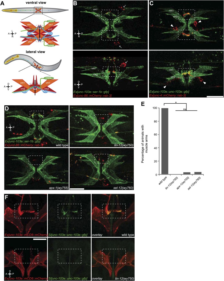
Figure 1. The muscle arms of type 2 vulval muscles (vm2) are missing in lin-12(wy750), apx-1(wy755) and sel-12(wy760) mutants.

(A) An illustration showing ventral (top) and lateral (bottom) views of C. elegans egg-laying circuit. vm1 (orange) and vm2 (red) are vulval muscles. HSN (green) and VC4/5 (blue) are presynaptic motoneurons. Each of the four vm2 cells extends a dendritic spine-like muscle arm laterally, on which it receives synapses with HSN and VC neurons (boxed areas). Top row of (AC) are ventral views; bottom row of (AC) are left lateral views. Anterior (A), posterior (P), left (L), right (R), dorsal (D), ventral (V). (B) Ventral (top) and lateral (bottom) views of vulval muscles labeled by SER-1B::GFP transgene in a young adult. HSN neurons are labeled by a mCherry::RAB-3 transgene. The boxed areas indicate synaptic regions. Arrows indicate the HSN cell bodies. Note that the laterally extended muscle arms on vm2 cells are colocalized with the HSN presynaptic specializations. (C) Ventral (top) and lateral (bottom) views of vulval muscles labeled by a UNC-103E::GFP transgene and VC4/5 neurons labeled by a mCherry::RAB-3 transgene. The boxed areas indicate synaptic regions. Arrowheads indicate cell bodies of VC4 and VC5. Note that the laterally extended muscle arms on vm2 cells are colocalized with the VC4/5 presynaptic specializations. Scale bar is 20 μm. (D) Ventral views of the vulval muscles and presynaptic regions of HSN neurons in wild-type, lin-12(wy750), apx-1(wy755) and sel-12(wy760) animals. The boxed areas indicate synaptic regions. Note the absence of vm2 muscle arms in mutants. Scale bar is 20 μm. (E) Quantification of the vm2 muscle arm defects in wild-type, lin-12(wy750), apx-1(wy755) and sel-12(wy760) animals. * p<0.0001, n.s. no significant difference, chi-squared test, n = 90–114 animals. (F) High magnification lateral views of the synaptic region in wild type and lin-12(wy750). vm2 muscle arms are visualized by a single-copy UNC-103E::GFP transgene. Vulval muscle morphology is labeled by mCD8::mCherry. The boxed areas indicate synaptic regions. Scale bar is 10 μm.

DOI: http://dx.doi.org/10.7554/eLife.00378.003

Figure 1.

Figure 1—figure supplement 1. vm2 muscle arms are the postsynaptic specializations.

Figure 1—figure supplement 1.

(A) Lateral views of the vulval muscles. The vulval muscles are labeled by mCD8::mCherry (top). vm2 muscles arms are labeled by a single-copy unc-103e::gfp transgene (middle). Note that the UNC-103E::GFP is enriched on the muscle arms. Boxed areas indicate synaptic regions. Anterior (A), posterior (P), dorsal (D), ventral (V). Scale bar is 20 μm.

Figure 1—figure supplement 2. vm2 muscle arm development is a guidance event independent of presynaptic specialization.

Figure 1—figure supplement 2.

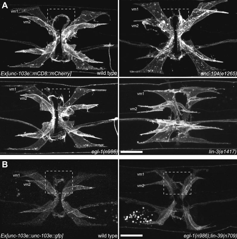
(A) Ventral views of the vulval muscle morphology in wild-type, unc-104(e1265), egl-1(n986) and lin-3(e1417) mutant animals. Boxed areas indicate synaptic regions. Scale bar is 20 μm. (B) Ventral views of the vulval muscle morphology in wild-type (left) and egl-1(n986); lin-39(n709) (right) mutant animals. Boxed areas indicate synaptic regions. Scale bar is 20 μm.

Figure 1—figure supplement 3. Schematic model of LIN-12/Notch signaling pathway.

Figure 1—figure supplement 3.

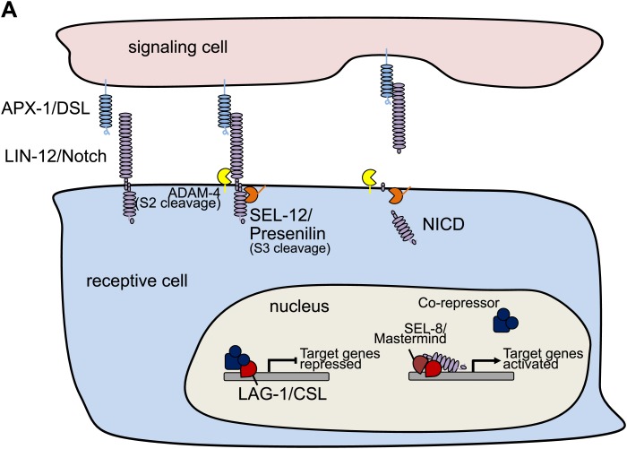
(A) Schematic model showing the core LIN-12/Notch signaling pathway. The binding of the ligand APX-1/DSL to the receptor LIN-12/Notch triggers proteolytic cleavages. SEL-12/Presenilin releases the Notch intracellular domain (NICD), which enters the cell nucleus and interacts with LAG-1/CSL and other transcription factors to activate target gene transcription.

Figure 1—figure supplement 4. lin-12(wy750) has specific vm2 muscle arm defects.

Figure 1—figure supplement 4.

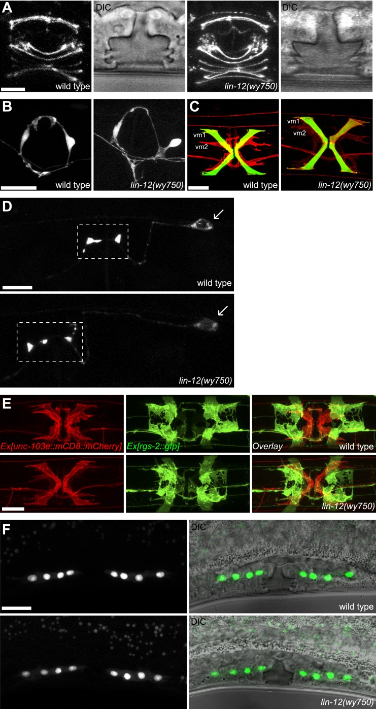
(A) Vulval morphology in wild-type (left two panels) and lin-12(wy750) (right two panels) animals. Vulval morphogenesis is examined by visualizing the apical boundaries of vulval epithelial cells with AJM-1::GFP or by DIC microscopy. Scale bar is 5 μm. (B) VC4 and VC5 axons in wild-type (left) and lin-12(wy750) (right) animals. VC axons are labeled with CAT-1::GFP. Scale bar is 20 μm. (C) Visualization of vm1 and vm2 in wild-type (left) and lin-12(wy750) (right) animals. Vulval muscles (both vm1 and vm2) are labeled with mCD8::mCherry. egl-15::GFP labels vm1 but not vm2 in both wild type and lin-12(wy750) animals. Scale bar is 20 μm. (D) HSN presynapstic regions in wild-type (top) and lin-12(wy750) (bottom) animals. HSN presynaptic regions are labeled by SNB-1::YFP. Boxed areas indicate synaptic regions. Arrows indicate the HSN cell bodies. Scale bar is 10 μm. (E) Visualizations of undifferentiated M lineage cells in wild-type (top) and lin-12(wy750) (bottom) mutants by hlh-8::GFP in mid-L4 animals (lateral view). Note that the pattern of the M lineage cells do not change in the lin-12(wy750) mutant. Scale bar is 20 μm. (F) Ventral view of vulval muscles and uterine muscles in wild-type (up) and lin-12(wy750) (down) animals. Vulval muscles (both vm1 and vm2) are labeled with mCD8::mCherry. rgs-2::GFP labels uterine muscles in both wild type and lin-12(wy750) animals. Note that the uterine muscle differentiation and morphology do not change in lin-12(wy750) mutant. Scale bar is 10 μm.

Figure 1—figure supplement 5. Schematic demonstrations of the quantitative measurements of vulval muscle morphology.

Figure 1—figure supplement 5.

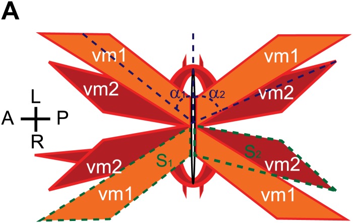
(A) Schematic demonstration of vulval muscles. The intersection angles between vulval opening and vulval muscles are shown with dashed blue lines. vm1 and vm2 are delineated by dashed green lines.

Figure 1—figure supplement 6. Quantifications of the vulval muscle morphology.

Figure 1—figure supplement 6.

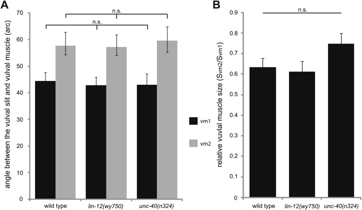
(A) Quantification of the intersection angles between vulval opening and vulval muscles with different genotypes indicated on the X-axis. n.s., no significant difference, t test, n = 48–80 pieces of vulval muscles. (B) Quantification of the relative size of vm2 to vm1 (Svm2/Svm1) with different genotypes indicated on the X-axis. n.s., no significant difference, t test, n = 48–80 pairs of vulval muscles.