Abstract
The purpose of this study was to evaluate the clinical efficacy of 3 new gingival retraction systems; Stay-put, Magic foam cord and expasyl, on the basis of their relative ease of handling, time taken for placement, hemorrhage control and the amount of gingival retraction. Thirty subjects were selected requiring fixed prosthesis. The 3 gingival retraction systems were used on the prepared abutments randomly. The time taken for placement of each retraction system was recorded. The vertical gingival retraction was measured before and after retraction using flexible measuring strip with 0.5 mm grading. The horizontal retraction was measured on polyether impressions made before the retraction and after retraction. Based on the results, magic foam cord retraction system can be considered more effective gingival retraction system among the three retraction systems used in the study.
Keywords: Gingival retraction, Stay-put, Magic foam cord, Expasyl
Introduction
The impression techniques used in the process of making fixed prostheses require the gingival tissue to be displaced to expose the finish lines on the prepared teeth. Therefore, effectively managing the gingiva prior to making an impression is a critical preliminary step in the process of fabricating restorations [1]. One of the most used methods to obtain gingival retraction is by means of cord packed into the sulcus [2]. Nonmedicated cords placed in the gingival sulcus are safe but have limited effect in controlling hemorrhage [3]. Medicated retraction cords are effective, however various studies in past have shown local and systemic side effects induced by medicaments used for gingival retraction [4–8]. The primary reason for not adequately capturing marginal detail is deficient gingival displacement technique [9]. To address these problems, 3 new retraction systems have been introduced, copper wire reinforced retraction cord (Stay-put; Roeko, Coltene/Whaledent), polyvinyl siloxane foam retraction system (Magic foam cord; Coltene/Whaledent Inc) and aluminum chloride paste retraction system (Expasyl; Kerr corporation).
There is no consensus cited in the literature regarding criteria for evaluation of the clinical efficacy with gingival retraction cords [10]. Previous studies have compared various gingival retraction methods like retraction cord, electrosurgery, and rotary gingival curettage [2, 11]. Till date, no studies exclusively done to compare the clinical efficacy of stay-put, magic foam cord and expasyl gingival retraction systems. The purpose of this study was to evaluate the efficacy of these three gingival retraction systems based on amount of gingival retraction attained, time taken for placement, hemorrhage control and relative ease of handling. Also, an effort was made to develop criteria for describing the clinical performance of gingival retraction systems.
Materials and Methods
Inclusion criteria: for the study are subjects with:
Thirty patients whose ages more than 18 years were selected requiring fixed prosthesis with minimum of two abutments
Clinically and radiographically healthy gingiva and periodontium around the abutments.
Abutment teeth of normal size and contour (no developmental anomaly or regressive age changes).
Exclusion criteria: Subjects with:
Age <18 years.
Gingival and periodontal disease.
Uncontrolled diabetes, hypertension, hyperthyroidism and other cardiovascular disorders.
The three gingival retraction systems were used (Table 1) on the prepared abutments randomly, such that each combination is repeated ten times. For example, in the first subject stay-put and expasyl were used for the 2 prepared abutments, in the second subject stay-put and magic foam cord were used and in third subject expasyl and magic foam cord were used for gingival retraction. The same order was followed for all the thirty subjects, so that all three retraction systems were compared with each other in group of two for ten times.
Table 1.
Gingival retraction systems tested
| Retraction systems | Composition | Manufacturer |
|---|---|---|
| Stay-put | Copper-wire reinforced retraction cord | Roeko, Coltene/Whaledent Inc, Cuyahoga Falls, Ohio |
| Expasyl | Aluminum Chloride retraction paste | Kerr corporation, Orange, California |
| Magic foam cord | Expanding type of Polyvinyl Siloxane | Coltene/Whaledent Inc, Cuyahoga Falls, Ohio |
The time taken for placement of each retraction system was recorded in seconds. Smooth rounded flexible measuring strip [12] with 0.5 mm grading (Fig. 1) was used to measure sulcus depth before retraction and after retraction. The measurements recorded in between two consecutive calibrations were considered as 0.25 mm. The horizontal sulcular width was measured indirectly using polyether (Impregnum Soft; 3 M ESPE AG, Germany) impressions of the prepared abutments, made before retraction and after retraction. The width of sulcular extension on the impressions was measured and compared using stereomicroscope (Fig. 2) and image analysis software (Image-Pro Express; Media Cybernetics, Silver Spring) with an accuracy of 1/10th of a micron. Sulcular depths and widths were measured at the mesiobuccal, midbuccal and distobuccal line angle regions. The hemorrhage scores (score# 0, 1, 2) were recorded immediately after removal of the retraction systems.
Fig. 1.
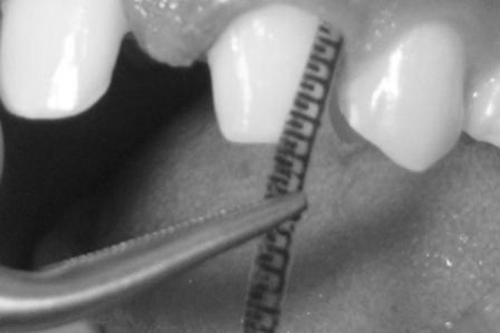
Use of flexible measuring strip to measure vertical gingival retraction
Fig. 2.
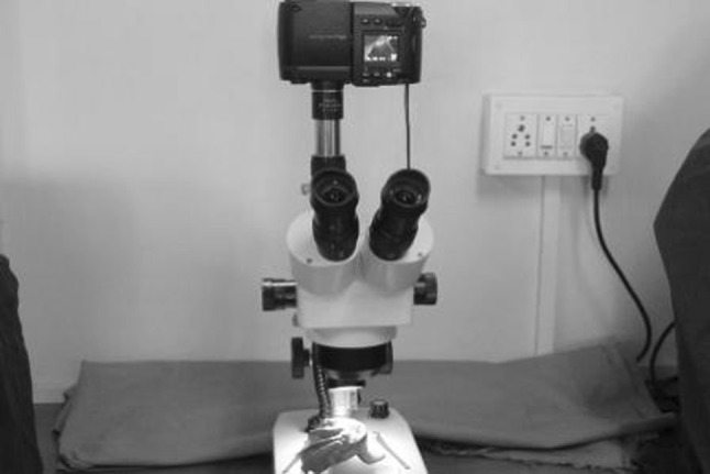
Stereomicroscope used to measure horizontal gingival retraction
The flexible measuring strips were fabricated by printing scale markings on the transparent plastic sheets to the accuracy of 0.5 mm (Fig. 1). The custom trays were fabricated by adapting two layers of softened base plate wax onto the diagnostic model to act as a spacer for the impression material.
Three retraction systems used in the study are listed in Table 1. The stay-put retraction cord of adequate size/width and length was cut and looped around the tooth. Cord packing was started from the mesial interproximal area by gently pushing the cord into the sulcus (Fig. 3). After 4 min the cord was removed.
Fig. 3.
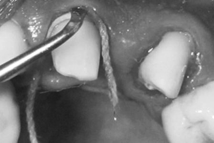
Stay-put retraction cord placement technique
The magic foam cord cartridge was attached to the auto-mixing gun and then the mixing syringe with intraoral tip was placed into the gingival sulcus and gingival retraction material was applied all around the tooth. After injecting the retraction material (Fig. 4) the corresponding comprecap was placed on to the abutment to push the material deep into the gingival sulcus (Fig. 5). After 4 min, the comprecap with the set retraction material attached to it was removed from the patient mouth.
Fig. 4.
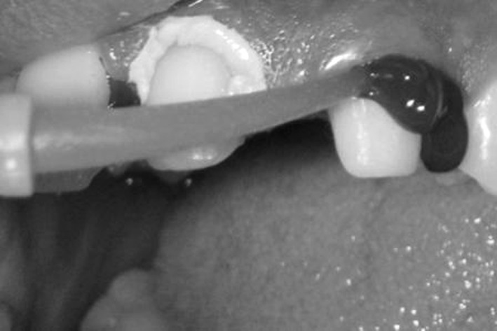
Magic foam cord retraction material injected around gingival sulcus
Fig. 5.
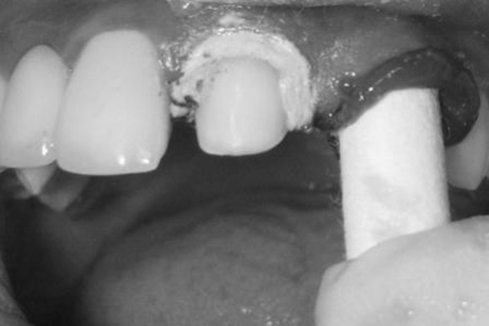
Comprecap placed on abutment to push magic foam cord retraction material into sulcus
The expasyl retraction paste [13] was injected slowly into the gingival sulcus with help of an applicator gun and cannula (Fig. 6). No pressure was applied on gingiva with the cannula. The paste is left in place for 4 min and then removed by rinsing.
Fig. 6.
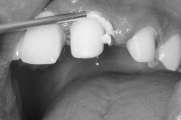
Expasyl retraction system placement technique
Time taken for the placement of each retraction system was recorded in seconds. Prior to the application of retraction system, with the help of flexible scale the sulcular depth at mesio-buccal, mid-buccal and disto-buccal regions were measured on both the abutment teeth. Similarly, the measurements were recorded after gingival retraction. The difference between the two readings was compared to obtain net amount of vertical gingival retraction. Although sulcular depth can be measured by using manual periodontal probe, but manual probing is invasive, which may cause patient discomfort [14].
To record the width of gingival sulcus before retraction, polyether impression of the prepared abutments were made using custom trays (Fig. 7). Monophase impression technique was used. Subsequently, these impressions were compared with the polyether impressions made after retraction using stereomicroscope. The stereomicroscopic images (10× resolution) of individual abutment teeth, on the polyether impressions made before retraction and after retraction were compared using image analysis software. The width of gingival sulcus was measured and compared at mesio-buccal, mid-buccal and disto-buccal regions of the sulcular extensions (Fig. 8). The image analysis measurements were in micrometer scale, which was later converted into millimeter grading. Previously studies have been conducted to measure the sulcular width on dies/cast [15], however; such measurements can be affected by the distortions due to pouring and setting of stone die. The amount of hemorrhage immediately after removal each retraction system was recorded in terms of scores 0–2 (Table 2). The ease of placement was assessed subjectively by the operator.
Fig. 7.
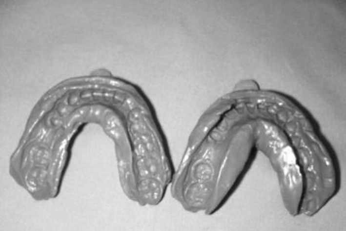
Polyether impressions made before retraction and after retraction
Fig. 8.
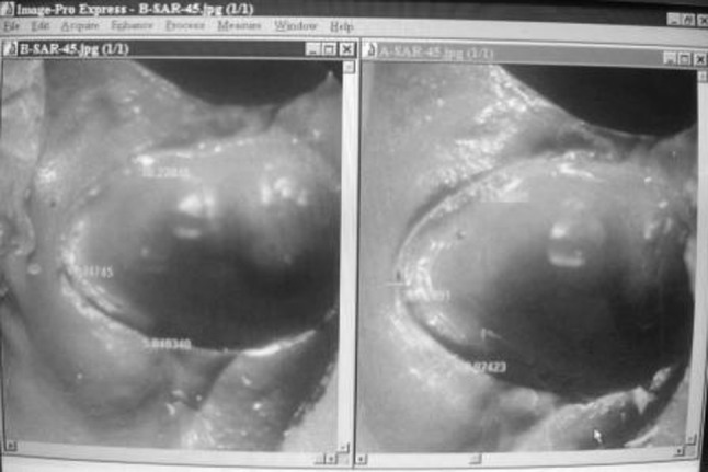
Stereomicroscopic and image-analysis of impressions made before retraction and after retraction to measure horizontal retraction (in one-tenth of microns)
Table 2.
Hemorrhage scores
| Score 0 | No bleeding |
| Score 1 | Bleeding controlled within 1 min |
| Score 2 | Bleeding not controlled within 1 min |
The mean time taken for placement of retraction system, the mean vertical retraction and the mean horizontal retraction attained from the three gingival retraction systems compared using one way ANOVA with the level of significance (P) set at 0.05. As two retraction systems were used on a patient, one on each prepared abutment, the Bonferroni test was conducted to find out which pair of retraction systems there exist a significant difference (P < 0.05). In order to compare the hemorrhage scores of the three systems the Kruskal–Wallis test was used followed by Mann–Whitney test to find out which pair, the significant difference exists (P < 0.05).
Results
The mean values with respect to time taken for placement, vertical retraction and horizontal retraction attained by using three gingival retraction systems are listed in Table 3. According to one way ANOVA, there were significant differences (P < 0.05) among the retraction systems in relation to mean time taken for placement, vertical retraction and horizontal retraction (Table 4). However, when set of two retraction systems were compared with each other using Bonferroni test (Table 5), no significant difference (P > 0.05) was found between stay-put and magic foam cord group with respect to mean vertical retraction and horizontal retraction. Significant differences (P < 0.05) were found between stay-put and expasyl group and also between magic foam and expasyl group.
Table 3.
Mean values obtained from three gingival retraction systems with relation to time taken, vertical retraction and horizontal retraction
| Parameters/retraction systems | N | Mean | SD | Minimum | Maximum |
|---|---|---|---|---|---|
| Time taken | |||||
| Stay-put | 20 | 215.1 s | 37.44 s | 135 s | 285 s |
| Expasyl | 20 | 75.15 s | 17.95 s | 45 s | 110 s |
| Magic foam cord | 20 | 79.75 s | 18.36 s | 45 s | 112 s |
| Vertical Retraction | |||||
| Stay-put | 20 | 1.0655 mm | 0.3851 mm | 0.42 mm | 2.17 mm |
| Expasyl | 20 | 0.484 mm | 0.195 mm | 0.17 mm | 1 mm |
| Magic foam cord | 20 | 0.8645 mm | 0.3029 mm | 0.33 mm | 1.42 mm |
| Horizontal Retraction | |||||
| Stay-put | 20 | 0.233 mm | 0.082 mm | 0.15 mm | 0.40 mm |
| Expasyl | 20 | 0.151 mm | 0.069 mm | 0.05 mm | 0.29 mm |
| Magic foam cord | 20 | 0.199 mm | 0.085 mm | 0.03 mm | 0.35 mm |
N number of abutments, SD standard deviation, s seconds, mm millimeters
Table 4.
Summary of ANOVA tests for determining any significant difference exists between three gingival retraction systems with relation to time taken, vertical retraction and horizontal retraction
| Parameters | Sum of squares | df | Mean square | F test | P value |
|---|---|---|---|---|---|
| Time taken (in seconds) | 252845.2 | 2 | 126422.62 | 183.97 | 0.001 |
| Vertical retraction (in mm) | 3.489 | 2 | 1.744 | 18.816 | 0.001 |
| Horizontal retraction (in mm) | 6.789E−02 | 2 | 3.395E−02 | 5.426 | 0.007 |
df degree of freedom
P value level of significance
Table 5.
Summary of Bonferroni Tests used to compare three gingival retraction systems with each other (multiple comparisons) with relation to time taken, vertical retraction and horizontal retraction
| Parameter/retraction systems (I) | (J) | Mean difference (I–J) | SE | P value | 95 % confidence interval | |
|---|---|---|---|---|---|---|
| Lower bound | Upper bound | |||||
| Time taken/ | ||||||
| Stay-put | Expasyl | 139.95* | 8.29 | 0.000 | 119.50 | 160.40 |
| Magic F | 135.35* | 8.29 | 0.000 | 114.90 | 155.80 | |
| Expasyl | Stay-put | −139.95* | 8.29 | 0.000 | −160.40 | −119.50 |
| Magic F | −4.60 | 8.29 | 1.000 | −25.05 | 15.85 | |
| Magic F | Stay-put | −135.35* | 8.29 | 0.000 | −155.80 | −114.90 |
| Expasyl | 4.60 | 8.29 | 1.000 | −15.05 | 25.05 | |
| Vertical retraction/ | ||||||
| Stay-put | Expasyl | 0.5815* | 9.628E−02 | 0.000 | 0.3440 | 0.8190 |
| Magic F | 0.2010 | 9.628E−02 | 0.124 | −3.6503E−02 | 0.4385 | |
| Expasyl | Stay-put | −0.5815* | 9.628E−02 | 0.000 | −0.8190 | -0.3440 |
| Magic F | −0.3805* | 9.628E−02 | 0.001 | −0.6180 | -0.1430 | |
| Magic F | Stay-put | −0.2010 | 9.628E−02 | 0.124 | −0.4385 | 3.6503E−02 |
| Expasyl | 0.3805* | 9.628E−02 | 0.001 | 0.1430 | 0.6180 | |
| Horizontal retraction/ | ||||||
| Stay-put | Expasyl | 8.200E−02* | 2.501E −02 | 0.005 | 2.030E−02 | 0.1437 |
| Magic F | 3.400E−02* | 2.501E−02 | 0.538 | −2.7696E−02 | −9.570E−02 | |
| Expasyl | Stay-put | −8.200E−02* | 2.501E−02 | 0.005 | −0.1437 | 2.030E−02 |
| Magic F | −4.800E−02 | 2.501E−02 | 0.180 | −0.1097 | −1.370E−02 | |
| Magic F | Stay-put | −3.400E−02* | 2.501E−02 | 0.538 | 9.5696E−02 | 22.770E−02 |
| Expasyl | 4.800E−02 | 2.501E−02 | 0.180 | −1.3696E−02 | 0.1097 | |
Magic F magic foam cord, SE standard error
*defines statistically significant value for a particular test
The hemorrhage scores on removal of each retraction system were compared using Kruskal–Wallis test (Table 6). The stay-put retraction cord induced maximal bleeding on removal. However, expasyl induced no bleeding on removal. The Mann–Whitney test (Table 7) showed that there was no significant difference (P > 0.05) in hemorrhage scores of magic foam cord and expasyl. Based on the author’s subjective analysis expasyl and magic foam cord were relatively easier to place than stay-put.
Table 6.
Kruskal–Wallis test used to evaluate hemorrhage scores of three retraction systems
| Retraction systems | Number of abutments (N) | Mean rank |
|---|---|---|
| Stay-put | 20 | 48.75 |
| Expasyl | 20 | 14.63 |
| Magic foam cord | 20 | 28.13 |
| Test statisticsa,b | |
|---|---|
| Hemorrhage | |
| Chi-square | 44.448 |
| df | 2 |
| Significance (P) | 0.000 |
df degree of freedom
P value = level of significance
aKruskal Wallis test
bGrouping variable: retraction systems
Table 7.
Mann–Whitney test used to compare three gingival retraction systems with each other (multiple comparisons) with relation to hemorrhage scores
| Retraction systems | Number of abutments | Mean rank | P value |
|---|---|---|---|
| Stay-put | 20 | 30.38 | <0.001* |
| Expasyl | 20 | 10.63 | |
| Stay-put | 20 | 28.88 | <0.001* |
| Magic foam cord | 20 | 12.13 | |
| Expasyl | 20 | 14.5 | <0.001* |
| Magic foam cord | 20 | 26.5 |
Discussion
All the measurements in the study were made by single operator to avoid inter-operator variability. The above mentioned results can be attributed to the following factors; stay-put cord is a “mechanical method” of the gingival displacement. The mechanical method involves physical displacement of the gingival tissue by placement of materials within the sulcus to obtain maximal gingival retraction [16]. Whereas, expasyl is a non-cord “mechanico-chemical” method of gingival displacement where the material is placed into the gingival sulcus with no pressure. Hence the amount of retraction observed may be less. It might be more effective under specific, limited conditions—when the sulcus is flexible and of sufficient depth. The magic foam cord is a “mechanical” gingival retraction system consisting of expanding type polyvinyl siloxane material. Hence, it might be the reason for getting better retraction from magic foam cord compared to expasyl retraction system. But the retraction was lesser than that from stay-put retraction cord where the cord was pushed mechanically into the gingival sulcus.
Based on the data collected, stay-put showed maximum bleeding on removal, followed by minimal bleeding on removal by magic foam cord. The expasyl retraction system induced no bleeding on removal. A study conducted by Weir and Williams, to compare the clinical effectiveness of mechanical–chemical tissue displacement methods showed that the maximum bleeding on removal was caused by dry retraction cords [3]. Also the placement of retraction cord into the gingival sulcus may cause injury to sulcular epithelium [8] and may induce bleeding on removal. The magic foam cord was potentially less traumatic as controlled pressure through comprecap was used, whereas expasyl was least traumatic and induced no bleeding as it contains aluminum chloride an astringent paste in its composition [9]. Among the three retraction systems compared in the present study, expasyl was relatively clinician friendly [13] and easy to place, as it was applied with an applicator gun directly into the gingival sulcus. The magic foam cord was also found easier to place and less time consuming than stay-put as it is injected with an automixing gun around the sulcus.
There are some limitations in this study; the influence of distendability of gingiva, gingival thickness, varied sulcus depth, location of the abutment teeth (anterior or posterior, maxillary or mandibular), and the visibility and accessibility on the gingival retraction were not considered. To standardize the variables and to minimize the errors, net amount of vertical and horizontal retraction was considered. Further, flexible measuring strips were used to measure sulcus depth (soft tissue), which may lead to some variations in the measured values. However, utmost care was taken to minimize these errors. Single retraction cord technique [9] was followed while using stay-put in all the cases, other retraction cord techniques such as double cord technique were not considered. Further, studies are required on three gingival retraction systems based on variables not considered in this study like location of abutment teeth (anterior or posterior, maxillary or mandibular), distendability of gingival and gingival thickness.
From the results and from the clinical point of view, the magic foam cord retraction system was found effective in almost all the variables considered in the present study. Finally, the choice of which gingival retraction system/technique to be used still depends on the clinical condition and operator’s preference [9].
Conclusions
Time taken for application of expasyl retraction system was significantly (P < 0.05) less compared to time taken for stay-put retraction cord.
The amount of vertical gingival retraction attained by using stay-put and magic foam cord retraction systems was significantly (P < 0.05) higher than expasyl.
The hemorrhage control with the expasyl retraction system was found better than hemorrhage control with the other two retraction system used in the study.
Expasyl and magic foam cord retraction system were found easier in placement compared to stay-put retraction cord.
Magic foam cord can be considered more effective among the three retraction systems used in this study, as it has taken less time and was easier in placement, attained good amount of retraction and induced minimal bleeding on removal compared to stay-put retraction cord.
References
- 1.Aimjirakul P, Masuda T, Takahashi H, Miura H. Gingival sulcus simulation model for evaluating the penetration characteristics of elastomeric impression materials. Int J Prosthodont. 2003;16:385–389. [PubMed] [Google Scholar]
- 2.Azzi R, Tsao TF, Carranza FA, Kennedy EB. Comparative study of gingival retraction methods. J Prosthet Dent. 1983;50:561–565. doi: 10.1016/0022-3913(83)90581-4. [DOI] [PubMed] [Google Scholar]
- 3.Weir DJ, Williams BH. Clinical effectiveness of mechanical–chemical tissue displacement methods. J Prosthet Dent. 1984;51:326–329. doi: 10.1016/0022-3913(84)90214-2. [DOI] [PubMed] [Google Scholar]
- 4.Woycheshin FF. An evaluation of the drugs used for gingival retraction. J Prosthet Dent. 1964;14:769–776. doi: 10.1016/0022-3913(64)90213-6. [DOI] [Google Scholar]
- 5.Buchanan WT, Thayer KE. Systemic effects of epinephrine-impregnated retraction cord in fixed partial denture prosthodontics. J Am Dent Assoc. 1982;104:482–484. doi: 10.14219/jada.archive.1982.0219. [DOI] [PubMed] [Google Scholar]
- 6.Shaw DH, Krejci RF, Cohen DM. Retraction cords with aluminum chloride: effect on the gingiva. Oper Dent. 1980;5:138–141. [PubMed] [Google Scholar]
- 7.Kopac I, Cvetko E, Marion L. Gingival Inflammatory response induced by chemical retraction agents in Beagle dogs. Int J Prosthodont. 2002;15:14–19. [PubMed] [Google Scholar]
- 8.Ferencz JL. Maintaining and enhancing gingival architecture in fixed prosthodontics. J Prosthet Dent. 1991;65:650–657. doi: 10.1016/0022-3913(91)90200-G. [DOI] [PubMed] [Google Scholar]
- 9.Donovan TE, Winston WL. Current concept in gingival displacement. Dent Clin N Am. 2004;48:433–444. doi: 10.1016/j.cden.2003.12.012. [DOI] [PubMed] [Google Scholar]
- 10.Jokstad A. Clinical trial of gingival retraction cords. J Prosthet Dent. 1999;81:258–261. doi: 10.1016/S0022-3913(99)70266-0. [DOI] [PubMed] [Google Scholar]
- 11.La Forgia A. Mechanical–chemical and electrosurgical tissue retraction for fixed prosthesis. J Prosthet Dent. 1964;14:1107–1114. doi: 10.1016/0022-3913(64)90180-5. [DOI] [Google Scholar]
- 12.Smith GR. A longitudinal study into the depth of clinical gingival sulcus of human canine teeth during and after eruption. J Periodont Res. 1982;17:427–433. doi: 10.1111/j.1600-0765.1982.tb01173.x. [DOI] [PubMed] [Google Scholar]
- 13.Smeltzer M. An alternative way to use gingival retraction paste. J Am Dent Assoc. 2003;134:1485. doi: 10.14219/jada.archive.2003.0078. [DOI] [PubMed] [Google Scholar]
- 14.Lynch JE, Hinders MK. Ultrasonic device for measuring periodontal attachment levels. Rev Sci Inst. 2002;73:2686–2693. doi: 10.1063/1.1484235. [DOI] [Google Scholar]
- 15.Bowles WH, Tardy SJ, Vahadi A. Evaluation of new gingival retraction agents. J Dent Res. 1991;70:1447–1449. doi: 10.1177/00220345910700111101. [DOI] [PubMed] [Google Scholar]
- 16.Darby H, Darby LH. Copper-band gingival retraction to produce void-free crown and bridge impressions. J Prosthet Dent. 1973;29:513–516. doi: 10.1016/0022-3913(73)90029-2. [DOI] [PubMed] [Google Scholar]


