Figure 5.
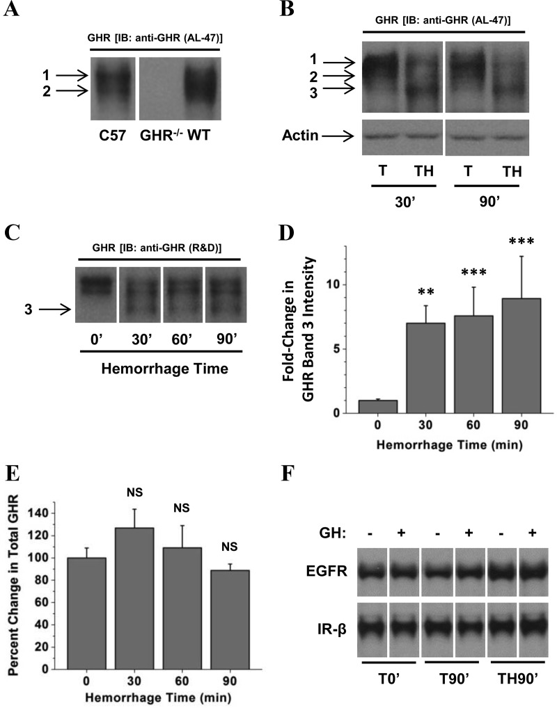
Effect of combined trauma and hemorrhage on hepatic GHR. Liver lysates were subjected to Western analysis and probed with GHR antiserum raised against the cytoplasmic domain (AL-47; A and B) or a rabbit polyclonal antibody raised against the extracellular domain of GHR (C). Representative lanes from the scanned image of a single film were chosen; bands of interest were cropped and rearranged to enhance clarity. In A–C, the numbers 1, 2, and 3 correspond to GHR bands 1, 2, and 3, respectively, as described in the text. Bands of interest were quantified by scanning densitometry. Data are presented as mean ± SEM fold change in GHR. D, Band 3 densitometric values were normalized to ERK, and this value was arbitrarily set to 1 for the 0′ hemorrhage group. E, Total GHR was calculated by adding the densitometric values for GHR bands 1, 2, and 3 in each lane and normalizing those to the total ERK signal for the same lane. F, Comparison of EGFR and IR-β levels in livers from mice subjected to trauma alone for 0 or 90 minutes or combined trauma and hemorrhage for 90 minutes and injection with GH or saline. The image in A was stretched vertically to improve clarity and to enhance band identification; this change was applied consistently to the entire image. The 0′ groups were arbitrarily set to 1 and 100% in D and E, respectively, and all other groups are expressed relative to 0′ groups in these panels. **P < .01 vs T0′; ***P < .001 vs T0′, both via 1-way ANOVA; NS, no statistically significant difference vs T0′; n = 4 or 7 mice per group.

