Abstract
Fibrocalcific pancreatic diabetes (FCPD) is a rare cause of diabetes (<1%) of uncertain etiology associated with >100-fold increased risk of pancreatic cancer. We present 3 patients of FCPD with pancreatic cancer who had long duration of diabetes (19 years, 25 years, and 28 years, respectively), all of whom presented with anorexia, weight loss, and worsened glycemic control. Patient-1 in addition presented with deep venous thrombosis. All the 3 patients had evidence of metastasis at the time of diagnosis. Computerized tomography (CT) abdomen revealed atrophic pancreas, dilated pancreatic ducts, and multiple calculi in the head, body, and tail of pancreas in all of them. Patient-1 had 38 mm × 38 mm × 32 mm mass in the tail of pancreas with multiple target lesions were seen in the right lobe of liver. Patient-2 had a mass in the tail of pancreas (46 × 34 × 31 mm) encasing the celiac plexus and superior mesenteric artery infiltrating the splenic hilum and splenic flexure of colon. Patient-3 also had a mass in the tail of pancreas (33 × 31 × 22 mm), with multiple target lesions in the liver, suggestive of metastasis. All patients had elevated serum CA19-9 (828.8, 179.65, and 232 U/L, respectively; normal <40 U/L). Patients of FCPD with anorexia, weight loss, worsening of glycemic control should be evaluated to rule out pancreatic cancer. Studies are warranted to evaluate CA19-9 as a screening tool for diagnosing pancreatic cancer at an earlier stage in FCPD.
Keywords: Carcinoma pancreas, calcific pancreatitis, diabetes, fibrocalcific pancreatic diabetes
INTRODUCTION
Tropical calcific pancreatitis (TCP) is chronic pancreatitis of uncertain etiology, usually presenting with the classical features of pain abdomen and steatorrhea in childhood or adolescence, with evidence of pancreatic calculi along with atrophic pancreas on imaging, found predominantly in the tropical and subtropical countries with the highest prevalence in the Indian states of Tamil Nadu and Kerala.[1] When associated with diabetes, representing a more advanced stage of the disease, it is known as fibrocalcific pancreatic diabetes (FCPD). FCPD constitutes <1% of all cases of diabetes.[2] It has been suggested that the risk of developing pancreatic cancer is the highest in patients with FCPD as compared to all other forms of pancreatitis and is believed to be 100-fold greater than those without FCPD.[3] This case series intends to highlight the increased occurrence of pancreatic cancer in FCPD, an observation not previously reported from this part of India. Pancreatic cancer was diagnosed in 3 patients during evaluation of patients with FCPD in the diabetic clinic of the Department of Endocrinology in the last 1-year.
Patient-1
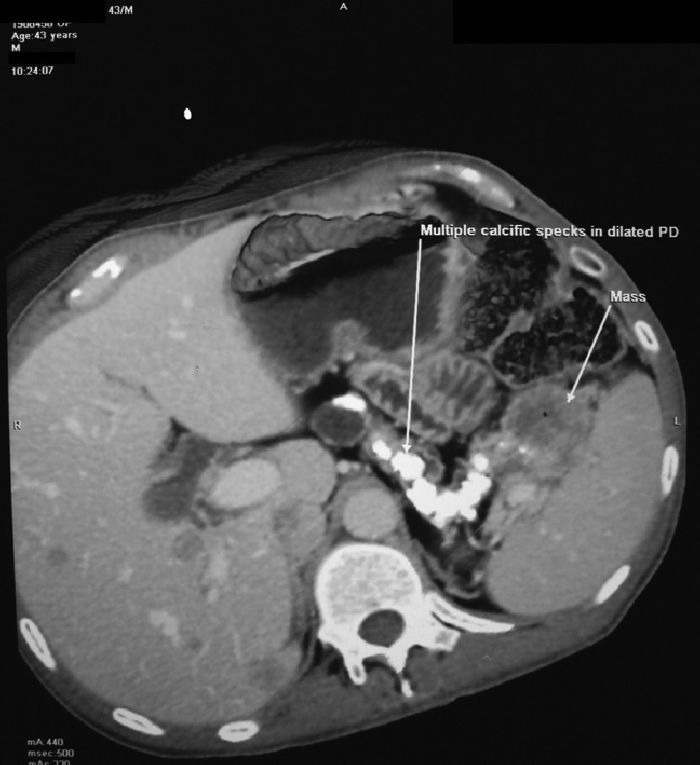
DP, 43-year-old man with diabetes since 24 years age, initially on oral hypoglycemic drugs with switch to premixed insulin use for last 2 years due to persistently uncontrolled blood glucose, presented to the diabetic clinic with anorexia, weight loss for 6 months, and painful swelling of left leg for last 7 days. There was no history of ketoacidosis. Examination was significant for low body mass index (BMI) 16.9 kg/m2, pallor, hepatomegaly, and non-pitting edema of left leg with erythema. Homan's and Moses's sign were positive. Ultrasonography (USG) Doppler of left leg confirmed thrombosis of popliteal, anterior tibial, posterior tibial, and common peroneal veins of the left leg. Ultrasonography abdomen revealed calcific pancreatitis with mass in the tail of pancreas. Computerized tomography (CT) abdomen revealed atrophic pancreas with dilated main and accessory pancreatic duct with multiple calculi in the head, body, and tail of pancreas along with 38 mm × 38 mm × 32 mm mass in the tail of pancreas [Figure 1]. Multiple target lesions were seen in the right lobe of liver, largest measuring 1.5 cm. He had no history of pain abdomen, steatorrhea, or chronic diarrhea. Serum CA19- 9 was elevated (828.8 U/L; normal <40). True-cut biopsy from right lobe of liver revealed moderately differentiated adenocarcinoma. A diagnosis of metastatic pancreatic carcinoma with deep venous thrombosis (DVT) was made in a patient with fibrocalcific pancreatic diabetes (FCPD). DVT improved with heparin and aspirin over 7 days. He died of cachexia and pneumonia 2 months later.
Figure 1.

Computerized tomography abdomen of patient-1 showing atrophic pancreas, dilated main pancreatic duct with multiple calculi in the head, body, and tail of pancreas with a 38 mm × 38 mm × 32 mm mass in the tail of pancreas. Multiple target lesions can also be seen in liver suggestive of metastasis
Patient-2
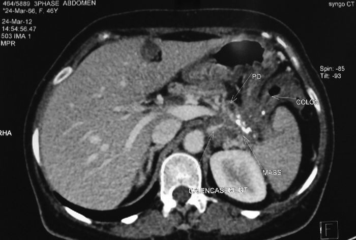
PB, 46-year-old lady with fibrocalcific pancreatic diabetes for last 25 years, on multiple subcutaneous insulin injections (MSII), was detected to have a pancreatic tail mass on USG abdomen done as a part of evaluation for severe pain abdomen, vomiting, weight loss, and worsened glycemic control for last 3 months. CT abdomen revealed dilated main pancreatic duct with multiple calculi, and a mass in the tail of pancreas (46 mm × 34 mm × 31 mm) encasing the celiac plexus, superior mesenteric artery, infiltrating the splenic hilum and splenic flexure of colon [Figure 2]. CA19-9 was 179.65 U/L (normal <40 U/L). The mass was considered inoperable. Patient refused chemotherapy. Patient was discharged after hemodynamic stabilization with analgesic coverage.
Figure 2.

Computerized tomography abdomen of patient-2 showing atrophic pancreas, multiple calculi in the tail of pancreas along with a 46 mm × 34 mm × 31 mm encasing the celiac plexus infiltrating the splenic hilum and splenic flexure of colon
Patient-3
SC, 54-year-old lady with FCPD for last 28 years, diagnosed during evaluation for pain abdomen with osmotic symptoms, on premixed insulin, was diagnosed to have mass in the tail of pancreas (33 mm × 31 mm × 22 mm), multiple calculi in the head and body of pancreas with multiple target lesion in the liver suggestive of metastasis on CT abdomen during evaluation for weight loss, fatigue, malaise along with worsened glycemic control for the last 1 year. Serum CA19-9 was elevated (232 U/L). She received gemcitabine for 4 months. She died of septicemia 4 months later.
DISCUSSION
Pancreatic adenocarcinoma is the fourth most common cause of cancer death worldwide with extremely poor prognosis (5-year survival <3%).[2,4] Association of diabetes with pancreatic cancer is controversial with some studies, suggesting a two-fold increase in patients of diabetes of >5 years duration and others suggesting diabetes to be protective against pancreatic cancer.[2,5]
All patients with pancreatic cancer in our series had a long duration of diabetes (19 years, 25 years, and 28 years, respectively). Anorexia and weight loss was a feature common in all the patients. One patient presented with deep venous thrombosis. All patients presented with metastasis highlighting the aggressive course of the disease. Also, all the patients had worsened glycemic control before the diagnosis of cancer. Pancreatic cancer has been reported to be associated with development of diabetes with improvement in glycemia following tumor resection.[6]
CA 19-9 was elevated in all of the patients of our series. CA19-9 is elevated in 70-80% of patients with pancreatic cancer, and elevated levels (at the time of diagnosis) are predictive of distant metastasis, survival, and disease recurrence (post surgery and/or chemotherapy).[4] Consensus statements have recommended screening for pancreatic cancer in any individual with an increased risk of >10-fold, which includes individuals with ≥3 first degree relatives with pancreatic cancer, Peutz Jegher syndrome, familial atypical multiple mole melanoma, and hereditary pancreatitis.[4] Since among all forms of pancreatitis, patients with FCPD have the highest risk of pancreatic cancer (absolute risk >100-fold), routine screening of all patients of FCPD with serum CA19-9 may help us in detecting the patients with pancreatic cancer at an earlier stage. Further studies are needed, however, in this direction before routine use of CA19-9 as a screening test for pancreatic cancer in FCPD.
Anorexia, weight loss, worsening of glycemic control in any patient with FCPD should be taken as warning signs, and these patients should be evaluated for any occult pancreatic malignancy, even in the absence of classical signs of pancreatic cancer like obstructive jaundice, palpable gall bladder, and pain abdomen.
Footnotes
Source of Support: Nil
Conflict of Interest: None declared
REFERENCES
- 1.Barman KK, Premalatha G, Mohan V. Tropical chronic pancreatitis. Postgrad Med J. 2003;79:606–15. doi: 10.1136/pmj.79.937.606. [DOI] [PMC free article] [PubMed] [Google Scholar]
- 2.Unnikrishnan R, Mohan V. Pancreatic diseases and diabetes. In: RIG H, Cockram C, Flybjerg T, Goldstein BJ, editors. Textbook of Diabetes. Vol. 18. Wiley Blackwell; 2004. p. 302. [Google Scholar]
- 3.Chari ST, Mohan V, Pitchumoni CS, Viswanathan M, Madanagopalan N, Lowenfels AB. Risk of pancreatic carcinoma in tropical calcifying pancreatitis: An epidemiologic study. Pancreas. 1994;9:62–6. doi: 10.1097/00006676-199401000-00009. [DOI] [PubMed] [Google Scholar]
- 4.Longo DL, Fauci AS, Kasper DL, Hauser SL, Jameson JL, Loscalzo J, editors. Harrison's Principle of Internal Medicine. 18th ed. McGraw Hill; 2012. Pancreatic cancer; p. 93. [Google Scholar]
- 5.Everhart J, Wright D. Diabetes mellitus as a risk factor for pancreatic cancer. A meta-analysis. JAMA. 1995;273:1605–9. [PubMed] [Google Scholar]
- 6.Gullo L. Diabetes and the risk of pancreatic cancer. Ann Oncol. 1999;10:79–81. [PubMed] [Google Scholar]


