Abstract
A middle-aged asymptomatic patient was referred to the eye clinic by her optician because of unusual optic nerve heads. She was found to have optic disc pits with bilateral serous retinal detachments which were non-progressive. She did not need any treatment and was safely followed up in the community. This uncommon condition is discussed along with possible pathophysiology and treatment.
Background
Uncommon disease
Age at presentation beyond which visual problems are unlikely to occur, so safe not to treat as course would be stable now.
Case presentation
A 53-year-old woman was referred to us by her optician because of suspicious looking optic nerve heads, with blurred margins inferiorly. The patient herself was asymptomatic.
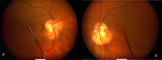
Examination revealed a best corrected visual acuity of 6/5 right and 6/9 left eye. Anterior segment examination was unremarkable with a normal intraocular pressure in both eyes. Fundal examination revealed bilateral asymmetric disc pits associated with a well-circumscribed area of retinal elevation inferiorly (figure 1).
Figure 1.

Colour fundus photos showing bilateral optic disc pits.
Investigations
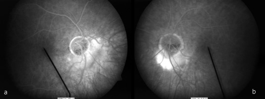
A fundus fluorescein angiogram showed these to be areas of peripapillary serous retinal detachment, which appeared to have sealed off (figure 2).
Figure 2.

Fundus fluorescein angiogram.
Outcome and follow-up
As the patient was asymptomatic, she was discharged with a copy of her fundus photograph for future comparison at yearly visits with her optician.
Discussion
Optic nerve head pits have a prevalence of 1/11 000. The usual age of onset is between 20 and 40 years. Optic disc pits are unilateral in 85–90% of cases.1 They are associated with serous retinal detachment in 52% of cases, increasing to 63% in temporally located pits.2 Various mechanisms have been reported to explain the source of this subretinal fluid, from vitreous and cerebrospinal fluid leakage through the optic pit into the subretinal space, to the retinal pigment epithelium itself.3 There has been one reported case of twins with a bilateral presentation of this anomaly in the literature; however, they were not associated with bilateral serous detachment.4
Management depends on clinical stage at presentation and the progression of visual loss. In case of the latter, a recent review has advocated surgical management with pars plana vitrectomy, with or without internal limiting membrane peel, with or without endolaser and gas endotamponade.5
We are unaware of a previously published case that reports bilaterality in both the optic disc pits and the serous detachments and could find no reference to it in a MEDLINE search. Our case also illustrates that these findings can be incidental in asymptomatic patients and might not require any treatment at all.
Learning points.
Uncommon disease, therefore important to be aware of its presentation and clinical signs in order to diagnose it.
Younger age group more likely to need/benefit from prophylactic treatment as serous macular detachments typically occurs in 30's and 40's.5
Asymptomatic disease with low risk of progression or visual loss may be just observed safely.
Visual field testing should be done as some patients may have nerve fibre layer defects in the area of the pit.
Footnotes
Competing interests: None.
Provenance and peer review: Not commissioned; externally peer reviewed.
References
- 1.Kolar P. Maculopathy in case of the pit of the disc. Cesk Slov Oftalmol 2005;61:330–6 [PubMed] [Google Scholar]
- 2.Brown GC, Shields JA, Goldberg RE. Congenital pits of the optic nerve head. II. Clinical Studies in Humans. Ophthalmology 1980;87:51–65 [DOI] [PubMed] [Google Scholar]
- 3.Johnson TM, Johnson MW. Pathogenic implications of subretinal gas migration through pits and atypical colobomas of the optic nerve. Arch Ophthalmol 2004;122:1793–800 [DOI] [PubMed] [Google Scholar]
- 4.Jonas JB, Freisler KA. Bilateral congenital optic nerve head pits in monozygotic siblings. Am J Ophthalmol 1997;124:844–6 [DOI] [PubMed] [Google Scholar]
- 5.Georgalas I, Ladas I, Georgopoulos G, et al. Optic disc pit: a review. Graefes Arch Clin Exp Ophthalmol 2011;249:1113–22 [DOI] [PubMed] [Google Scholar]


