Abstract
Tendinopathy and small partial-thickness tears of the rotator cuff tendon are common presentations in sports medicine. No promising treatment has yet been established. Corticosteroid injections may improve symptoms in the short term but do not primarily treat the tendon pathology. Ultrasound-guided autologous tenocyte implantation (ATI) is a novel bioengineered treatment approach for treating tendinopathy. We report the first clinical case of ATI in a 20-year-old elite gymnast with a rotator cuff tendon injury. The patient presented with 12 months of increasing pain during gymnastics being unable to perform most skills. At 1 year after ATI the patient reported substantial improvement of clinical symptoms. Pretreatment and follow-up MRIs were reported and scored independently by two experienced musculoskeletal radiologists. Tendinopathy was improved and the partial-thickness tear healed on 3 T MRI. The patient was able to return to national-level competition.
Background
In sports medicine, rotator cuff tendinopathy and small partial-thickness tears are difficult to treat. Rest, physiotherapy, non-steroidal anti-inflammatory drugs and corticosteroid injections have formed the mainstay of treatment. Surgery is reserved for large partial and full thickness tears or in patients with an unfavourable acromion. No non-surgical regenerative treatment for this spectrum of rotator cuff pathology has yet been clinically introduced. Platelet-rich plasma has been injected into other tendons1 and been used as an adjuvant treatment in rotator cuff surgical repairs, but the benefits still lack scientific validation.2–4 Rotator cuff tendon tear pathology studies demonstrate tenocyte depletion and dysfunction,5 which underline the need for biological or other cell therapies. Autologous tenocyte implantation (ATI) is a novel bioengineered treatment for rotator cuff injuries. It is expected to be safe, regenerative and has yielded promising results in animal experiments.6 7 We present the first clinical application of ATI for rotator cuff pathology in an elite gymnast with clinical and 3 T MRI follow-up.
Case presentation
A 20-year-old gymnast who has been competing at national level for 3 years on rings and parallel bars was referred to an orthopaedic shoulder specialist for the treatment of ongoing left shoulder pain in August 2011. Symptoms began more than 12 months prior to the initial presentation. There was no specific trauma, but symptoms were gradually progressive despite physiotherapy. In April 2011, the pain had become more severe. The athlete described the symptoms as moderate pain in the left shoulder when supporting the entire bodyweight on his arms and severe when hanging and swinging from his arms at full extension on rings and parallel bars. No night pain was experienced. In June 2011, a subacromial corticosteroid injection became necessary to enable the athlete to participate at national championships. However, shoulder symptoms recurred and precluded continuation of regular training sessions.
Investigations
A 3 Tesla MRI arthrogram was undertaken (August 2011) which showed focal signal increase on T2-weighted images and increased tendon thickness associated with a 50–100% partial-thickness supraspinatus rim-rent tear with fluid signal not created by contrast as proven by different weightings (figure 1).
Figure 1.
The pre-autologous tenocyte implantation T2 image (Pre ATI) shows increased tendon thickness with focal signal increase and a 50–100% rim-rent tear of the anterior supraspinatus insertion. Follow-up MRI at 4 months shows reduction of increased tendon thickness and decreased focal signal increase with infill and healing at the tear site. At 10 months there is recurrence of tendinopathy, but at the initial tear site no tear is detectable.
The acromion morphology was type 1. No labral tear was noted.
Treatment
The tendon injury was considered not amenable to surgery. As symptoms were recalcitrant to physiotherapy and conventional injection, the decision was made for treatment with ATI. This technology was nationally approved for clinical application as a tendon regenerative technology by the Therapeutic Goods Administration in 2010.
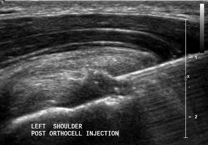
In September 2011, tenocytes were harvested from the patient's patella tendon under local anaesthesia. Cells were cultivated in a GMP laboratory. At 3 weeks, 2 ml of tenocyte suspension containing two million cultured tenocytes were injected under ultrasound-guidance into the tendinopathic tendon and partial-thickness tear (see figure 2). The procedure was performed by a senior interventional musculoskeletal radiologist.
Figure 2.
Ultrasound-guided autologous tenocyte implantation.
Outcome and follow-up
The athlete was completely rested from all training for 4 weeks after ATI. Light training then began and at 12 weeks after ATI, the athlete resumed a full 26 h/week of gymnastic training. At 4 months after ATI, the athlete reported no pain when supporting full body weight on his arms and either no pain or a mild level of pain when hanging and swinging from his arms at full extension or hyperextension.
At 10 months clinical symptoms were stable. Function scores were recorded (VAS pain, 1/10; Oxford shoulder score, 47/48; Quick DASH, 13/55 with sports module, 6/20). At 4 and 10 months post-ATI, 3 Tesla MRIs were reported and scored for rotator cuff tendinopathy, partial tear thickness and anteroposterior (AP) tear size by two independent senior musculoskeletal radiologists. Tendinopathy (tendon thickening with persistent focal signal increase) improved at 4 months, but was persistent at 10 months. The partial-thickness rim-rent tear had filled in and was not detectable at 4 and 10 months. These findings were independently reported and confirmed by a second-senior musculoskeletal radiologist (figure 1, 4 and 10 months).
Discussion
Regenerative approaches for tendinopathy include treatments with autologous blood or plasma or cells such as autologous mesenchymal stem cells, skin fibroblasts or tenocytes.
For human rotator cuff partial-thickness tears and tendinopathy no non-surgical tendon regenerative treatment has yet been reported.
Surgical rotator cuff repairs have been augmented with platelet-rich plasma (PRP)2 3 and stem cells8 during open or arthroscopic surgery. The treatments were reported to be safe, but outcome benefits have not been scientifically proven.
In the patella tendon, PRP and other cell based approaches have been used for the treatment of tendinopathies.1 9 In a recent level-1 randomised controlled trial, collagen-producing skin-derived tenocyte-like cells have been shown to be superior regarding recovery time, pain and function compared to plasma injections alone.9
Despite these promising results, instability of the phenotype is a concern as skin-derived cells are non-homologous to tendon tissue.
ATI uses a patient's own tenocytes to restore cell density in the injured tendon. In our animal studies, we have shown increased type 1 collagen production following ATI and tendon healing with regeneration of a normal tendon structure in acute and chronic rotator cuff lesions.6 7 With this case, we report the first clinical application of ultrasound-guided ATI for rotator cuff pathology. In an elite gymnast marked improvement in symptoms and sporting function was observed with MRI evidence of rotator cuff tendon infill and healing without the requirement for surgical intervention.
Learning points.
Autologous tenocyte implantation (ATI) can be applied non-surgically as an ultrasound-guided intratendinous injection.
Indications are small partial-thickness rotator cuff tears with or without tendinopathy.
It can be a safe and effective treatment option for the elite athlete.
Regeneration of the tendon with MRI signal intensity improvements and infill can occur.
Footnotes
Competing interests: None.
Patient consent: Obtained.
Provenance and peer review: Not commissioned; externally peer reviewed.
References
- 1.Finnoff JT, Fowler SP, Lai JK, et al. Treatment of chronic tendinopathy with ultrasound-guided needle tenotomy and platelet-rich plasma injection. PM R 2011;3:900–11 [DOI] [PubMed] [Google Scholar]
- 2.Castricini R, Longo UG, De Benedetto M, et al. Platelet-rich plasma augmentation for arthroscopic rotator cuff repair: a randomized controlled trial. Am J Sports Med 2011;39:258–65 [DOI] [PubMed] [Google Scholar]
- 3.Randelli P, Arrigoni P, Ragone V, et al. Platelet rich plasma in arthroscopic rotator cuff repair: a prospective RCT study, 2-year follow-up. J Shoulder Elbow Surg 2011;20:518–28 [DOI] [PubMed] [Google Scholar]
- 4.Nguyen RT, Borg-Stein J, McInnis K. Applications of platelet-rich plasma in musculoskeletal and sports medicine: an evidence-based approach. PM R 2011;3:226–50 [DOI] [PubMed] [Google Scholar]
- 5.Wu B, Dela Rosa T, Yu Q, et al. Cellular response and extracellular matrix breakdown in rotator cuff rupture. Arch Orthop Trauma Surg 2010;131:405–11 [DOI] [PubMed] [Google Scholar]
- 6.Chen J, Yu Q, Wu B, et al. Autologous tenocyte therapy for experimental Achilles tendinopathy in a rabbit model. Tissue Eng Part A 2011;17:2037–48 [DOI] [PubMed] [Google Scholar]
- 7.Chen JM, Willers C, Xu J, et al. Autologous tenocyte therapy using porcine-derived bioscaffolds for massive rotator cuff defect in rabbits. Tissue Eng 2007;13:1479–91 [DOI] [PubMed] [Google Scholar]
- 8.Ellera Gomes JL, da Silva RC, Silla LMR, et al. Conventional rotator cuff repair complemented by the aid of mononuclear autologous stem cells. Knee Surg Sports Traumatol Arthrosc 2012;20:373–7 [DOI] [PMC free article] [PubMed] [Google Scholar]
- 9.Clarke AW, Alyas F, Morris T, et al. Skin derived tenocyte like cells for the treatment of patella tendonopathy. Am J Sports Med 2010;39:614–23 [DOI] [PubMed] [Google Scholar]


