

Abstract
We report three cases of adolescent boys with mild diplegic cerebral palsy (CP) who suffered disruption of the knee extensor mechanism. Two had fractures of the patella and the third a fracture avulsion of the tibial tubercle combined with an undisplaced fracture of the patella. All three had gait analysis prior to sustaining the fractures and were known to have mild knee crouch. Each participated in sport including football. Each suffered an acute deterioration in gait resulting in a referral for repeat gait analysis, and x-ray of the affected knee. With the increased involvement of children with CP in sporting activities, especially children with mild knee crouch, we caution that knee extensor rupture might be an increasing problem.
Background
Fractures involving the knee extensor mechanism, fracture of the patella and avulsion of the tibial tubercle, are rare injuries in paediatrics.1 2 When they occur they usually do owing to an acute incident of forced knee flexion in exercise such as a mistimed jump. Similar injuries occur in children with diplegic CP though we consider the mechanism of injury is due to an increased moment arm across the knee joint caused by knee crouch.3 Some consider these fractures to be of little consequence in children with CP4–6; however, we have documented the deterioration in gait that was suggested to be associated with these lesions.3 We have also described the diagnostic gait pattern of the disruption of the knee extensor mechanism that these fractures cause.3 In this report, we draw attention to the fact that these injuries occur in children with mild CP diplegia with proven mild knee crouch. These three adolescents presented over a three-years period, and the incidence of diplegic CP in Ireland is between 30 and 35 per year.
Case presentation
Case reports
Case 1
A 15-years-old boy who was known to have asymmetric spastic diplegia, left side more affected, with a gross motor function classification system (GMFCS) level 1, presented with a 2-week history of limping. He played competitive taekwondo and swimming as well as football. His gait had been regularly assessed in the gait laboratory and it showed mild bilateral mid-stance knee crouch (figure 1A). His recent gait analysis, carried out after the recent deterioration in gait, demonstrated significant deterioration in his left knee pattern since his previous analysis (figure 1B). He was in fixed crouch of between 40° and 50° with no shock absorption or mid-stance extension of the left knee. Clinically, he had evidence of patella alta and a fixed knee flexion contracture of 18° with very tight hamstrings. He also had pain on palpation around the left patellar tendon. Plain radiographs (figure 2) of the left knee confirmed the diagnosis of complete extensor disruption with a fracture of the inferior pole of the patella.
Figure 1.

Graphs of single gait cycles. The vertical lines separate stance from swing for each leg. (A) shows the patient base-line sagittal kinematic analysis of both knees (left—blue line, right—red line) which demonstrates very mild crouch gait. (B) Kinematic analysis 2 years later shows significant crouch with loss of shock absorption and a complete failure to return to extension of the left knee (blue line). The shaded area outlines the normal values.
Figure 2.
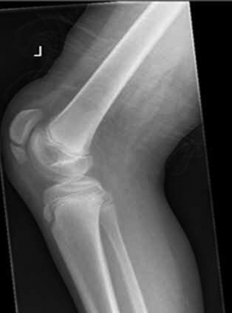
Lateral view of the left knee showing avulsion fracture of the inferior pole of the patella.
Case 2
An active 14-years-old boy who was known to have asymmetric diplegic cerebral palsy, GMSCF level 1, was checked regularly with gait analysis. His gait analysis demonstrated mild left knee crouch (figure 3) for which he was treated with botulinum toxin injections to his left hamstrings. He played school football. He was referred for repeat gait analysis when noticed to be limping. This showed features typical of extensor mechanism failure. X-rays of his left knee demonstrated a fracture of his left patella (figure 4). He complained of anterior knee pain which was not well localised.
Figure 3.

Sagittal kinematics of knees demonstrating pre-morbid mild left knee crouch (blue lines) over two gait cycles.
Figure 4.

Lateral radiograph of the left knee showing a transverse fracture at the inferior pole of the patella and patella alta.
Case 3
An active 12-years-old boy who is known to have spastic diplegia, GMSCF level 1, attended for repeat gait analysis owing to a recent deterioration in his gait. He was known to have bilateral knee crouch with midstance knee flexion of 20°. He had a left hamstring lengthening performed 2 years back, however he had continued to walk in crouch at both knees. He played school football and other sports. On his most recent analysis, there was an obvious deterioration of his left knee crouch with kinematic features typical of knee extensor disruption. X-rays of his knees showed a fracture of the distal pole of the left patella and a displaced avulsion fracture of the tibial tuberosity and patella alta (figure 5).
Figure 5.

Lateral radiograph of the left knee showing fracture of the distal pole of the patella and displaced avulsion fracture of the tibial tuberosity and patella alta.
Outcome and follow-up
Only one, case 2, was treated surgically while the other two cases were treated by casting. There has been no recovery to full sporting activities by any case with an average follow-up of 2 years.
Discussion
With the great success of the London Paralympic games it is likely that many children with diplegic CP will be encouraged to participate in sporting activities. We report three cases of diplegic CP boys with GMFCS of one (minimally affected) who suffered fractures of the knee extensor mechanism. All three patients were known to have preexisting mild knee crouch. We performed base-line gait analysis upon request on all three boys prior to them sustaining their fractures but their degree of knee crouch was not considered severe enough to advise limitation to their activities. In our laboratory, we perform gait analysis on a patient at a self-determined walking speed and we do not induce fatigue. It is possible that during exercise such as football, knee crouch could worsen owing to muscle fatigue thereby increasing the flexion moment arm around the knee. In these circumstances the extreme forces endured by the knee extensors cause them to fail without a major traumatic event. Fracture involving the knee extensor mechanism is not a condition confined to severely involved CP patients, but occurs in children with mild diplegia with knee crouch as demonstrated by these three cases. This must be kept in mind when supervising these children in sport and any complaint of knee pain or a noticeable deterioration of gait should raise the suspicion of knee extensor disruption.
Learning points.
Fractures of the knee extensor mechanism are relatively common in diplegic cerebral palsy (CP).
They occur in adolescents with mild diplegic CP who are active in sport.
These fractures are stress-related and occur because of knee crouch.
The presentation may be silent with a noticeable deterioration of gait.
Footnotes
Competing interests: None.
Patient consent: Obtained.
Provenance and peer review: Not commissioned; externally peer reviewed.
References
- 1.Maguire JK, Canale ST. Fractures of the patella in children and adolescents. J Pediatr Orthop 1993;13:567–71 [PubMed] [Google Scholar]
- 2.Grogan DP, Carey TP, Leffers D, et al. Avulsion fractures of the patella. J Pediatr Orthop 1990;10:721–30 [DOI] [PubMed] [Google Scholar]
- 3.O'Sullivan R, Walsh M, Kiernan D, et al. The knee kinematic pattern associated with disruption of the knee extensor mechanism in ambulant patients with diplegic cerebral palsy. Clin Anat 2010;23:586–92 [DOI] [PubMed] [Google Scholar]
- 4.Rosenthal RK, Levine DB. Fragmentation of the distal pole of the patella in spastic cerebral palsy. J Bone Joint Surg Am 1977;59:934–9 [PubMed] [Google Scholar]
- 5.Topoleski TA, Kurtz CA, Grogan DP. Radiographic abnormalities and clinical symptoms associated with patella alta in ambulatory children with cerebral palsy. J Pediatr Orthop 2000;20:636–9 [DOI] [PubMed] [Google Scholar]
- 6.Lloyd-Roberts GC, Jackson AM, Albert JS. Avulsion of the distal pole of the patella in cerebral palsy. A cause of deteriorating gait. J Bone Joint Surg Br 1985;67:252–4 [DOI] [PubMed] [Google Scholar]