Abstract
An 8-year-old boy came with a chief complaint of an abnormally shaped tooth situated in upper front teeth region. On examination a supernumerary tooth with multiple lobes was present palatally to the maxillary right permanent central incisor. The morphology of the tooth crown was found to be unusual due to the presence of five lobes in the crown portion. Because of the supernumerary tooth, the permanent right central incisor was displaced labially. Radiographic examination showed a completely formed supernumerary tooth with dilacerated root. On the basis of clinical and radiographic examination, the supernumerary tooth was diagnosed as multilobed mesiodens. Since patient expressed dissatisfaction with the presence of supernumerary tooth, it was decided to extract this mesiodens followed by orthodontic treatment for alignment of labially placed maxillary right permanent central incisor.
Background
Mesiodens may affect the dentition in various ways. Some of the problems that can be caused by this type of tooth are crowding or abnormal diastema, displacement and/or rotation of adjacent teeth, failure of eruption, hindrance to orthodontic movement, enlargement of the follicle and possible cystic change, root abnormalities like dilacerations of the developing root, occlusal interference, aesthetic impairment, carious developmental grooves, periodontal problems, irritation of the tongue and diagnostic problems.
Therefore, the presence of mesiodens and its morphology in young patients is of great concern, and early diagnosis is crucial to minimise these complaints.
Case presentation
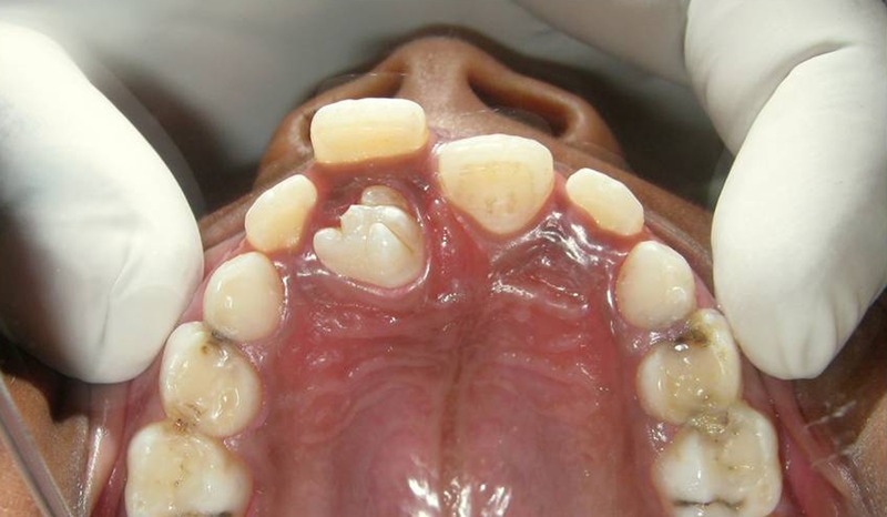
An 8-year-old boy reported with chief complaint of abnormally shaped upper front tooth. The family and medical history were not relevant. On intraoral examination supernumerary tooth was noted palatally to maxillary right permanent central incisor (figure 1). Other permanent and primary teeth were normal with no abnormalities. No signs of any syndrome were present in the child. No history of trauma to teeth was present.
Figure 1.

Occlusal view of multilobed supernumerary tooth in relation to maxillary right permanent central incisor.
Morphology of the crown of mesiodens was found to be unusual. Crown of mesiodens was grossly triangular in outline from occlusal view and had five lobes which were separated by developmental grooves. Grossly, three lobes were located labially and two lobes were present palatally. There were no signs of decay in the grooves.
The patient did not experience pain or discomfort during biting but it has affected the normal position of the maxillary right permanent central incisor.
Investigations
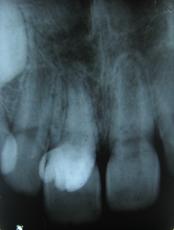
Intraoral periapical radiograph showed a completely formed supernumerary tooth with dilacerated root (figure 2).
Figure 2.

Radiographic image of a multilobed mesiodens.
Treatment
Since the patient was concerned with supernumerary tooth and malaligned maxillary anterior teeth, it was decided to extract multilobed mesiodens followed by orthodontic correction of malaligned teeth as a treatment plan. Oral prophylaxis was done followed by extraction of mesiodens under local anaesthesia. Postextraction instructions were given to the child as well as parents. The patient was recalled after a week for follow-up and treatment.
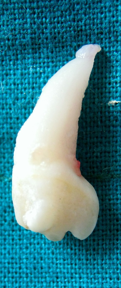
After extraction, the mesiodens was examined anatomically. Incisally mesiodens was triangular in shape with apex located labially and base towards palatal side. Mesiolabial, distolabial and palatal lobes were sharp and located at periphery of the occlusal table while two central lobes were located between lines joining the three lobes. Three developmental grooves were passing between these lobes which were caries free. The root of mesiodens was dilacerated from cervix (figure 3).
Figure 3.

Extracted multilobed mesiodens.
Outcome and follow-up
Unfortunately, the patient did not return for orthodontic treatment.
Discussion
It was originally postulated that mesiodens represented a phylogenetic relic of extinct ancestors who had three central incisors.1 A second theory known as dichotomy suggests that the tooth bud is split to create two teeth, one of which is the mesiodens.2 The third theory involving hyperactivity of the dental lamina is the most widely supported.3 According to this theory, remnants of the dental lamina or palatal offshoots of active dental lamina are induced to develop into an extra tooth bud which results in a supernumerary tooth and because of hyperactivity of dental lamina it could be possible reason for development of multilobed mesiodens.
There are several possible aetiological factors for dilaceration of the tooth. The most widely accepted factor for dilaceration of the tooth is mechanical trauma to the primary predecessor tooth. Other possible factors that have been reported in a literature are developmental anomalies of primary tooth germ, advanced root canal infections, ectopic development of the tooth germ and lack of space and hereditary factors.4 Probably in this case ectopic development of tooth and lack of space in the arch may be the possible aetiological factor for dilaceration of the multilobed mesiodens.
The prevalence of hyperdontia is reported between 0.15% and 3.9%.5 The prevalence of mesiodens varies between 0.09% and 2.05% in different studies and it is reported to be more common in males than in females. The literature reports that 80–90% of all supernumerary teeth occur in the maxilla.6 Von Arx1 reported that majority of supernumerary teeth lay palatal to the central incisors.
Primosch classified supernumerary teeth into supplemental and rudimentary, according to its shape. Supplemental or eumorphic refers to supernumerary teeth of normal size and shape, may also be termed incisiform. Rudimentary or dysmorphic defines teeth of abnormal shape and smaller size, including conical, tuberculate and molariform types. A conical mesiodens is small, peg-shaped (coniform) teeth with normal root; a tuberculate mesiodens is short, barrel-shaped with normal appearing crown or invaginated having rudimentary, incompletely developed root.3
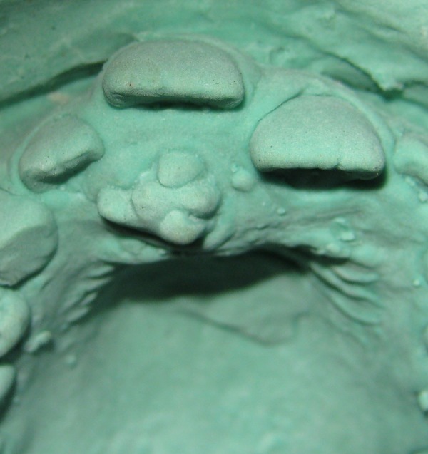
Koch et al7 have classified mesiodens as 56% conical, 12% tuberculate, 11% supplemental and 12% other configurations which includes molariform or multilobed mesiodens. In this case, the mesiodens belongs to the other configurations of the Koch's classification (figure 4).
Figure 4.

Study model of maxillary teeth showing multilobed mesiodens.
The presence of mesiodens may be seen as an isolated finding or frequently associated with various craniofacial anomalies, including cleidocranial dysostosis, Gardner's syndrome, cleft lip and palate, Fabry-Anderson's syndrome or chondro ectodermal dysplasia (Ellis-van Greveld syndrome).8
Similar type of case report of multilobed mesiodens was published by Nagaveni et al.9 In this case report the morphology of the tooth crown was found to be unusual with three lobes separated by non-carious developmental grooves. It was found that the extra tooth also had a palatal talon cusp.
Mesiodens may lead to several local problems such as diastema, displacement or rotation of adjacent teeth, development of dentigerous cyst, compromised aesthetics, resorption of neighbouring roots, crowding and dilacerations of permanent teeth.3 7 10 Therefore, the presence of supernumerary tooth in young patients is of great concern, and early diagnosis and treatment is crucial to minimise these complaints.
Learning points.
Early detection and management of supernumerary teeth with abnormalities is a necessary part of preventive dentistry.
Supernumerary teeth may occur in isolation or as part of a syndrome, such as cleidocranial dysplasia, Gardner's syndrome, cleft lip and palate. A careful check for a family history of supernumerary teeth could point to the presence of a genetically determined syndrome.
This type of abnormal teeth with dental anomalies hamper the routine activity of the patients by causing pain and discomfort while eating or closing the mouth and/or may cause malocclusion, so it should be treated as soon as possible to build better oral hygiene of the patient.
Footnotes
Competing interests: None.
Patient consent: Obtained.
Provenance and peer review: Not commissioned; externally peer reviewed.
References
- 1.Von Arx T. Anterior maxillary supernumerary teeth: a clinical and radiographic study. Aus Dent J 1992;37:189–95 [DOI] [PubMed] [Google Scholar]
- 2.Sedano HO, Gorlin RJ. Familial occurrence of mesiodens. Oral Surg Oral Med Oral Pathol 1969;27:360–1 [DOI] [PubMed] [Google Scholar]
- 3.Primosch RE. Anterior supernumerary teeth-assessment and surgical intervention in children. Pediatr Dent 1981;3:204–15 [PubMed] [Google Scholar]
- 4.Jafrazadeh H, Abbott PV. Dilaceration: review of an endodontic challenge. J Endod 2007;33:1025–31 [DOI] [PubMed] [Google Scholar]
- 5.Luten JR., Jr The prevalence of supernumerary teeth in primary and mixed dentitions. J Dent Child 1967;34:346–53 [PubMed] [Google Scholar]
- 6.Meighani P. Mesiodens: diagnosis and management of supernumerary (mesiodens): a review of the literature. J Dent Tehran Univ Med Sci 2010;7:41–9 [PMC free article] [PubMed] [Google Scholar]
- 7.Koch H, Schwartz O, Klausen B. Indications for surgical removal of supernumerary teeth in the premaxilla. Int J Maxillofac Surg 1986;15:273–81 [DOI] [PubMed] [Google Scholar]
- 8.Rajab LD, Hamdan MA. Supernumerary teeth: review of the literature and a survey of 152 cases. Int J Paediatr Dent 2002;12:244–54 [DOI] [PubMed] [Google Scholar]
- 9.Nagaveni NB, Umashankara KY, Sreedevi Reddy BP, et al. Multi-lobed mesiodens with a palatal talon cusp—a rare case report. Braz Dent J 2010;21:375–8 [DOI] [PubMed] [Google Scholar]
- 10.Hattab FN, Yassin OM, Rawashdeh MA. Supernumerary teeth: report of three cases and review of the literature. J Dent Child 1994;61:382–94 [PubMed] [Google Scholar]


