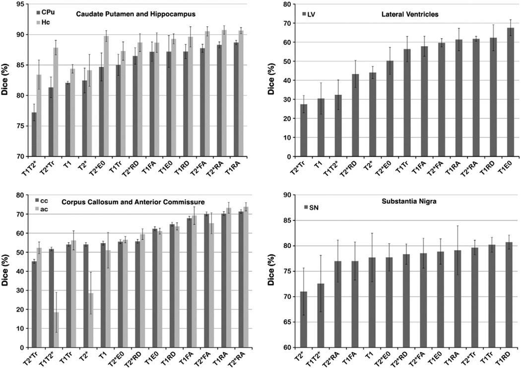
Fig. 5.
Dice coefficients indicate that combining information from T1 or T2*w images with information from DTI parameter images improves the accuracy of registration-based segmentation. The improvement between single channel (T1w or T2*w), and T1RA or T2*RA combined channel segmentation was >6% for gray matter structures like CPu and Hc. As expected, accuracy improved for white matter structures, >26% for ac and cc in T1RA, T2*RA relative to T1T2*, and 16% relative to T1 or T2*. Dice coefficients also increased for small nuclei such as SN (~10% higher in T1RD and T1Tr than in T2*, 3% higher in T1RD than in T1), as well as for ventricles (>16% higher in T2RD than in T2*, 30% higher in T2Tr relative to T1). While for SN the combination T1RD and T1Tr produced the most accurate segmentation (80 ± 1%), and T1E0 for LV (68 ± 4%), the combination of T2*RA produced the most accurate segmentation for most structures (88 ± 2% for CPu, and 90 ± 1% for Hc).

