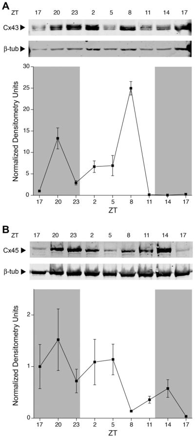
Fig. 4.
Changes in connexin proteins expressed in the rat OB membrane across light and dark phases in the rat OB. (A) Representative membrane proteins harvested over various time points (ZT), separated by SDS–PAGE, and electro-transferred to nitrocellulose. Nitrocellulose blots were probed with anti-Cx43 (1:500) and anti-β-III-tubulin (1:2000). Arrows indicate expected size in kilodaltons (Mr = 43 kDa and 50 kDa, respectively). The corresponding line plot represents the quantitative densitometry of Cx43 in pixel density normalized to β-III-tubulin and the 0 time point. Data are mean +/− SEM for 5–7 animals per time point. Cx43 protein changes were statistically significant across the 24-h (Kruskal–Wallis H test, P < 0.05) and rhythmic (harmonic regression analysis, P < 0.05). (B) Same as (A) but probed with anti-Cx45 (1:250) and anti-β-III-tubulin (1:2000). Arrows indicate expected size in kDa (Mr = 45 kDa and 50 kDa, respectively). Cx45 protein changes were statistically significant across the 24-h (Kruskal–Wallis H test, P < 0.05) but arrhythmic (harmonic regression analysis, P > 0.05). Cx43 and Cx45 protein changes were statistically significant for JTK_CYCLE analysis (P < 0.05).

