Abstract
Objective
To investigate the correlation between CD133-positive gastric cancer and clinicopathological features and its impact on survival.
Methods
A search in the Medline and Chinese CNKI (up to 1 Dec 2011) was performed using the following keywords gastric cancer, CD133, AC133, prominin-1 etc. Electronic searches were supplemented by hand searching reference lists, abstracts and proceedings from meetings. Outcomes included overall survival and various clinicopathological features.
Results
A total of 773 gastric cancer patients from 7 studies were included. The median rate of CD133 expression by immunohistochemistry (IHC) was 44.8% (15.2%–57.4%) from 5 studies, and that by reverse transcription polymerase chain reaction (RT-PCR) was 91.3% (66.7%–100%) from 4 studies. The accumulative 5-year overall survival rates of CD133-positive and CD133-negative patients were 21.4% and 55.7%, respectively. Meta-analysis showed that CD133-positive patients had a significant worse 5-year overall survival compared to the negative ones (OR = 0.20, 95% CI 0.14–0.29, P<0.00001). With respect to clinicopathological features, CD133 overexpression by IHC method was closely correlated with tumor size, N stage, lymphatic/vascular infiltration, as well as TNM stage.
Conclusion
CD133-positive gastric cancer patients had worse prognosis, and was associated with common clinicopathological poor prognostic factors.
Introduction
Gastric cancer (GC) is the fourth most common cancer worldwide [1]. Following only lung cancer, GC is also the second leading cause of cancer-related death in Asia. Although underwent radical resection and postoperative adjuvant therapy, most of GC patients will die of recurrence and metastasis, with 5-year overall survival no more than 50% for resectable patients in China [2].
The advancement in survival of GC patients in past few decades was relatively small, due to a lack of deep understanding the molecular mechanism of cancer. Recently, a rare subpopulation cancer cells, termed cancer stem cells (CSC) have been thought to be responsible for the initial, progression, metastasis and ultimately recurrence of cancer, for they have the exclusive properties of self-renew and could giving rise to all the heterogeneous lineages of cancer cells that eventually constitute tumor bulk [3]. CD133 is a trans-membrane glycoprotein, its expression in cell surface down-regulates quickly as cell differentiated [4]. CD133 has been used widely as a marker to identify CSC in colon, lung, brain, pancreatic cancer and so on [5]–[8]. Its prognostic value for cancer patients has also been found in many cancers [9]–[12].
With respect to gastric cancer, the correlation between CD133 and clinicopothological features of GC and its prognostic value is relatively unclear. Thus a systematic review of published literatures was conducted to clarify the relationship between CSC marker CD133 and GC based on current evidences.
Methods
Literature Search Strategy
A comprehensive literature search of electronic databases PubMed and Chinese CNKI was performed up to December 1, 2011. Search strings of PubMed was (((“cd133” [Title/Abstract]) OR “ac133” [Title/Abstract]) OR “prominin 1” [Title/Abstract]) AND (((“stomach neoplasms” [MeSH Terms]) AND “carcinoma” [MeSH Terms]) OR “gastric cancer” [Title/Abstract]). The reference lists of relative articles were also screened to further identify potential studies.
Selection Criteria
Diagnosis of gastric cancer was proven by histopathological methods. Studies of CD133 expression based on primary gastric cancer tissue (via either surgical or biopsy), rather than serum or any other kinds of specimen were included. Expression of CD133 was detected by any method. All studies on the correlation of CD133 overexpression with clinicopathological markers and the association of CD133 overexpression on disease-free and overall survival of gastric cancer were included. There was no limitation on language as well as the minimum patients of every single study. When there were multiple articles by the same group based on similar patients and using same detection methods, only the largest or the most recently article was included.
Data Extraction
Data tables were made to extract all relevant data from texts, tables and figures of each included studies, including author, year, country, patient number, detection method, clinicopathological features, positive rates of CD133 overexpression, as well as the expression-related survival. In case the prognosis was only plotted as Kaplan-Meier curve in some articles, the software GetData Graph Digitizer 2.24 (http://getdata-graph-digitizer.com/) was applied to digitize and extract the data.
Statistical Analysis
Meta-analysis was supplemented when applicable; otherwise, outcomes were presented in a narrative way. Software RevMan 5.0 (the Cochrane Collaboration, Copenhagen) was employed for data analysis. Comparisons of dichotomous measures were performed by pooled estimates of odds ratios (OR), as well as their 95% confidence intervals (CI). P value<0.05 was considered as statistical significance. Fixed or Random model was used depending on heterogeneity analysis. Statistical heterogeneity was tested using a Chi-square test with significance being set at p<0.10, the total variation among studies was estimated by I-square [13].
Results
Literatures Information
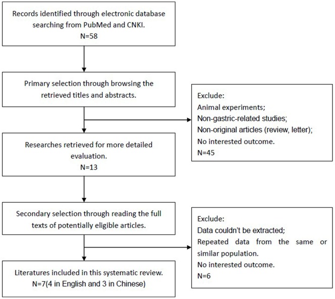
Fifty-eight articles were identified initially using the search strategy above. Forty-five of those were excluded due to non-human experiments, non-gastric-related studies, non-original articles (review, letter), through reading title and abstract. After excluded those data couldn’t be extracted and repeated ones by reading full text [14]–[19], eventually, there were 7 studies (4 in English and 3 in Chinese) included in the present Meta-analysis [20]–[26] (Figure 1).
Figure 1. Flow chart for selection of studies.
Study Characteristics
The 7 included studies were all based on Asian population, including 1 from Japan, 1 from Singapore and the rest 5 from China. A total of 773 patients with a median of 110 (ranged from 31 to 336) were included, most of which were male patients (74.8% from 6 studies). The median age ranged from 58 to 65 years old. In regarding to TNM stage, a median of 43.2% (22.6%–72.2%) patients were stage I or II, while the other 56.8% (27.8%–77.4%) were stage III or IV. Differentiated grading of tumor was reported in 5 studies and among those, roughly two thirds were poorly differentiated. Around 67.8% (58.8%–78.9%) of reported patients were identified as metastatic lymph node status.
CD133 Expression Status Stratification
All detected specimen were derived from gastric cancer tissues by means of surgical resection. Two different methods were employed for CD133 overexpression status screening. In detailed, immunohistochemistry (IHC) on membrane protein level is involved in 3 included studies, reverse transcription polymerase chain reaction (RT-PCR) on mRNA level in 2 studies and the both in the rest two studies. The median rate of CD133 overexpression by IHC from 5 studies was 44.8% (15.2%–57.4%). CD133 mRNA overexpression rates in gastric cancer tissue were much higher, with a median rate of 91.3% (66.7%–100%), which was of significant differences compared to normal gastric tissue in all 4 studies according to RT-PCR results (Table 1).
Table 1. General characteristics of included studies.
| Studies | Year | Cases | Method | CD133 expression rate (%) | 5-year OS (%) | P value | |
| CD133 (+) | CD133 (−) | ||||||
| Wang T [26] | 2011 | 103 | IHC | 44 | 20.8 | 43.4 | <0.01 |
| Zhao P [23] | 2010 | 336 | IHC | 57.4 | 22.8 | 57.3 | <0.01 |
| Ishigumin S [24] | 2010 | 97 | IHC | 28 | 29 | 78 | <0.01 |
| Yu JW [25] | 2010 | 99 | IHC | 29.3 | 0 | 40.4 | <0.01 |
| 31 | RT-PCR | 100 | – | – | – | ||
| Xue YM [22] | 2011 | 33 | IHC | 15.2 | – | – | – |
| 33 | RT-PCR | 100 | – | – | – | ||
| Tang BF [20] | 2008 | 38 | RT-PCR | 100 | – | – | – |
| Yan JL [21] | 2011 | 36 | RT-PCR | 66.7 | – | – | – |
| Summary | – | 742 | IHC | 44.8% | 21.4 | 55.7 | – |
| RT-PCR | 91.3% | ||||||
Abbreviations: OS, over survival; GC, gastric cancer; IHC, immunohistochemistry; RT-PCR, reverse transcription polymerase chain reaction.
CD133 Overexpression and 5-year Overall Survival
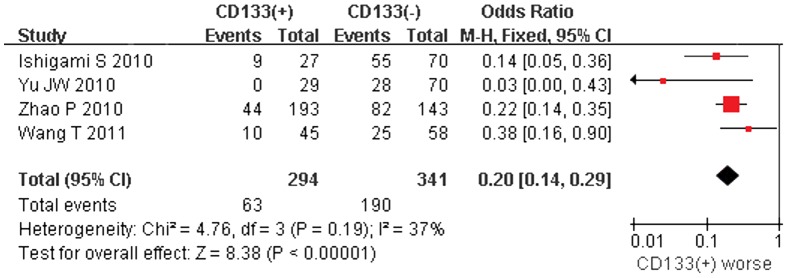
5-year overall survival rate was extracted from 4 studies, all of which relied on IHC solution. The accumulative 5-year overall survival rates of CD133-positive and CD133-negative GC patients were 21.4% (63/294) and 55.7% (190/341), respectively. (Table 1) Meta-analysis indicated that patients with CD133-positive suffered with a significant poor prognosis in comparing to CD133-negative ones (OR = 0.20, 95% CI: 0.14–0.29, P<0.00001, Fixed Model). In fact, 3 out of 4 studies have also concluded CD133 overexpression as a poor prognostic factor in gastric cancer patients (Figure 2).
Figure 2. Meta-analysis of 5-year overall survival between CD133(+) and CD133(−) gastric cancer patients.

CD133 Overexpression and Clinicopothological Features
All seven studies presented data on clinicopothological features. However, two different screening solutions (IHC and RT-PCR) are applied and variance also appeared in criterions, i.e., for tumor size, Zhao et al. taken 4 cm as borderline while Yu et al. used 5 cm, therefore, systematic review was conducted in a narrative way instead of meta-analysis.
In gastric cancer patients, there was no clear correlation between CD133 overexpression and ages (5 out of 5 studies), sex (4 out of 4 studies), tumor depth (5 out of 7 studies) and tumor grades (5 out of 7 studies). However, CD133 overexpression was likely to associate with tumor size (4 out of 5 studies), lymph node metastasis (5 out of 7 studies) as well as lymphatic vessel/vascular infiltration (3 out of 3 studies). With regard to the relationship between TNM stages and CD133 overexpression, unexpected, an opposite trend has been found in two different screening solutions. Three out of four studies by IHC revealed a positive correlation between CD133 and tumor stages whereas 4 studies by RT-PCR suggested none (Table 2).
Table 2. Narrative review of clinicopathological features with CD133 overexpression.
| Items | Significant correlation (p<0.05) | Non-significant correlation (p≥0.05) |
| Age | – | IHC: [23], [25], [26] RT-PCR: [21], [22], [25] |
| Sex | – | IHC: [25], [26] RT-PCR: [21], [22], [25] |
| Tumor Size | IHC: [23], [25] RT-PCR: [20], [22] | RT-PCR: [21] |
| Depth | IHC: [23], [24] | IHC: [25], [26] RT-PCR: [20], [21], [22], [25] |
| TNM stage | IHC: [23], [24], [25] | IHC: [26] RT-PCR: [20], [21], [22], [25] |
| Tumor grade | IHC: [23] RT-PCR: [21] | IHC: [24], [25], [26] RT-PCR: [20], [22], [25] |
| Lymph node metastasis | IHC: [23], [24], [25] RT-PCR: [20], [22], [25] | IHC: [26] RT-PCR: [21] |
| TNM stage | IHC: [23], [24], [25] | IHC: [26] RT-PCR: [20], [21], [22], [25] |
| Lymphatic vessel/vascular infiltration | IHC: [24], [25] RT-PCR: [25] | – |
Discussion
With a poor prognosis, gastric cancer poses a great burden particular in eastern Asia countries. [27]. However, hitherto there is no clinically approved biomarker so far for an early intervention [28]. CD133, as a potential prognostic marker has been identified in a number of cancers [29]. In the present study, CD133 in GC patients appeared to have a median expression rate of 44.8% (15.2%–57.4%) by IHC solution based on Asian patients. With reference to clinicopathological features, CD133 expression was closely associated with tumor size, N stage, lymphatic/vascular infiltration, as well as TNM stage (by IHC method). It is believed that the correlation of lymph node metastasis and TNM stage were of high prognostic value [30]–[31]. In accordance with some other types of malignances, CD133 overexpressed GC patients was of lower 5-year overall survival in comparing to negative ones.
CD133, also termed prominin-I in a family of 5-transmembrane glycoprotein, was the first to be identified in mice by Weigmann in 1997 [32]. It was originally classified as a marker of primitive haematopoietic and neural stem cells. Also, CD133 are widely used as a candidate of cancer stem cell marker in gastrointestinal tumors. It is intriguing that cultured in serum-free medium supplemented with EGF and bFGF, one CD133-positve colon cancer cell is able to form a cancer sphere however CD133-negative ones are not. While injected to severe combined immune deficiency (SCID) mouse, only CD133-positive colon cancer cells could form tumors [33]. This exclusive self-renew and tumorgenecity capacity strongly suggested it to be a potential colon cancer stem cell marker. Caner stem cell is believed to play a key role in initiating, progression, metastasis and recurrence of cancer, and eventually influence the overall survival of cancer patients. Study by Horst et al. found that overexpression of CD133 was detected in 28.4% of stage II-A colon cancer [34]. Combined analysis of CD133 and β-catenin identified an elevated risk in stage II-A colon cancer patients. For those cases characterized by lack of nuclear β-catenin regulation and high CD133 expression, a vastly declined cancer-specific and disease-free survivals were found compared to their counter partners. Although the relationship between CD133 and gastric cancer stem cell remains obscure, emerging evidence have disclosed the potential correlation [35].
In line with Tratuzumab targeted therapy, the ToGA trial indicated a Combination of tranterzumab with chemotherapy significantly improved the overall survival of HER2-positive gastric cancer patients, compared with chemotherapy alone [36]. The treatment is based on the rationale that approximately 20% GC patients with HER2 overexpression [37]. Cancer stem cell could be a potential target for therapeutics [38]. Given the insights from the strong correlations between CD133 and prognosis/clinicopathological features, it could help in the development of strategies to gastric cancer. And in vitro test have shown that, cytotoxic drug is able to induce cell apoptosis by specifically combined to surface antigen CD133, and therefore inhibit the growth of gastric cell line [39]. Nevertheless the clinically translational potentials warrant further investigation.
Though the results from our study pointed to a positive correlation between CSC marker CD133 and overall survival, there are still some controversies. First of all, CD133 is still a candidate but not a definite CSC marker, for example, results from Rocco A [40] showed that neither CD133 nor CD44 can be an eligible marker to isolate cancer stem cells; some other studies revealed that not only CD133-positive, but also CD133-negative ones can initiate a tumor [41]–[42]. And many scientists insist on combined markers for the identification of CSC now, i.e., CD44+CD24- for breast CSC, CD44+CD54- for gastric CSC [43]–[44]. Secondly, for gastric cancer and some other cancer patients whose tumor tissue over express a CSC marker, their recurrence rate or overall survival was not always worse than the negative ones [45]–[47]. Thus more prospective studies are needed to draw a definite conclusion.
This systematic review has some limitations. First, the number of included studies, as well as the included GC patients in each study, is relatively small. Secondly, all those seven studies are based on Asian population, including 1 from Japan, 1 from Singapore and the rest 5 from China. It is believed that distinct site difference exists in gastric tumor between western and eastern populations. In Asia, the majority gastric cancer present in the distal stomach whereas cardia cancer is the predominately cancer type in western countries. The etiology, biology features, and prognosis are of significant difference between the two categories. As a result, whether the overexpression rate of CD133 as well as its function in Western patients is identical with Asian ones is still unknown, because there isn’t any study on this topic based on Western populations until known. Higher quality studies with more volume of patients are needed to clarify the clinicopathological factors associated with CD133-positive gastric cancer and its impact on survival outcomes. More importantly, an improved knowledge in cancer biology and cancer stem cell can further potentiate the induction of new targeted therapeutics.
Acknowledgments
The authors thank the substantial work of the Volunteer Team of Gastric Cancer Surgery (VOLTGA) of West China Hospital, Sichuan University, China. We also thank to Cheng Zhou from Heidelberg medical school for his great help in the linguistic modification.
Funding Statement
Domestic support from (1) National Natural Science Foundation of China (no. 81071777); (2) Outstanding Young Scientific Scholarship Foundation of Sichuan University, from the Fundamental Research Funds for the Central Universities of China (no. 2011SCU04B19); and (3) New Century Excellent Talents in University support program, Ministry of Education of China (2012SCU-NCET-11-0343). The funders had no role in study design, data collection and analysis, decision to publish, or preparation of the manuscript.
References
- 1. Bertuccio P, Chatenoud L, Levi F, Praud D, Ferlay J, et al. (2009) Recent patterns in gastric cancer: A global overview. Int J Cancer 125: 666–673. [DOI] [PubMed] [Google Scholar]
- 2.Ji JF (2003) Several surgical problems in the treatment of gastric cancer. Journal of Practical Oncology 18: 341–346. Chinese.
- 3. Clarke MF, Dick JE, Dirks PB, Eaves CJ, Jamieson CH, et al. (2006) Cancer stem cells: Perspectives on current status and future directions–AACR Workshop on Cancer Stem Cells. Cancer Res 66: 9339–9344. [DOI] [PubMed] [Google Scholar]
- 4. Peichev M, Naiyer AJ, Pereira D, Zhu Z, Lane WJ, et al. (2000) Expression of VEGFR-2 and AC133 by circulating human CD34 (+) cells identifies a population of functional endothelial precursors. Blood 95: 952–958. [PubMed] [Google Scholar]
- 5. O’Brien CA, Pollett A, Gallinger S, Dick JE (2007) A human colon cancer cell capable of initiating tumour growth in immunodeficient mice. Nature 445: 106–110. [DOI] [PubMed] [Google Scholar]
- 6. Eramo A, Lotti F, Sette G, Pilozzi E, Biffoni M, et al. (2008) Identification and expansion of the tumorigenic lung cancer stem cell population. Cell Death Differ 15: 504–514. [DOI] [PubMed] [Google Scholar]
- 7. Singh SK, Clarke ID, Terasaki M, Bonn VE, Hawkins C, et al. (2003) Identification of a cancer stem cell in human brain tumors. Cancer Res 63: 5821–5828. [PubMed] [Google Scholar]
- 8. Hermann PC, Huber SL, Herrler T, Aicher A, Ellwart JW, et al. (2007) Distinct populations of cancer stem cells determine tumor growth and metastatic activity in human pancreatic cancer. Cell Stem Cell 1: 313–323. [DOI] [PubMed] [Google Scholar]
- 9. Horst D, Kriegl L, Engel J, Kirchner T, Jung A (2008) CD133 expression is an independent prognostic marker for low survival in colorectal cancer. Br J Cancer 99: 1285–1289. [DOI] [PMC free article] [PubMed] [Google Scholar]
- 10. Zeppernick F, Ahmadi R, Campos B, Dictus C, Helmke BM, et al. (2008) Stem cell marker CD133 affects clinical outcome in glioma patients. Clin Cancer Res 14: 123–129. [DOI] [PubMed] [Google Scholar]
- 11. Song W, Li H, Tao K, Li R, Song Z, et al. (2008) Expression and clinical significance of the stem cell marker CD133 in hepatocellular carcinoma. Int J Clin Pract 62: 1212–1218. [DOI] [PubMed] [Google Scholar]
- 12. Shimada M, Sugimoto K, Iwahashi S, Utsunomiya T, Morine Y, et al. (2010) CD133 expression is a potential prognostic indicator in intrahepatic cholangiocarcinoma. J Gastroenterol 45: 896–902. [DOI] [PubMed] [Google Scholar]
- 13.Higgins JPT, Green S. Cochrane Handbook for Systematic Reviews of Interventions Version 5.0.2 [updated September 2009]. The Cochrane Collaboration. 2008. The Cochrane Collaboration website. Available: http://www.cochrane-handbook.org/. Accessed 15 January 2011.
- 14. Maeda S, Shinchi H, Kurahara H, Mataki Y, Maemura K, et al. (2008) CD133 expression is correlated with lymph node metastasis and vascular endothelial growth factor-C expression in pancreatic cancer. Br J Cancer 98: 1389–1397. [DOI] [PMC free article] [PubMed] [Google Scholar]
- 15. Hibi K, Sakata M, Kitamura YH, Sakuraba K, Shirahata A, et al. (2010) Demethylation of the CD133 gene is frequently detected in early gastric carcinoma. Anticancer Res 30: 1201–1203. [PubMed] [Google Scholar]
- 16.Li YZ, Wang FH, Zhao P (2008) Expressions of CD133 and Ki-67 in gastric carcinoma and their clinicopathologic significances. World Chinese Journal of Digestology 16: 3167–3171. Chinese.
- 17.Zhang P, Wu JG, Yu JW, Ni XC, Wu HB, et al.. (2011) Expression of CD133 protein in primary lesions of gastric cancer and its clinical significance. Chin J Bases Clin General Surg 18: 281–285. Chinese.
- 18.Zhang P, Wu JG, Wu HB, Wang ST, Yu JW, et al.. (2010) Expression of CD133 mRNA in tissues of gastric cancer and its relationship with clincopathological features. Journal of Shanghai Jiaotong University (Medical Science) 30: 213–217. Chinese.
- 19. Boegl M, Prinz C (2009) CD133 expression in different stages of gastric adenocarcinoma. Br J Cancer 100: 1365–1366. [DOI] [PMC free article] [PubMed] [Google Scholar]
- 20.Tang BF, Ma LY, Zhai YJ, Yu YW, Zhang MF, et al.. (2008) Expression and clinical significance of cancer stem cellmarker CD133 in gastric cancer tissues. Chinese Clinical Oncology 13: 495–498. Chinese.
- 21.Yan JL, Qian H, Zhu W, Yan YM, Cao HL, et al.. (2011) Detection and diagnostic significance of cancer stem cell marker CD133 for gastric cancer. Chin J Clin Lab Sci 29: 112–114. Chinese.
- 22.Xue YM, Sun PC, Wu G, Zhu YZ, Liao XN, et al.. (2011) Expression of CD133 in tissue of gastric cancer and its clinical significance. Journal of Chinese Practical Diagnosis and Therapy 25: 355–357. Chinese.
- 23. Zhao P, Li Y, Lu Y (2010) Aberrant expression of CD133 protein correlates with Ki-67 expression and is a prognostic marker in gastric adenocarcinoma. BMC Cancer 10: 218–223. [DOI] [PMC free article] [PubMed] [Google Scholar]
- 24. Ishigami S, Ueno S, Arigami T, Uchikado Y, Setoyama T, et al. (2010) Prognostic impact of CD133 expression in gastric carcinoma. Anticancer Res 30: 2453–2457. [PubMed] [Google Scholar]
- 25. Yu JW, Zhang P, Wu JG, Wu SH, Li XQ, et al. (2010) Expressions and clinical significances of CD133 protein and CD133 mRNA in primary lesion of gastric adenocarcinoma. J Exp Clin Cancer Res 29: 141–150. [DOI] [PMC free article] [PubMed] [Google Scholar]
- 26. Wang T, Ong CW, Shi J, Srivastava S, Yan B, et al. (2011) Sequential expression of putative stem cell markers in gastric carcinogenesis. Br J Cancer 105: 658–665. [DOI] [PMC free article] [PubMed] [Google Scholar]
- 27. Chen XZ, Jiang K, Hu JK, Zhang B, Gou HF, et al. (2008) Cost-effectiveness analysis of chemotherapy for advanced gastric cancer in China. World J Gastroenterol 14: 2715–2722. [DOI] [PMC free article] [PubMed] [Google Scholar]
- 28. Chen XZ, Zhang WK, Yang K, Wang LL, Liu J, et al. (2012) Correlation between serum CA724 and gastric cancer: multiple analyses based on Chinese population. Mol Biol Rep 39: 9031–9039. [DOI] [PubMed] [Google Scholar]
- 29. Yang K, Chen XZ, Zhang B, Yang C, Chen HN, et al. (2011) Is CD133 a biomarker for cancer stem cells of colorectal cancer and brain tumors? A meta-analysis. Int J Biol Markers 26: 173–180. [DOI] [PubMed] [Google Scholar]
- 30. Siewert JR, Böttcher K, Stein HJ, Roder JD (1998) Relevant prognostic factors in gastric cancer: ten-year results of the German Gastric Cancer Study. Ann Surg 228: 449–461. [DOI] [PMC free article] [PubMed] [Google Scholar]
- 31. Kim JP, Lee JH, Kim SJ, Yu HJ, Yang HK (1998) Clinicopathologic characteristics and prognostic factors in 10 783 patients with gastric cancer. Gastric Cancer 1: 125–133. [DOI] [PubMed] [Google Scholar]
- 32. Weigmann A, Corbeil D, Hellwig A, Huttner WB (1997) Prominin, a novel microvilli-specific polytopic membrane protein of the apical surface of epithelial cells, is targeted to plasmalemmal protrusions of non-epithelial cells. Proc Natl Acad Sci U S A 94: 12425–12430. [DOI] [PMC free article] [PubMed] [Google Scholar]
- 33. Vermeulen L, Todaro M, de Sousa Mello F, Sprick MR, Kemper K, et al. (2008) Single-cell cloning of colon cancer stem cells reveals a multi-lineage differentiation capacity. Proc Natl Acad Sci U S A 105: 13427–13432. [DOI] [PMC free article] [PubMed] [Google Scholar]
- 34. Horst D, Kriegl L, Engel J, Jung A, Kirchner T (2009) CD133 and nuclear beta-catenin: the marker combination to detect high risk cases of low stage colorectal cancer. Eur J Cancer 45: 2034–2040. [DOI] [PubMed] [Google Scholar]
- 35.Lu RQ, Wu JG, Zhou GC, Jiang HG, Ni XC, et al.. (2011) Study on biological characteristics of CD133 positive population in gastric cancer cells by magnetic activated cell sorting in vitro. Clin J Bases Clin General Surg 18: 1265–1270. Chinese.
- 36. Bang YJ, Van Cutsem E, Feyereislova A, Chung HC, Shen L, et al. (2010) Trastuzumab in combination with chemotherapy versus chemotherapy alone for treatment of HER2-positive advanced gastric or gastro-oesophageal junction cancer (ToGA): a phase 3, open-label, randomised controlled trial. Lancet 376: 687–697. [DOI] [PubMed] [Google Scholar]
- 37. Yu GZ, Chen Y, Wang JJ (2009) Overexpression of Grb2/HER2 signaling in Chinese gastric cancer: their relationship with clinicopathological parameters and prognostic significance. J Cancer Res Clin Oncol 135: 1331–1339. [DOI] [PMC free article] [PubMed] [Google Scholar]
- 38. Todaro M, Francipane MG, Medema JP, Stassi G (2010) Colon cancer stem cells: promise of targeted therapy. Gastroenterology 138: 2151–2162. [DOI] [PubMed] [Google Scholar]
- 39. Smith LM, Nesterova A, Ryan MC, Duniho S, Jonas M, et al. (2008) CD133/prominin-1 is a potential therapeutic target for antibody-drug conjugates in hepatocellular and gastric cancers. Br J Cancer 99: 100–109. [DOI] [PMC free article] [PubMed] [Google Scholar]
- 40. Rocco A, Liguori E, Pirozzi G, Tirino V, Compare D, et al. (2012) CD133 and CD44 Cell surface markers do not identify cancer stem cells in primary human gastric tumors. J Cell Physiol 227: 2686–2693. [DOI] [PubMed] [Google Scholar]
- 41. Shmelkov SV, Butler JM, Hooper AT, Hormigo A, Kushner J, et al. (2008) CD133 expression is not restricted to stem cells, and both CD133+ and CD133− metastatic colon cancer cells initiate tumors. J Clin Invest 118: 2111–2020. [DOI] [PMC free article] [PubMed] [Google Scholar]
- 42. Yang K, Chen XZ, Zhang B, Yang C, Chen HN, et al. (2011) Is CD133 a biomarker for cancer stem cells of colorectal cancer and brain tumors? A meta-analysis. Int J Biol Markers 26: 173–180. [DOI] [PubMed] [Google Scholar]
- 43. Al-Hajj M, Wicha MS, Benito-Hernandez A, Morrison SJ, Clarke MF (2003) Prospective identification of tumorigenic breast cancer cells. Proc Natl Acad Sci U S A 100: 3983–3988. [DOI] [PMC free article] [PubMed] [Google Scholar]
- 44. Chen T, Yang K, Yu J, Meng W, Yuan D, et al. (2012) Identification and expansion of cancer stem cells in tumor tissues and peripheral blood derived from gastric adenocarcinoma patients. Cell Res 22: 248–258. [DOI] [PMC free article] [PubMed] [Google Scholar]
- 45. Yong CS, Ou Yang CM, Chou YH, Liao CS, Lee CW, et al. (2012) CD44/CD24 Expression in recurrent gastric cancer: a retrospective analysis. BMC Gastroenterol 12: 95–101. [DOI] [PMC free article] [PubMed] [Google Scholar]
- 46. Fernandez JC, Vizoso FJ, Corte MD, Gava RR, Corte MG, et al. (2004) CD44s expression in resectable colorectal carcinomas and surrounding mucosa. Cancer Invest 22: 878–885. [DOI] [PubMed] [Google Scholar]
- 47. Ngan CY, Yamamoto H, Seshimo I, Ezumi K, Terayama M, et al. (2007) A multivariate analysis of adhesion molecules expression in assessment of colorectal cancer. Journal of Surgical Oncology 95: 652–662. [DOI] [PubMed] [Google Scholar]


