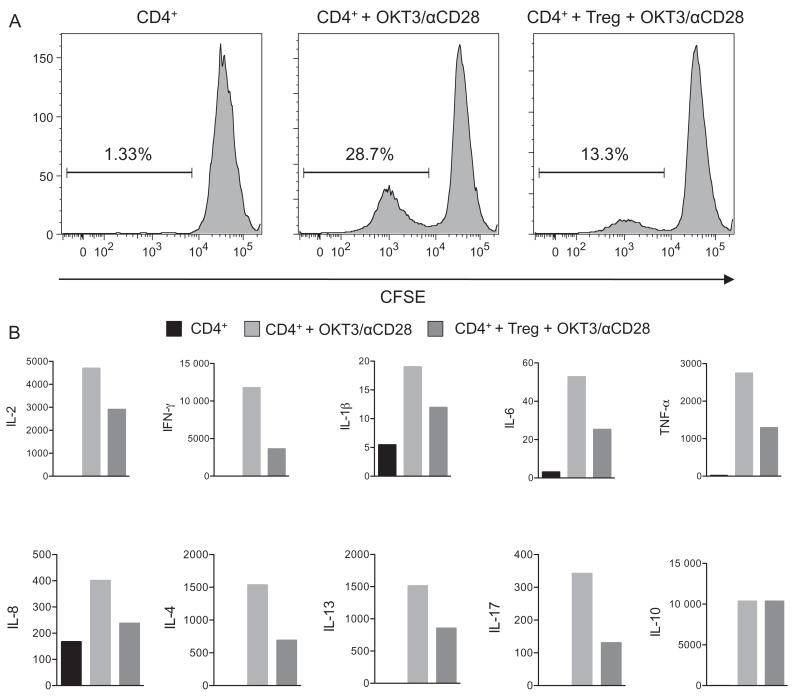
Figure 2.
Lamina propria Treg suppress proliferation and cytokine production of activated CD4 T cells. Sorted lamina propria Treg (4×10⁴) isolated from a surgical ileum specimen were co-cultured with 2×10⁵ magnetically sorted peripheral blood CD4 T cells from a healthy donor. Responder CD4 T cells were labelled with carboxyfluorescein diacetate succinimidyl ester (CFSE) and stimulated with OKT-3 antibody and anti-CD28. Activated and non-activated CD4 T cells without Treg were used as controls. (A) The proliferation of responder T cells as indicated by CFSE dilution was analysed by flow cytometry on day 5. Treg presence suppressed the percentage of proliferating cells by 50% compared with responder cells without Treg. (B) Culture supernatant cytokine concentrations on day 2 were determined using Milliplex Map multiplex magnetic bead-based immunoassay kits on a Luminex Flexmap 3D Platform. Except for IL10, Th1 and Th2 cytokine production by responder cells was suppressed by approximately 50%. IL, interleukin, TNFα, tumour necrosis factor α.

