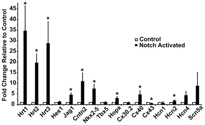
Figure 3.
Transient Notch Activation Converts the Transcriptional Profile of Newborn Cardiomyocytes to a Conduction-like Phenotype. RT-qPCR analysis of induced gene expression changes for canonical Notch target genes and conduction system-enriched genes after activation of Notch signaling (black bars) for 48 hours in cultured perinatal ventricular cardiomyocytes. All fold changes are normalized to control cells infected with a GFP-expressing adenovirus (white bars). Notch activation up-regulates known Notch targets, as well as Purkinje fiber-enriched transcripts. n=7 replicates. Data are expressed as mean ± SEM. Group comparison was performed using a Student’s unpaired 2-tailed t-test. *p<0.05

