Abstract
Background
Genome-wide association studies (GWAS) identify regions of the genome that are associated with particular traits, but do not typically identify specific causative genetic elements. For example, while a large number of single nucleotide polymorphisms associated with type 2 diabetes (T2D) and related traits have been identified by human GWAS, only a few genes have functional evidence to support or to rule out a role in cellular metabolism or dietary interactions. Here, we use a recently developed Drosophila model in which high-sucrose feeding induces phenotypes similar to T2D to assess orthologs of human GWAS-identified candidate genes for risk of T2D and related traits.
Results
Disrupting orthologs of certain T2D candidate genes (HHEX, THADA, PPARG, KCNJ11) led to sucrose-dependent toxicity. Tissue-specific knockdown of the HHEX ortholog dHHEX (CG7056) directed metabolic defects and enhanced lethality; for example, fat-body-specific loss of dHHEX led to increased hemolymph glucose and reduced insulin sensitivity.
Conclusion
Candidate genes identified in human genetic studies of metabolic traits can be prioritized and functionally characterized using a simple Drosophila approach. To our knowledge, this is the first large-scale effort to study the functional interaction between GWAS-identified candidate genes and an environmental risk factor such as diet in a model organism system.
Keywords: Genome-wide association study; Drosophila melanogaster; Diabetes mellitus, type 2; Hyperglycemia; Dyslipidemias; Phylogeny; Reverse genetics; High-throughput screening assays; HHEX protein, Human
Author summary
The search for genetic risk factors for common human diseases often relies on the use of linkage and association studies to establish correlation between genomic markers and disease risk. These studies require additional functional evaluation of candidate genes, including their possible interaction with diet and environment. The number of candidate genes is typically large and the development of appropriate genetic tools in mammalian systems is slow. By contrast, large-scale genetic screens, using widely available genetic tools, are routinely conducted in the fruit fly Drosophila melanogaster. In this study, we used Drosophila to screen candidate genes identified in human genome-wide scans as associated with risk of metabolic abnormalities such as type 2 diabetes. We show that a number of human candidate genes have fly orthologs that play an important role in Drosophila tolerance to high dietary sucrose. We further explored some of the specific metabolic abnormalities that can result when these genes’ activities are reduced in flies, focusing on a gene we call dHHEX (CG7056), the fly ortholog of human HHEX.
Background
Type 2 diabetes (T2D), a disease state characterized by impaired insulin sensitivity and hyperglycemia, is one of the world’s leading causes of mortality and morbidity [1-3]. In recent years, genome-wide association studies (GWAS) have had success in identifying susceptibility loci for type 2 diabetes and related traits in humans [4,5]. These studies establish associations between markers, such as single nucleotide polymorphisms (SNPs), and disease. However, they typically lack the resolution needed to identify causal variants, because SNPs may exist in linkage disequilibrium with multiple protein-coding loci as well as with non-coding gene-regulatory elements that can act over a long distance [6,7]. Mouse models of diabetes and obesity can serve as convenient platforms to functionally probe a small number of candidate genes [8], but this approach is expensive and slow, limiting the number of genes that can be readily assessed.
The biochemical pathways involved in growth and metabolism are ancient and well conserved across the animal kingdom from C. elegans and Drosophila to rodents and humans [9]. Analogous to insulin and glucagon in vertebrates, Drosophila insulin-like peptides (dILPs) and adipokinetic hormone (Akh) regulate circulating glucose homeostasis. In addition, many tissues known to be important in type 2 diabetes have functional analogs in Drosophila including blood, adipose tissue and liver, skeletal muscle, pancreatic beta cells, brain, and kidney [9]. Indeed, Drosophila melanogaster raised on diets high in sugar exhibit hallmark features of type 2 diabetes including insulin resistance, fasting hyperglycemia, and increased fat storage [10].
Off-the-shelf genetic tools in Drosophila, including mutations and inducible RNA interference (RNAi), allow the functions of specific genes to be rapidly queried; a Drosophila genetic approach has recently been used to follow up a small-scale GWAS for Alzheimer pathology [11]. Here we make use of the advantages of Drosophila as a model system for exploration of whole-animal metabolism. Starting from a subset of previously published human SNPs and regions associated with disease risk, we utilize Drosophila to screen candidate genes in each region in an unbiased manner. We provide functional evidence that disruption of some of these genes can predispose the flies to dietary sucrose-induced lethality. We show in one region that HHEX may contribute to type 2 diabetes phenotypes including hyperglycemia and insulin insensitivity; in addition, our data suggests two neighboring genes may also contribute to the risk identified by GWAS. Thus, in addition to implicating specific genes as disease drivers, our work demonstrates the power of Drosophila to provide rapid, functional, diet- and environment-sensitive assays for GWAS follow-up studies and to deconvolute regions that contain multiple risk loci.
Results and discussion
Fly orthologs of human genes in disease risk-associated regions
We focused on a set of 38 human genomic regions in which SNPs have been associated with type 2 diabetes disease status [12-14] as well as related quantitative traits (QTs), including levels of fasting blood glucose [15,16], triglycerides [17-19], low-density lipoprotein (LDL) [17,18], and high-density lipoprotein (HDL) [17,18]. We included the latter SNPs since there is considerable overlap between mechanisms that regulate lipid and glucose metabolism. Beginning with the 130 human genes located within approximately 100 kb of each SNP, we identified fly orthologs as inferred by Ensembl’s phylogenetic analyses (release 43) [20,21] and found that 71 of the 130 candidate human genes—within 33 of the 38 human genomic regions—have fly orthologs with one-to-one, one-to-many, many-to-one, or many-to-many orthology relationships. In total, we identified 83 fly orthologs corresponding to 71 human genes under consideration. Human GWAS traits, associated index SNPs, genomic regions, candidate genes, and fly orthologs are listed in Table 1 and Additional file 1: Table S1.
Table 1.
Human genes identified by GWAS
| Region | Trait | Genes |
|---|---|---|
| 1p32.3 |
LDL |
TMEM61, BSND, PCSK9, USP24 |
| 1p31.3 |
TG |
ANGPTL3*, DOCK7, ATG4C** |
| 1p13.3 |
LDL |
KIAA1324, SARS, CELSR2**, PSRC1, MYBPHL, SORT1 |
| 1p12 |
T2D |
ADAM30, NOTCH2 |
| 1q42.13 |
TG, HDL |
GALNT2 |
| 2p24.1 |
LDL |
APOB |
| 2p23.3 |
T2D |
NRBP1**, KRTCAP3, IFT172, FNDC4, GCKR, C2orf16, ZNF512 |
| 2p21 |
T2D |
THADA**, PLEKHH2 |
| 2q24.3 |
FG |
NOSTRIN*, SPC25, G6PC2, ABCB11** |
| 3p25.2 |
T2D |
PPARG** |
| 3p14.1 |
T2D |
ADAMTS9* |
| 3q27.3 |
T2D |
IGF2BP2*, C3orf65 |
| 5q13.3 |
LDL |
HMGCR, COL4A3BP |
| 6p22.3 |
T2D |
CDKAL1 |
| 6p21.32 |
LDL |
HLA-DPA1, COL11A2, RXRB**, SLC39A7, HSD17B8, MIR219-1, RING1**, VPS52, RPS18 |
| 7p15.2 |
T2D |
JAZF1 |
| 7p13 |
FG |
AEBP1, MIR4649, POLD2, MYL7, GCK**, YKT6, CAMK2B |
| 7q11.23 |
TG |
BAZ1B, BCL7B, TBL2, MLXIPL**, VPS37D, DNAJC30*, WBSCR22, STX1A |
| 8p21.3 |
TG, HDL |
LPL** |
| 8q24.11 |
T2D |
SLC30A8* |
| 8q24.13 |
TG |
TRIB1* |
| 9p21.3 |
T2D |
MTAP, CDKN2A, CDKN2B |
| 9q31.1 |
HDL |
ABCA1 |
| 10p13 |
T2D |
NUDT5, CDC123, CAMK1D |
| 10q23.33 |
T2D |
IDE*, KIF11*, HHEX** |
| 10q25.2 |
T2D |
TCF7L2** |
| 11p15.1 |
T2D |
NUCB2*, NCR3LG1, KCNJ11**, ABCC8* |
| 11p11.2 |
T2D |
EXT2, ALX4 |
| 11q23.3 |
TG |
BUD13, ZNF259, APOA5, APOA4, APOC3, APOA1, SIK3 |
| 12q15 |
T2D |
TSPAN8, LGR5 |
| 12q23.3 |
HDL |
MYO1H, KCTD10*, UBE3B, MMAB, MVK** |
| 15q21.3 |
HDL |
LIPC** |
| 16q12.1 |
T2D |
RPGRIP1L, FTO |
| 16q12.2 |
HDL |
CETP, NLRC5, SLC12A3**, HERPUD1, MIR138-2 |
| 18q21.1 |
HDL |
LIPG**, ACAA2 |
| 19p13.2 |
LDL |
SMARCA4, LDLR, SPC24, KANK2 |
| 19p13.11 |
TG, LDL |
GATAD2A, TSSK6, NDUFA13**, YJEFN3, CILP2, PBX4, LPAR2, GMIP, ATP13A1** |
| 19q13.33 | LDL | BCL3, CBC, BCAM, PVRL2, TOMM40**, APOE, APOC1, APOC4, APOC2, CLPTM1, RELB** |
List of all human genes located near the SNPs considered in the study. Asterisks indicate genes with Drosophila orthologs that function in sucrose tolerance in Drosophila; doubled asterisks indicate strong hits. Italicized genes were not evaluated in the Drosophila sucrose-intolerance screen, mostly because they lack Drosophila orthologs. References and more detailed experimental results are in Additional file 1: Table S1.
Screen for modifiers of sucrose tolerance
We and others have previously shown that flies fed high-sugar diets, including a 1.0 M sucrose diet, exhibit diabetes-like phenotypes [10,22]. Additionally, flies die as larvae when fed very high levels of dietary sucrose (above 1.25 M), but survival to pupariation is comparable between flies fed 0.15 M and 1.0 M sucrose diets (Additional file 1: Figure S1). We hypothesized that knocking down a gene that mediates sucrose tolerance would affect larval viability differently on high- vs. low-sucrose diets. Such a gene would be required for survival to pupariation on a 1.0 M sucrose diet, but may prove dispensable on a control 0.15 M sucrose diet.
To test this hypothesis and to identify modifiers of the sucrose intolerance phenotype, we screened knockdowns of the selected genes (Table 1) using the now-classic GAL4/UAS system [23]. Fly lines containing inducible RNA-interference (UAS-RNAi) elements were acquired for most of these genes; multiple fly lines were available for many loci, and a total of 137 RNAi fly lines were tested. We used tubP-GAL4 to direct broad expression of GAL4, which in turn induces broad expression of the RNAi-encoding transgene. Sucrose tolerance was then assessed by (i) scoring pupariation rates relative to non-RNAi controls and (ii) comparing high-sucrose vs. low-sucrose feeding. An important strength of this approach is that we can distinguish between sucrose-dependent and sucrose-independent toxicity. The results of the screen are detailed in Additional file 1: Table S1 and summarized in Figures 1 and 2 and Table 1.
Figure 1.

Effect of sucrose on survival of RNAi candidates. Ranked estimates and confidence intervals (orange/purple: 95%, gray: Bonferroni-adjusted, N = 113) for the ln(OR) of knockdown vs. non-knockdown sibling pupariation on 1.0 M sucrose vs. 0.15 M sucrose. For each human gene, the cross with the most dramatic effect is shown; crosses that were lethal independent of dietary sucrose are omitted. Bars are labeled with the names of the human orthologs corresponding to each RNAi line; the names of the RNAi lines themselves are in Additional file 1: Table S1. We considered crosses whose 95% confidence intervals exclude zero as hits, and we considered crosses whose Bonferroni-adjusted confidence intervals exclude zero as strong hits. In a few cases, a gene was a hit in both directions; in such cases, both crosses are shown and the gene name is marked with an asterisk. More complete details are in Additional file 1: Tables S1 and S2.
Figure 2.
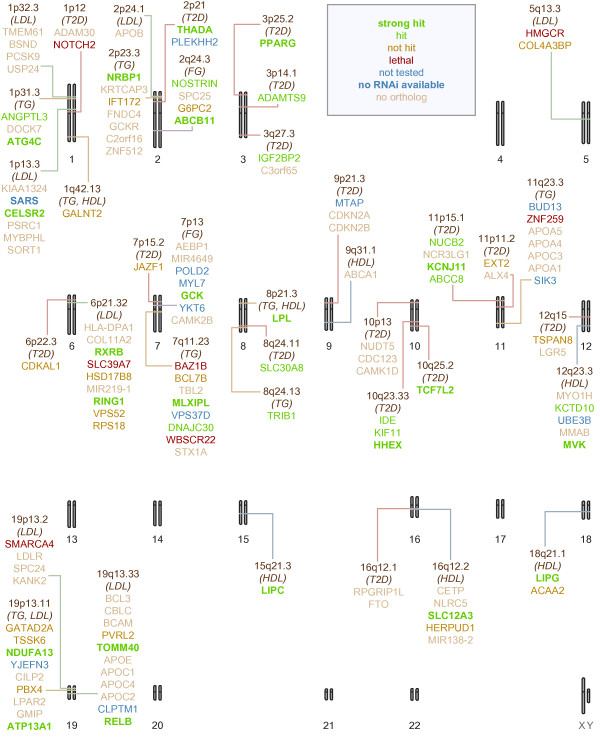
Graphical summary of the Drosophila sucrose-intolerance screen. The 38 regions of interest, and the human genes located in them, are marked on a schematic karyogram of the human genome. The regions are labeled with the metabolic traits with which they are associated. The gene names are color-coded to indicate their outcome in our screen. More complete details are in Additional file 1: Table S1.
We defined hits as crosses in which knockdown resulted in statistically significant sucrose-sensitive toxicity at a 0.05 threshold. We defined strong hits as results that had p-values less than 4.42 × 10⁻⁴, corresponding to a Bonferroni adjustment for the total of 113 lines for which sucrose tolerance could be assessed (we excluded crosses where sucrose tolerance could not be assessed due to sucrose-independent lethality). 47 of the screened lines were hits, corresponding to 34 human genes; 29 of these were strong hits, corresponding to 22 human genes. As a control, we verified that the relative survival to pupariation was similar for flies fed 1.0 M sucrose vs. 0.15 M sucrose food for the GD and KK genetic backgrounds (without RNAi transgenes) used in the study (Additional file 1: Table S2, Figure 1).
Regions containing a single candidate gene
The approximately 100 kb radius window we applied included only a single human gene at 12 regions. At each of these regions, it seems plausible that variants in this single gene are causative for the associated traits. 10 of these regions were tested with fly orthologs (two regions did not contain fly orthologs). Seven fly orthologs were identified as hits in at least one cross. Strong hits corresponded to PPARG and TCF7L2 (T2D candidates), LIPC (HDL candidate), and LPL (TG and HDL candidate); orthologs of ADAMTS9 and SLC30A8 (T2D candidates) and TRIB1 (TG candidate) were also hits. The 7/10 hit rate at single-gene regions indicates that our model likely reflects at least some important aspects of human T2D and related QTs, and that our assay can provide useful modifier data.
Not all single-gene regions tested positive in our Drosophila assay: orthologs of GALNT2, CDKAL1, and JAZF1 failed to display modifier activity (Figures 1 and 2, Table 1, Additional file 1: Table S1). This may be due to several factors. First, the RNAi constructs and insertions used work with differing efficacy, at least in part likely due to their random insertion sites within the genome [24]. Consistent with this phenomenon, some RNAi lines targeting the same Drosophila gene gave discordant results in our assay; most commonly, one line showed modifier activity and another did not. Second, phylogenetic inference of orthology may not be correct. Indeed, Ensembl’s inferences were refined while our study was underway. Third, the GWAS result may be a false positive, or the true causative variant may lie outside of the window we initially selected or may be undetected within the region. However, GALNT2 acts in metabolic pathways [25] and a coding mutation in CDKAL1 has been closely correlated with T2D risk in humans [26]. Fourth, our screening approach may not identify loci that are risk factors due to upregulation. Lastly, failure of Drosophila to confirm modifier status for several of these regions may reflect limitations of using flies to explore GWAS.
Regions containing multiple candidate genes
At the remaining regions, our approximately 100 kb radius window defined more than one candidate human gene, many with fly orthologs. The region near rs4607517 contains the gene GCK, encoding glucokinase, which is required for glucose-stimulated insulin secretion and proper glucose metabolism. GCK mutations are causative alleles in a monogenic form of diabetes [27], making it a strong candidate to further validate our approach. Indeed, sucrose-specific toxicity was strongly enhanced by knockdown of all but one of the four putative fly orthologs of GCK (Figure 1, Additional file 1: Table S1).
For other regions we tested orthologs of multiple human genes, and at six of the remaining regions our sucrose toxicity screen implicated a single human gene ortholog. These genes were THADA and IGF2BP2 (T2D candidates), CELSR2 (LDL candidate), NRBP1 (TG candidate), SLC12A3 and LIPG (HDL candidates). At six regions our screen did not identify any hits. While this may be explained in part by potential lack of sensitivity in our system, in all cases these regions included other genes we were not able to test.
At nine regions, our screen implicated more than one human gene. This may reflect a lack of specificity of this assay, perhaps due to off-target effects of the RNAi constructs or differences in insect and mammalian physiology. On the other hand, these results highlight the fact that model organism screening has the ability to identify multiple modifier loci within a single region and at the level of individual genes. Of particular utility, genes that are tightly linked in humans can be subjected to individual functional testing using specific RNAi knockdown. A striking example of this is a region at 10q23.33 that contains IDE, KIF11, and HHEX, three genes with clear one-to-one fly orthologs. Genome-wide scans [12,13,28] have consistently reported SNP signals associated with T2D near this region. Attention has focused on HHEX, which has been most closely linked with these signals [29], and which encodes a metabolism-related HOX-class transcription factor. Indeed, our screening identified CG7056—which we refer to here as dHHEX—as the most robust modifier of sucrose-mediated lethality in our study (Figure 1, Additional file 1: Table S1).
We also identified Drosophila orthologs of the neighboring genes IDE and KIF11—Drosophila genes ide and klp61F—as modifiers (Figures 1 and 2, Additional file 1: Table S1). Intriguingly, IDE knockout mice exhibit hyperglycemia and insulin insensitivity in an age-dependent manner [30,31]. KIF11 has also been knocked out in mice but is embryonic lethal; metabolic effects of partial loss of KIF11 have not been characterized [32]. Our data suggest that IDE and KIF11 may contribute to patients’ metabolic risk. Their role may be obscured by the high risk conferred by neighboring HHEX locus; the three loci may act independently, or perhaps variations in copy number affect the three loci to increase patient risk.
Further characterization of dHHEX: diet
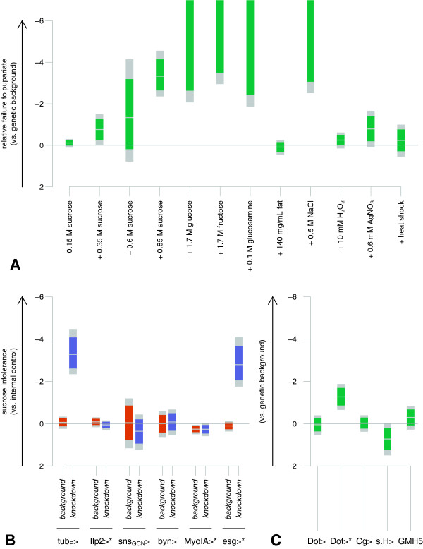
We next used Drosophila to further explore aspects of dHHEX’s role in the response to high dietary sucrose. Raised on a variety of feeding conditions (Additional file 1: Table S3, Figure 3A) tubP>RNAidHHEX flies remained comparable to wild type on a number of stressful diets including diets containing hydrogen peroxide and silver nitrate and, notably, a high-fat diet. We observed elevated lethality when tubP>RNAidHHEX flies were raised on high-salt diets (slightly hyperosmolar relative to 1.0 M sucrose food). This result suggests an impaired ability to respond to hyperosmolar conditions that may contribute to sucrose-dependent lethality. To better understand the role of dHHEX specifically in glucose metabolism, we used dietary glucosamine to explore the hexosamine biosynthetic pathway (HBP), a primary pathway of glucose metabolism. The HBP has been implicated in mechanisms of glucose toxicity [33] and dietary glucosamine increases HBP flux in flies [34]. tubP>RNAidHHEX flies proved glucosamine-intolerant: the addition of 0.1 M glucosamine to a control diet resulted in failure to pupariate. The ability of low levels of glucosamine to cause lethality suggests that glucose metabolism is indeed a primary effector of dietary sucrose toxicity in tubP>RNAidHHEX flies.
Figure 3.
Diet-specific and tissue-specific lethality of dHHEX knockdown. A. tubP>RNAidHHEX-V15721 flies tolerate many non-ideal diets, but are sensitive to high sucrose, as well as to glucosamine and to high salt. Estimates and confidence intervals (green: 95%, gray: Bonferroni-adjusted, N = 12) of ln(OR) are shown comparing odds of knockdown vs. non-knockdown animals pupariating compared to the genetic background control on that diet. More complete details are in Additional file 1: Table S3. B–C. tubP-GAL4, Dot-GAL4, and esg-GAL4 driving dHHEX knockdown using UAS-RNAidHHEX-V15721 confer lethality on high sucrose, but a number of other drivers do not. Estimates and confidence intervals (colored: 95%, gray: Bonferroni-adjusted, N = 11) of ln(OR) are shown for survival of knockdown vs. non-knockdown animals, compared to the genetic background control. Pupariation (unmarked) or eclosion (asterisks) or both were scored. More complete details are in Additional file 1: Tables S4 and S5.
Further characterization of dHHEX: tissues
We next used targeted knockdown to determine which tissues require normal dHHEX activity in the face of high dietary sucrose (Additional file 1: Table S4, Figure 3B–C). Mammalian studies have implicated HHEX function in diverse tissues and organs including liver, heart, pancreas, thyroid, and hematopoietic cells [35-39]. We used a panel of tissue-selective GAL4 lines to direct expression of the UAS-RNAidHHEX transgene in specific tissues: dilp2-GAL4 targets insulin producing cells (beta cell analogs), Cg-GAL4 targets several tissues including the fat body (functional analog of mammalian liver and adipose tissue), Dot-GAL4 targets several tissues including nephrocytes (functional analogs of glomerular podocytes) and gut, GMH5 targets heart, srp.Hemo-GAL4 targets hemocytes (phagocytic blood cell analogs) as well as nephrocytes, snsGCN-GAL4 targets nephrocytes, esg-GAL4 targets several cell types including midgut stem cells, MyoIA-GAL4 targets differentiated midgut, and byn-GAL4 targets hindgut. Flies carrying a GAL4 driver plus UAS-RNAidHHEX were raised on high-sucrose diets along with control animals, and the number of knockdown to non-knockdown animals surviving to pupariation or eclosion on high or low sucrose was scored. Of the three distinct UAS-RNAidHHEX lines we used in the initial screen, we focused on the V15721 line, which exhibited the most survival of knockdown flies on 0.15 M sucrose when driven by tubP-GAL4.
Knockdown of dHHEX in the heart, hemocytes, nephrocytes, hindgut, and differentiated midgut did not affect viability on high-sucrose feeding compared to low-sucrose feeding, and perhaps surprisingly, neither did knockdown in the insulin producing cells or the fat body (Figure 3B–C). However, knockdown driven by either Dot-GAL4 or esg-GAL4 did affect survival to eclosion in a sugar-sensitive manner (although, interestingly, knockdown driven by Dot-GAL4 did not affect survival to pupariation). Both of these drivers express in a range of tissues but one point of overlap in their domains is in the midgut stem cells.
Further characterization of dHHEX: metabolics
Given the fat body’s important role in metabolism, we profiled hemolymph (blood) glucose, body size, and whole-animal triglyceride levels in Cg>RNAidHHEX-V15721 wandering third-instar and adult flies reared on 1.0 M sucrose. Dicer-2 (Dcr-2) overexpression was included to enhance RNAi efficacy [24]. Female flies were studied because this experimental genotype is male-lethal.
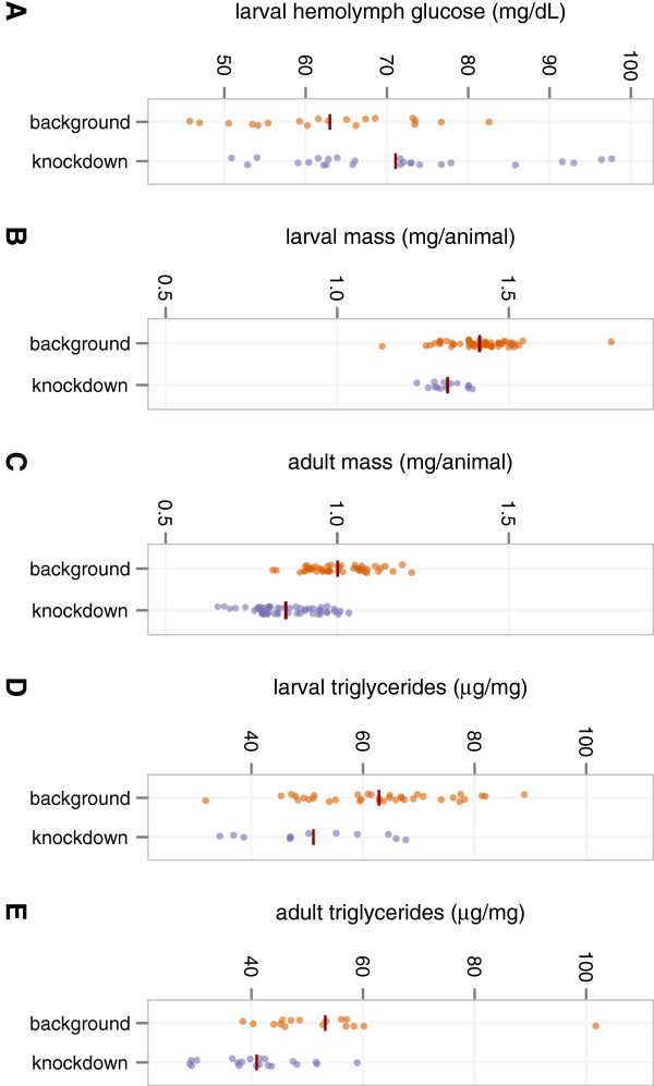
Both larval and adult Cg>RNAidHHEX-V15721, Dcr-2 flies were significantly smaller than controls when reared on a high-sucrose diet throughout development (p < 0.001, Additional file 1: Table S6, Figure 4B–C). Since flies have a single receptor that is orthologous to both human insulin and human IGF1 receptors, and since this receptor controls both glucose homeostasis and growth [40-42], the smaller body size of knockdown animals suggests that Cg>RNAidHHEX-V15721, Dcr-2 flies have decreased insulin signaling activity and hence increased insulin resistance when confronted with high dietary sucrose. Consistent with this view, wandering (non-feeding) Cg>RNAidHHEX-V15721, Dcr-2 larvae were hyperglycemic after high-dietary-sucrose rearing (p < 0.029, Additional file 1: Table S6, Figure 4A). Intriguingly, in both Cg>RNAidHHEX-V15721, Dcr-2 larvae and adults, hyperglycemia and reduced body size were accompanied by significantly lower triglyceride levels when compared to controls (p < 0.007, Additional file 1: Table S6, Figure 4D–E).
Figure 4.
Metabolic profiling of Cg>RNAidHHEX-V15721, Dcr-2. A. Wandering third-instar females of Cg>RNAidHHEX-V15721, Dcr-2 are hyperglycemic. Each measurement is made from a pooled sample of hemolymph from 5–8 animals; dark red line segments show mean values. p < 0.029. B–C. Wandering third-instar and newly eclosed adult females of Cg>RNAidHHEX-V15721, Dcr-2 have reduced body size. Each measurement is the mean per-animal mass of a group of 6–10 animals; dark red line segments show mean values. p < 0.001 for both larvae and adults. D–E. Wandering third-instar and newly eclosed adult females of Cg>RNAidHHEX-V15721, Dcr-2 have reduced triglyceride levels. Each measurement is made from a pooled sample of whole-animal homogenate from 6–10 animals; dark red line segments show mean values. p < 0.007 for both larvae and adults (p < 0.002 for adults when one extreme outlier is excluded). All p-values for metabolic data were calculated using a bidirectional t-test without assuming equal variances. More complete details are in Additional file 1: Tables S6–S8.
Conclusions
To date, genome-wide scans for genes and alleles that contribute to metabolic disease risk have identified numerous candidates, but functional follow-up studies have been more difficult to perform in mammalian systems. We hypothesized that human genes involved in susceptibility to developing T2D and related traits can be prioritized by knocking down their fly orthologs and assaying sucrose-sensitive lethality. Evidence in support of our hypothesis includes the fact that this assay identifies GCK, a gene known to cause a monogenic form of diabetes, and the fact that sucrose-sensitive lethality correlates with metabolic abnormalities in the flies. To our knowledge, this is the first large-scale functional study of metabolic-trait candidate genes identified by GWAS analysis, and the first to specifically address an interaction between genes and environment.
Through more detailed analysis of the function of the Drosophila HHEX ortholog, we have shown that this gene plays an important role in whole-animal metabolism in this system through its effects in the fat body—a functional analog of mammalian liver and adipose tissue. Loss of dHHEX results in insulin resistance and hyperglycemia and, interestingly, a reduction of whole-animal triglyceride levels in this system. It has been proposed that the conversion of fatty acids into triglycerides may protect against tissue lipotoxicity [43]; the hyperglycemia observed in Cg>RNAidHHEX-V15721 flies suggests dHHEX may play a role in determining the capacity of the fly to store energy as triglycerides. We additionally showed that there are multiple other candidate genes for T2D and related QTs (fasting glucose, triglycerides, LDL, and HDL) that have diet-dependent roles in overall organismal viability. Further systematic study of these genes, including T2D candidate genes such as PPARG, IDE, and KIF11, may help elucidate their molecular functions in their respective pathways. Since many fundamental aspects of metabolism have been conserved during evolution, it is reasonable to hypothesize that these functions may be similar in humans as in flies; whether this is true will, of course, have to be determined case by case.
In general, Drosophila offers a rich resource for providing rapid, inexpensive, whole-animal tests of gene function. In addition to screening candidate genes identified by GWAS approaches, this same approach could prove useful, as whole genome sequencing becomes more common, for identifying specific mutations that are causative rather than simply correlated. Perhaps its most important advantage is the ability to assess all candidates in an unbiased manner, identifying surprising hits and untangling complex regions.
Methods
Fly stocks
RNAi stocks (listed in Additional file 1: Table S1) were acquired from the Vienna Drosophila Resource Center, as well as genetic background controls w1118 (for GD lines, VDRC #60000) and y– w1118; P{attP, y+, w3’}VIE-260B (for KK lines, VDRC #60100) [24]. tubP-GAL4 (BDSC #5138) [44], Cg-GAL4 (BDSC #7011) [45], dilp2-GAL4 (BDSC #37516) [46], esg-GAL4 (BDSC #26816), Dot-GAL4 (BDSC #6903) [47,48], UAS-Dcr-2 (BDSC #24648) [24], and TM6B, Tb1 (BDSC #120) are available from the Bloomington Drosophila Stock Center. MyoIA-GAL4 (DGRC-K #112001) is available from the Drosophila Genetic Resource Center, Kyoto. Additional fly stocks were generously provided by the Drosophila community: GMH5 by Rolf Bodmer [49], snsGCN-GAL4 by Susan Abmayr [50], srp.Hemo-GAL4 by Katja Brückner [51,52], byn-GAL4 by Volker Hartenstein [53,54], and a T(2;3) balancer by Larry Zipursky.
Fly media
We modified a commonly used Drosophila semi-defined medium [55] as previously described [10]. Briefly, we replaced all added sugars in the recipe (glucose and sucrose) with 51.3 g/L sucrose (to yield 0.15 M sucrose) plus any other desired components. The primary screen was carried out on 0.15 M sucrose (low sucrose) and 1.0 M sucrose (high sucrose) foods.
Scoring and statistics
In tubP-GAL4, byn-GAL4, and snsGCN-GAL4 studies, flies carrying a GAL4-encoding transgene and a balancer as the homologous chromosome were crossed to flies carrying a RNAi-encoding transgene, and survival to pupariation was scored by counting non-tubby (driver>RNAi) and tubby (UAS-RNAi; TM6B or a T(2;3) balancer) pupae, except for a small number of exceptions, where survival to adulthood of non-curly (tubP>RNAi) compared to curly (tubP-GAL4; CyO) or non-stubble (tubP>RNAi) compared to stubble (tubP-GAL4; TM3, Sb–) animals was scored instead. In studies of other drivers, either a similar cross was performed and survival to adulthood was scored by counting non-curly (driver>RNAi) and curly (driver; CyO) animals, or else rates of eclosion were compared for a fixed number of knockdown embryos compared to a fixed number of control embryos possessing the GAL4 insertion and the proper genetic background, but lacking the RNAi-encoding insertion. Counts that were extremely different from experimental replicates were excluded from analysis (3 out of over 800 replicates were excluded in this way). If fewer than 5 knockdown animals survived in all experimental replicates for a given comparison, then we considered the knockdown to be generally toxic and did not assess sucrose intolerance.
For each comparison, we used Fisher’s exact test to assess whether our data were consistent with the null hypothesis that relative survival of knockdown flies was the same on high- and low-sucrose feeding, as well as to compute point estimates and confidence intervals for the natural logarithm of the odds ratio (ln(OR)) for survival of the knockdown and control genotypes on high and low sucrose. We considered crosses to be hits when statistically significant with a 0.05 threshold, and we considered crosses to be strong hits when statistically significant with a Bonferroni-adjusted threshold. For the RNAi screen for sucrose sensitivity, this threshold was 4.42 × 10⁻⁴, corresponding to 113 crosses tested (excluding crosses that were lethal on both diets, since no determination about sucrose sensitivity can be made for these crosses). For the diet survey, this threshold was 4.17 × 10⁻³, corresponding to 12 diets tested. For the driver survey, this threshold was 4.55 × 10⁻³, corresponding to 11 driver-phenotype pairs tested. We used confidence intervals for hypothesis testing; they could in principle also be used for effect-size comparison and equivalence testing.
Metabolic parameters were comparing using two-sided unpaired t-tests without assuming equal variances.
Computations were performed in R, a language and environment for statistical computing, version 2.15.1. Plots were generated in R, some using the ggplot2 package. Some tables were constructed using XeLaTeX and the longtable and booktabs packages.
Metabolic studies
Hemolymph glucose and whole-animal triglycerides were measured as previously described [10]. Briefly, to collect hemolymph, wandering third-instar larvae were lanced and hemolymph from 5–8 larvae was pooled to collect 1 μL. Glucose levels were measured using the Infinity Glucose Hexokinase Reagent kit (Thermo Fisher #TR15421). Triglycerides were measured using the Infinity Triglycerides Reagent kit (Thermo Fisher #TR22321) on whole-animal homogenates of groups of 6–10 animals. Per-animal mass was measured by weighing groups of 6–10 animals.
Abbreviations
GWAS: Genome-wide association study; HBP: Hexosamine biosynthesis pathway; HDL: High-density lipoprotein; LDL: Low-density lipoprotein; QT: Quantitative trait; SNP: Single nucleotide polymorphism; T2D: Type 2 diabetes.
Competing interests
RLC and TJB are co-founders of Medros, Inc., which uses models of human disease for drug development.
Authors’ contributions
RLC, TJB, and FSC conceived of the project, and NN and FSC identified human and fly genes of interest. All authors contributed to the design of the experiments. PVR conducted hemolymph glucose measurements, JLF conducted mass and triglyceride measurements, and JP and JN conducted all other experiments. JP designed and conducted the statistical analysis. JP, PVR, NN, and TJB wrote this manuscript, all authors contributed to completing and revising it, and all authors read and approved the final manuscript.
Supplementary Material
Includes Figure S1 and Tables S1–S8.
Contributor Information
Jay Pendse, Email: jay.pendse@mssm.edu.
Prasanna V Ramachandran, Email: pvramach@bcm.edu.
Jianbo Na, Email: jianbo.na@mssm.edu.
Narisu Narisu, Email: narisu@mail.nih.gov.
Jill L Fink, Email: jfink@wustl.edu.
Ross L Cagan, Email: ross.cagan@mssm.edu.
Francis S Collins, Email: collinsf@od.nih.gov.
Thomas J Baranski, Email: baranski@wustl.edu.
Acknowledgments
We thank Vivek A. Rudrapatna, Ruth I. Johnson, Susumu Hirabayashi, Erdem Bangi, Tirtha Kamal Das, Laura Palanker Musselman, Dac Anh Nguyen, Lori Bonnycastle, Michael Stitzel, Michael Erdos, and Laura Scott for advice and helpful discussions, and countless members of the free and open source software community for valuable guidance and code samples. This research was supported by NIH grants R21 DK069940 (RLC), P60 DK20579 (Washington University DRTC, TJB), and P20 RR020643 (TJB), as well as by grants from the NephCure Foundation (JN and RLC) and from the American Diabetes Association (JN and RLC). JP was supported in part by NIGMS Training Grant T32 GM-62754, NIH. NN and FSC were supported by the intramural program of NHGRI, NIH.
References
- Zimmet P, Alberti KGMM, Shaw J. Global and societal implications of the diabetes epidemic. Nature. 2001;414:782–787. doi: 10.1038/414782a. [DOI] [PubMed] [Google Scholar]
- Roglic G, Unwin N. Mortality attributable to diabetes: estimates for the year 2010. Diabetes Res Clin Pract. 2010;87:15–19. doi: 10.1016/j.diabres.2009.10.006. [DOI] [PubMed] [Google Scholar]
- Alwan A, MacLean DR, Riley LM, d’Espaignet ET, Mathers CD, Stevens GA, Bettcher D. Monitoring and surveillance of chronic non-communicable diseases: progress and capacity in high-burden countries. Lancet. 2010;376:1861–1868. doi: 10.1016/S0140-6736(10)61853-3. [DOI] [PubMed] [Google Scholar]
- McCarthy MI. Genomics, type 2 diabetes, and obesity. N Engl J Med. 2010;363:2339–2350. doi: 10.1056/NEJMra0906948. [DOI] [PubMed] [Google Scholar]
- Hindorff LA, Sethupathy P, Junkins HA, Ramos EM, Mehta JP, Collins FS, Manolio TA. Potential etiologic and functional implications of genome-wide association loci for human diseases and traits. Proc Natl Acad Sci U S A. 2009;106:9362–9367. doi: 10.1073/pnas.0903103106. [DOI] [PMC free article] [PubMed] [Google Scholar]
- Lettice LA, Heaney SJH, Purdie LA, Li L, De Beer P, Oostra BA, Goode D, Elgar G, Hill RE, De Graaff E. A long-range shh enhancer regulates expression in the developing limb and fin and is associated with preaxial polydactyly. Hum Mol Genet. 2003;12:1725–1735. doi: 10.1093/hmg/ddg180. [DOI] [PubMed] [Google Scholar]
- Spilianakis CG, Lalioti MD, Town T, Lee GR, Flavell RA. Interchromosomal associations between alternatively expressed loci. Nature. 2005;435:637–645. doi: 10.1038/nature03574. [DOI] [PubMed] [Google Scholar]
- Cox RD, Church CD. Mouse models and the interpretation of human GWAS in type 2 diabetes and obesity. Dis Model Mech. 2011;4:155–164. doi: 10.1242/dmm.000414. [DOI] [PMC free article] [PubMed] [Google Scholar]
- Baker KD, Thummel CS. Diabetic larvae and obese flies—emerging studies of metabolism in drosophila. Cell Metab. 2007;6:257–266. doi: 10.1016/j.cmet.2007.09.002. [DOI] [PMC free article] [PubMed] [Google Scholar]
- Palanker Musselman L, Fink JL, Narzinski K, Ramachandran PV, Sukumar Hathiramani S, Cagan RL, Baranski TJ. A high-sugar diet produces obesity and insulin resistance in wild-type Drosophila. Dis Model Mech. 2011;4:842–849. doi: 10.1242/dmm.007948. [DOI] [PMC free article] [PubMed] [Google Scholar]
- Shulman JM, Chipendo P, Chibnik LB, Aubin C, Tran D, Keenan BT, Kramer PL, Schneider JA, Bennett DA, Feany MB, De Jager PL. Functional screening of Alzheimer pathology genome-wide association signals in Drosophila. Am J Hum Genet. 2011;88:232–238. doi: 10.1016/j.ajhg.2011.01.006. [DOI] [PMC free article] [PubMed] [Google Scholar]
- Sladek R, Rocheleau G, Rung J, Dina C, Shen L, Serre D, Boutin P, Vincent D, Belisle A, Hadjadj S, Balkau B, Heude B, Charpentier G, Hudson TJ, Montpetit A, Pshezhetsky AV, Prentki M, Posner BI, Balding DJ, Meyre D, Polychronakos C, Froguel P. A genome-wide association study identifies novel risk loci for type 2 diabetes. Nature. 2007;445:881–885. doi: 10.1038/nature05616. [DOI] [PubMed] [Google Scholar]
- Scott LJ, Mohlke KL, Bonnycastle LL, Willer CJ, Li Y, Duren WL, Erdos MR, Stringham HM, Chines PS, Jackson AU, Prokunina-Olsson L, Ding C-J, Swift AJ, Narisu N, Hu T, Pruim R, Xiao R, Li X-Y, Conneely KN, Riebow NL, Sprau AG, Tong M, White PP, Hetrick KN, Barnhart MW, Bark CW, Goldstein JL, Watkins L, Xiang F, Saramies J. A genome-wide association study of type 2 diabetes in Finns detects multiple susceptibility variants. Science. 2007;316:1341–1345. doi: 10.1126/science.1142382. [DOI] [PMC free article] [PubMed] [Google Scholar]
- Zeggini E, Weedon MN, Lindgren CM, Frayling TM, Elliott KS, Lango H, Timpson NJ, Perry JRB, Rayner NW, Freathy RM, Barrett JC, Shields B, Morris AP, Ellard S, Groves CJ, Harries LW, Marchini JL, Owen KR, Knight B, Cardon LR, Walker M, Hitman GA, Morris AD, Doney ASF, McCarthy MI, Hattersley AT. Replication of genome-wide association signals in UK samples reveals risk loci for type 2 diabetes. Science. 2007;316:1336–1341. doi: 10.1126/science.1142364. [DOI] [PMC free article] [PubMed] [Google Scholar]
- Chen W-M, Erdos MR, Jackson AU, Saxena R, Sanna S, Silver KD, Timpson NJ, Hansen T, Orrù M, Grazia Piras M, Bonnycastle LL, Willer CJ, Lyssenko V, Shen H, Kuusisto J, Ebrahim S, Sestu N, Duren WL, Spada MC, Stringham HM, Scott LJ, Olla N, Swift AJ, Najjar S, Mitchell BD, Lawlor DA, Smith GD, Ben-Shlomo Y, Andersen G, Borch-Johnsen K. Variations in the G6PC2/ABCB11 genomic region are associated with fasting glucose levels. J Clin Invest. 2008;118:2620–2628. doi: 10.1172/JCI34566. [DOI] [PMC free article] [PubMed] [Google Scholar]
- Dupuis J, Langenberg C, Prokopenko I. New genetic loci implicated in fasting glucose homeostasis and their impact on type 2 diabetes risk. Nat Genet. 2010;42:105–116. doi: 10.1038/ng.520. [DOI] [PMC free article] [PubMed] [Google Scholar]
- Willer CJ, Sanna S, Jackson AU, Scuteri A, Bonnycastle LL, Clarke R, Heath SC, Timpson NJ, Najjar SS, Stringham HM, Strait J, Duren WL, Maschio A, Busonero F, Mulas A, Albai G, Swift AJ, Morken MA, Narisu N, Bennett D, Parish S, Shen H, Galan P, Meneton P, Hercberg S, Zelenika D, Chen W-M, Li Y, Scott LJ, Scheet PA. Newly identified loci that influence lipid concentrations and risk of coronary artery disease. Nat Genet. 2008;40:161–169. doi: 10.1038/ng.76. [DOI] [PMC free article] [PubMed] [Google Scholar]
- Kathiresan S, Melander O, Guiducci C, Surti A, Burtt NP, Rieder MJ, Cooper GM, Roos C, Voight BF, Havulinna AS, Wahlstrand B, Hedner T, Corella D, Tai ES, Ordovas JM, Berglund G, Vartiainen E, Jousilahti P, Hedblad B, Taskinen M-R, Newton-Cheh C, Salomaa V, Peltonen L, Groop L, Altshuler DM, Orho-Melander M. Six new loci associated with blood low-density lipoprotein cholesterol, high-density lipoprotein cholesterol or triglycerides in humans. Nat Genet. 2008;40:189–197. doi: 10.1038/ng.75. [DOI] [PMC free article] [PubMed] [Google Scholar]
- Kooner JS, Chambers JC, Aguilar-Salinas CA, Hinds DA, Hyde CL, Warnes GR, Pérez FJG, Frazer KA, Elliott P, Scott J, Milos PM, Cox DR, Thompson JF. Genome-wide scan identifies variation in MLXIPL associated with plasma triglycerides. Nat Genet. 2008;40:149–151. doi: 10.1038/ng.2007.61. [DOI] [PubMed] [Google Scholar]
- Hubbard TJP, Aken BL, Ayling S, Ballester B, Beal K, Bragin E, Brent S, Chen Y, Clapham P, Clarke L, Coates G, Fairley S, Fitzgerald S, Fernandez-Banet J, Gordon L, Graf S, Haider S, Hammond M, Holland R, Howe K, Jenkinson A, Johnson N, Kahari A, Keefe D, Keenan S, Kinsella R, Kokocinski F, Kulesha E, Lawson D, Longden I. Ensembl 2009. Nucleic Acids Res. 2009;37:D690–D697. doi: 10.1093/nar/gkn828. [DOI] [PMC free article] [PubMed] [Google Scholar]
- Vilella AJ, Severin J, Ureta-Vidal A, Heng L, Durbin R, Birney E. EnsemblCompara GeneTrees: complete, duplication-aware phylogenetic trees in vertebrates. Genome Res. 2009;19:327–335. doi: 10.1101/gr.073585.107. [DOI] [PMC free article] [PubMed] [Google Scholar]
- Pasco MY, Léopold P. High Sugar-induced insulin resistance in Drosophila relies on the lipocalin neural lazarillo. PLoS One. 2012;7:e36583. doi: 10.1371/journal.pone.0036583. [DOI] [PMC free article] [PubMed] [Google Scholar]
- del Valle RA, Didiano D, Desplan C. Power tools for gene expression and clonal analysis in Drosophila. Nat Methods. 2012;9:47–55. doi: 10.1038/nmeth.1800. [DOI] [PMC free article] [PubMed] [Google Scholar]
- Dietzl G, Chen D, Schnorrer F, Su K-C, Barinova Y, Fellner M, Gasser B, Kinsey K, Oppel S, Scheiblauer S, Couto A, Marra V, Keleman K, Dickson BJ. A genome-wide transgenic RNAi library for conditional gene inactivation in Drosophila. Nature. 2007;448:151–156. doi: 10.1038/nature05954. [DOI] [PubMed] [Google Scholar]
- Bennett EP, Weghuis DO, Merkx G, van Kessel AG, Eiberg H, Clausen H. Genomic organization and chromosomal localization of three members of the UDP-N-acetylgalactosamine: polypeptide N-acetylgalactosaminyltransferase family. Glycobiology. 1998;8:547–555. doi: 10.1093/glycob/8.6.547. [DOI] [PubMed] [Google Scholar]
- Steinthorsdottir V, Thorleifsson G, Reynisdottir I, Benediktsson R, Jonsdottir T, Walters GB, Styrkarsdottir U, Gretarsdottir S, Emilsson V, Ghosh S, Baker A, Snorradottir S, Bjarnason H, Ng MCY, Hansen T, Bagger Y, Wilensky RL, Reilly MP, Adeyemo A, Chen Y, Zhou J, Gudnason V, Chen G, Huang H, Lashley K, Doumatey A, So W-Y, Ma RCY, Andersen G, Borch-Johnsen K. A variant in CDKAL1 influences insulin response and risk of type 2 diabetes. Nat Genet. 2007;39:770–775. doi: 10.1038/ng2043. [DOI] [PubMed] [Google Scholar]
- Vionnet N, Stoffel M, Takeda J, Yasuda K, Bell GI, Zouali H, Lesage S, Velho G, Iris F, Passa P. Nonsense mutation in the glucokinase gene causes early-onset non-insulin-dependent diabetes mellitus. Nature. 1992;356:721–722. doi: 10.1038/356721a0. [DOI] [PubMed] [Google Scholar]
- Saxena R, Voight BF, Lyssenko V, Burtt NP, de Bakker PIW, Chen H, Roix JJ, Kathiresan S, Hirschhorn JN, Daly MJ, Hughes TE, Groop L, Altshuler D, Almgren P, Florez JC, Meyer J, Ardlie K, Bengtsson Boström K, Isomaa B, Lettre G, Lindblad U, Lyon HN, Melander O, Newton-Cheh C, Nilsson P, Orho-Melander M, Råstam L, Speliotes EK, Taskinen M-R, Tuomi T. Genome-wide association analysis identifies loci for type 2 diabetes and triglyceride levels. Science. 2007;316:1331–1336. doi: 10.1126/science.1142358. [DOI] [PubMed] [Google Scholar]
- Voight BF, Scott LJ, Steinthorsdottir V. Twelve type 2 diabetes susceptibility loci identified through large-scale association analysis. Nat Genet. 2010;42:579–589. doi: 10.1038/ng.609. [DOI] [PMC free article] [PubMed] [Google Scholar]
- Farris W, Mansourian S, Chang Y, Lindsley L, Eckman EA, Frosch MP, Eckman CB, Tanzi RE, Selkoe DJ, Guenette S. Insulin-degrading enzyme regulates the levels of insulin, amyloid beta-protein, and the beta-amyloid precursor protein intracellular domain in vivo. Proc Natl Acad Sci U S A. 2003;100:4162–4167. doi: 10.1073/pnas.0230450100. [DOI] [PMC free article] [PubMed] [Google Scholar]
- Abdul-Hay SO, Kang D, McBride M, Li L, Zhao J, Leissring MA. Deletion of insulin-degrading enzyme elicits antipodal, age-dependent effects on glucose and insulin tolerance. PLoS One. 2011;6:e20818. doi: 10.1371/journal.pone.0020818. [DOI] [PMC free article] [PubMed] [Google Scholar]
- Chauvière M, Kress C, Kress M. Disruption of the mitotic kinesin Eg5 gene (Knsl1) results in early embryonic lethality. Biochem Biophys Res Commun. 2008;372:513–519. doi: 10.1016/j.bbrc.2008.04.177. [DOI] [PubMed] [Google Scholar]
- Hanover JA, Lai Z, Lee G, Lubas WA, Sato SM. Elevated O-linked N-acetylglucosamine metabolism in pancreatic beta-cells. Arch Biochem Biophys. 1999;362:38–45. doi: 10.1006/abbi.1998.1016. [DOI] [PubMed] [Google Scholar]
- Na J, Musselman LP, Pendse J, Baranski TJ, Bodmer R, Ocorr K, Cagan R. A Drosophila model of high sugar diet-induced cardiomyopathy. PLoS Genet. 2013;9:e1003175. doi: 10.1371/journal.pgen.1003175. [DOI] [PMC free article] [PubMed] [Google Scholar]
- Bort R, Martinez-Barbera JP, Beddington RSP, Zaret KS. Hex homeobox gene-dependent tissue positioning is required for organogenesis of the ventral pancreas. Development. 2004;131:797–806. doi: 10.1242/dev.00965. [DOI] [PubMed] [Google Scholar]
- Bort R, Signore M, Tremblay K, Martinez Barbera JP, Zaret KS. Hex homeobox gene controls the transition of the endoderm to a pseudostratified, cell emergent epithelium for liver bud development. Dev Biol. 2006;290:44–56. doi: 10.1016/j.ydbio.2005.11.006. [DOI] [PubMed] [Google Scholar]
- Hallaq H, Pinter E, Enciso J, McGrath J, Zeiss C, Brueckner M, Madri J, Jacobs HC, Wilson CM, Vasavada H, Jiang X, Bogue CW. A null mutation of Hhex results in abnormal cardiac development, defective vasculogenesis and elevated Vegfa levels. Development. 2004;131:5197–5209. doi: 10.1242/dev.01393. [DOI] [PubMed] [Google Scholar]
- Martinez Barbera JP, Clements M, Thomas P, Rodriguez T, Meloy D, Kioussis D, Beddington RS. The homeobox gene Hex is required in definitive endodermal tissues for normal forebrain, liver and thyroid formation. Development. 2000;127:2433–2445. doi: 10.1242/dev.127.11.2433. [DOI] [PubMed] [Google Scholar]
- Paz H, Lynch MR, Bogue CW, Gasson JC. The homeobox gene Hhex regulates the earliest stages of definitive hematopoiesis. Blood. 2010;116:1254–1262. doi: 10.1182/blood-2009-11-254383. [DOI] [PMC free article] [PubMed] [Google Scholar]
- Chen C, Jack J, Garofalo RS. The Drosophila insulin receptor is required for normal growth. Endocrinology. 1996;137:846–856. doi: 10.1210/en.137.3.846. [DOI] [PubMed] [Google Scholar]
- Brogiolo W, Stocker H, Ikeya T, Rintelen F, Fernandez R, Hafen E. An evolutionarily conserved function of the Drosophila insulin receptor and insulin-like peptides in growth control. Curr Biol. 2001;11:213–221. doi: 10.1016/S0960-9822(01)00068-9. [DOI] [PubMed] [Google Scholar]
- Shingleton AW, Das J, Vinicius L, Stern DL. The temporal requirements for insulin signaling during development in Drosophila. PLoS Biol. 2005;3:e289. doi: 10.1371/journal.pbio.0030289. [DOI] [PMC free article] [PubMed] [Google Scholar]
- Listenberger LL, Han X, Lewis SE, Cases S, Farese RV, Ory DS, Schaffer JE. Triglyceride accumulation protects against fatty acid-induced lipotoxicity. Proc Natl Acad Sci U S A. 2003;100:3077–3082. doi: 10.1073/pnas.0630588100. [DOI] [PMC free article] [PubMed] [Google Scholar]
- Lee T, Luo L. Mosaic analysis with a repressible cell marker for studies of gene function in neuronal morphogenesis. Neuron. 1999;22:451–461. doi: 10.1016/S0896-6273(00)80701-1. [DOI] [PubMed] [Google Scholar]
- Asha H, Nagy I, Kovacs G, Stetson D, Ando I, Dearolf CR. Analysis of Ras-induced overproliferation in Drosophila hemocytes. Genetics. 2003;163:203–215. doi: 10.1093/genetics/163.1.203. [DOI] [PMC free article] [PubMed] [Google Scholar]
- Rulifson EJ, Kim SK, Nusse R. Ablation of insulin-producing neurons in flies: growth and diabetic phenotypes. Science. 2002;296:1118–1120. doi: 10.1126/science.1070058. [DOI] [PubMed] [Google Scholar]
- Kimbrell DA, Hice C, Bolduc C, Kleinhesselink K, Beckingham K. The Dorothy enhancer has Tinman binding sites and drives hopscotch-induced tumor formation. Genesis. 2002;34:23–28. doi: 10.1002/gene.10134. [DOI] [PubMed] [Google Scholar]
- Yi P, Han Z, Li X, Olson EN. The mevalonate pathway controls heart formation in Drosophila by isoprenylation of Ggamma1. Science. 2006;313:1301–1303. doi: 10.1126/science.1127704. [DOI] [PubMed] [Google Scholar]
- Wessells RJ, Fitzgerald E, Cypser JR, Tatar M, Bodmer R. Insulin regulation of heart function in aging fruit flies. Nat Genet. 2004;36:1275–1281. doi: 10.1038/ng1476. [DOI] [PubMed] [Google Scholar]
- Zhuang S, Shao H, Guo F, Trimble R, Pearce E, Abmayr SM. Sns and Kirre, the Drosophila orthologs of Nephrin and Neph1, direct adhesion, fusion and formation of a slit diaphragm-like structure in insect nephrocytes. Development. 2009;136:2335–2344. doi: 10.1242/dev.031609. [DOI] [PMC free article] [PubMed] [Google Scholar]
- Brückner K, Kockel L, Duchek P, Luque CM, Rørth P, Perrimon N. The PDGF/VEGF receptor controls blood cell survival in Drosophila. Dev Cell. 2004;7:73–84. doi: 10.1016/j.devcel.2004.06.007. [DOI] [PubMed] [Google Scholar]
- Das D, Aradhya R, Ashoka D, Inamdar M. Post-embryonic pericardial cells of Drosophila are required for overcoming toxic stress but not for cardiac function or adult development. Cell Tissue Res. 2007;331:565–570. doi: 10.1007/s00441-007-0518-z. [DOI] [PubMed] [Google Scholar]
- Iwaki DD, Lengyel JA. A Delta-Notch signaling border regulated by Engrailed/Invected repression specifies boundary cells in the Drosophila hindgut. Mech Dev. 2002;114:71–84. doi: 10.1016/S0925-4773(02)00061-8. [DOI] [PubMed] [Google Scholar]
- Takashima S, Mkrtchyan M, Younossi-Hartenstein A, Merriam JR, Hartenstein V. The behaviour of Drosophila adult hindgut stem cells is controlled by Wnt and Hh signalling. Nature. 2008;454:651–655. doi: 10.1038/nature07156. [DOI] [PubMed] [Google Scholar]
- Backhaus B, Sulkowski E, Schlote FW. A semi-synthetic, general-purpose medium for D. melanogaster. D. I. S. (not peer-reviewed) 1984;60:210–212. [Google Scholar]
Associated Data
This section collects any data citations, data availability statements, or supplementary materials included in this article.
Supplementary Materials
Includes Figure S1 and Tables S1–S8.


