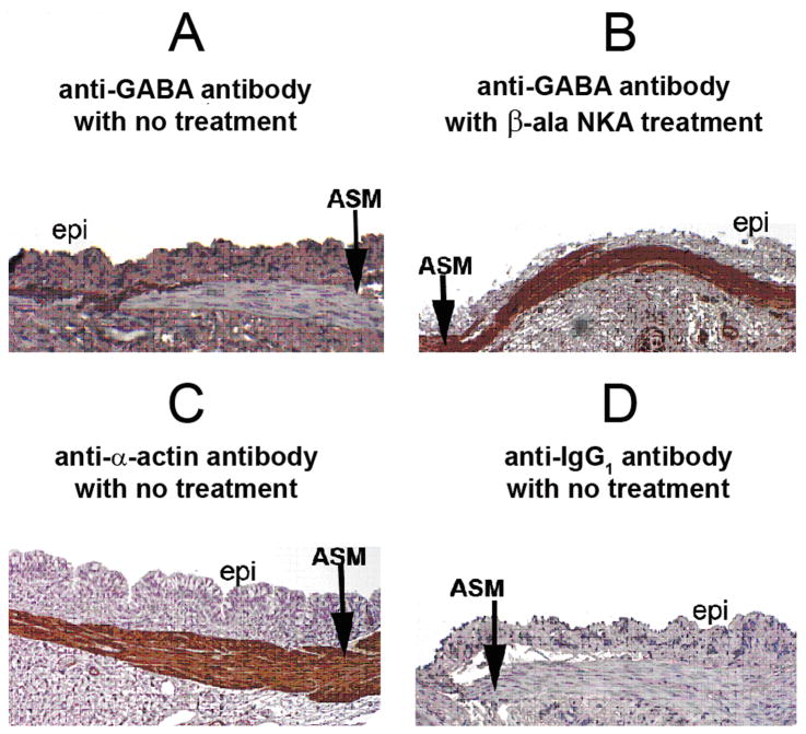
Figure 1. Immunohistochemical staining for γ-aminobutyric acid (GABA) in guinea pig tracheal rings.
Representative immunohistochemical GABA staining in guinea pig tracheal rings. (A) Untreated tracheal segment illustrates limited staining for GABA (brown) in airway smooth muscle (ASM), with the majority of staining localized to the interface between the ASM and adjacent epithelium (epi). (B) Following 15 min treatment with a neurokinin 2 agonist (10uM β-ala neurokinin A fragment 4–10) a dramatic increase in GABA staining occurs over airway smooth muscle. (C) Immunohistochemical staining for α–actin confirms the identity of the airway smooth muscle layer. (D) Antibody negative control: Isotype specific (IgG1) negative control antibody for the mouse anti-GABA antibody used in panels A and B reveals no staining in airway smooth muscle.

