

Abstract
Objective
To determine the major changes in the microstructure of Candida albicans (C. albicans) after treatment with Euphorbia hirta (E. hirta) L. leaf extract.
Methods
Transmission electron microscopy was used to study the ultrastructural changes caused by E. hirta extract on C. albicans cells at various exposure time.
Results
It was found that the main abnormalities were the alterations in morphology, lysis and complete collapse of the yeast cells after 36 h of exposure to the extract. Whereas the control cultures showed a typical morphology of Candida with a uniform central density, typically structured nucleus, and a cytoplasm with several elements of endomembrane system and enveloped by a regular, intact cell wall.
Conclusions
The significant antifungal activity shown by this methanol extract of E. hirta L. suggests its potential against infections caused by C. albicans. The extract may be developed as an anticandidal agent.
Keywords: Candida albicans, Euphorbia hirta L., Transmission electron microscopy, Anticandidal agent
1. Introduction
Candida albicans (C. albicans) is a dimorphic organism that commonly inhabits in oral and vaginal mucosa and gastrointestinal tract of human beings as one of the commensal organisms. It causes opportunistic infections in immunocompromised patients, and produces allergic reactions[1]. It also causes a variety of infections that range from non-life threatening mucosal candidiasis like vaginal yeast infections, thrush, skin and diaper rash to lethal disseminated candidiasis in those with compromised immune systems who have an implantable medical device such as a pacemaker or artificial joint, or who use broad-spectrum antibiotics[2]–[4]. C. albicans infections faces a number of problems including limited number of effective antifungal agents, toxicity of the available antifungal agents, resistance of Candida to commonly used antifungals, relapse of Candida infections and non-cost effective antifungal agents[5]. Therefore, new agents from natural resources that can inhibit the growth of C. albicans are greatly needed and would enhance the number of effective therapeutic alternatives.
Euphorbia hirta (E. hirta) L. belongs to the family Euphorbiaceae. It is a small annual herb common to tropical countries. It is usually erect, slender-stemmed, spreading up to 80 cm tall, though sometimes it can be seen lying down. The plant is an annual broad-leaved herb that has a hairy stem with many branches from the base to the top. The leaves are opposite, elliptical, oblong or oblong-lanceolate, with a faintly toothed margin and darker on the upper surface. The flowers are small, numerous and crowded together in dense cymes (dense clusters in upper axils) about 1 cm in diameter. The stem and leaves produce a white or milky juice when cut. It is frequently seen occupying open waste spaces, banks of watercourses, grasslands, road sides, and pathways[6]–[8].
E. hirta L. is a very popular herb amongst practitioners of traditional medicine, widely used as a decoction or infusion to treat various ailments including intestinal parasites, diarrhoea, peptic ulcers, heartburn, vomiting, amoebic dysentery, asthma, bronchitis, hay fever, laryngeal spasms, emphysema, coughs, colds, kidney stones, menstrual problems, sterility and venereal diseases. Moreover, the plant is also used to treat affections of the skin and mucous membranes, including warts, scabies, tinea, thrush, aphthae, fungal afflictions, measles, guinea worm and as an antiseptic to treat wounds, sores and conjunctivitis. The plant has a reputation as an analgesic to treat severe headache, toothache, rheumatism, colic and pains during pregnancy. It is used as an antidote and pain relief of scorpion stings and snakebites. The use of the latex to facilitate removal of thorns from the skin is common[6],[9].
Our previous studies showed that the methanol extract of E. hirta L. possessed a good in vitro antifungal activity against C. albicans with 21 mm zone of inhibition, a minimum inhibition concentration (MIC) value of 3.125 mg/mL, and the time kill study revealed a fungicidal effect with 1 and 2 MIC values of the plant extract[6]. On the other hand, leaf extract exhibited significant antioxidant activities when evaluated by DPPH, ferric reducing power assays and also found to possess noticeable content of phenolic and flavonoid compounds. The present study was carried out to further substantiate the anticandidal activity of the leaf extract at utrastructural level by using electron microscopy.
2. Materials and methods
2.1. Plant collection
The fresh plant was harvested from various areas in Universiti Sains Malaysia, in June 2009. The taxonomic identity of the plant was confirmed by the botanist of School of Biological Sciences at Universiti Sains Malaysia. The plant materials were washed under tap water and separated into leaves, flowers, stems and roots. The separated parts were air dried in shade for ten days and then in oven at 60 °C for one to two days, then grinded to fine powder by using electric blender and stored in clean labelled airtight bottles.
2.2. Preparation of the plant extract
A hundred grams of each part powder was extracted by maceration in 400 mL of methanol for 14 days with frequent agitation. The mixture was filtered through clean muslin cloth followed by double filtration with Whatman No. 1 filter paper and the filtrate was concentrated by rotary evaporator with vacuum at 50 °C, poured in glass Petri dishes and brought to dryness at 60 °C oven. The percentage yield of the crude extract was determined for each part and was 11.1%, 7.3%, 4.7% and 4.1% for leaves, stems, flowers and roots, respectively. The obtained paste like mass was then stored in parafilm sealed Petri-dishes in dark cabinet. The extracts were reconstituted by dissolving in methanol to the required concentrations. The reconstituted extracts were maintained at (2-8) °C.
2.3. Transmission electron microscope (TEM)
The preparation procedure of C. albicans cultures for TEM was the same as described previously in SEM section. The scratched samples were fixed in in McDowell-Trump fixative prepared in 0.1 M phosphate buffer for 24 h at 4 °C, washed twice in the same buffer, postfixed in 1% osmium tetroxide in 0.1 M phosphate buffer for 1 h at 4 °C, then double washed in distilled water. The samples were blocked in warm agar (50 °C), dehydrated in ethanol series (50%, 75%, 95% and double 100%) and embedded in Spurr's resin. Finally, glass knives were used to obtain ultra thin sections of 90 nm that were stained with uranyl acetate and lead citrate for observations under TEM (LIBRA 120- ZEISS)[10].
3. Results
Control (untreated) C. albicans cell as examined by TEM showed typical morphology of Candida with a uniform central density, typically structured nucleus, and a cytoplasm with several elements of endomembrane system and enveloped by a regular, intact cell wall (Figure 1). After 12 h of treatment with E. hirta L. leaf extract (Figure 2), the cell appeared very dense with the vesicles and membranous bodies dispositioned within the cell. The cytoplasmic volume started to decrease. After 24 h of exposure (Figure 3) the cells exhibited notable alterations in the cell membrane and the cell wall. The cytoplasmic volume decreased more with notable structural disorganization within the cell cytoplasm. Figure 4 showed the effect of E. hirta extract on the yeast cells after 36 h of exposure. A certain number of cells apparently underwent drastic shape changes and some lysed and collapsed. It seems that the plant extract induced dysfunctions of the cell membrane and the cell lost its metabolic functions at this stage.
Figure 1. TEM images of the untreated cells of C. albicans (after 24 h).
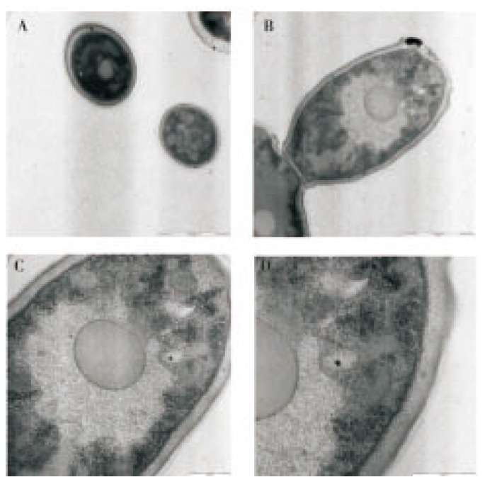
A: 4 000×; B: 5 000×; C: 10 000×; D: 16 000×.
Figure 2. TEM images of C. albicans cells after 12 h treatment with E. hirta L. leaf extract.
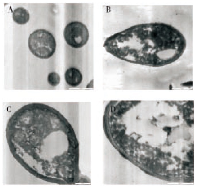
A: 4 000 ×; B: 5 000 ×; C: 10 000 ×; D: 16 000 ×.
Figure 3. TEM images of C. albicans cells after 24 h treatment with E. hirta L. leaf extract.
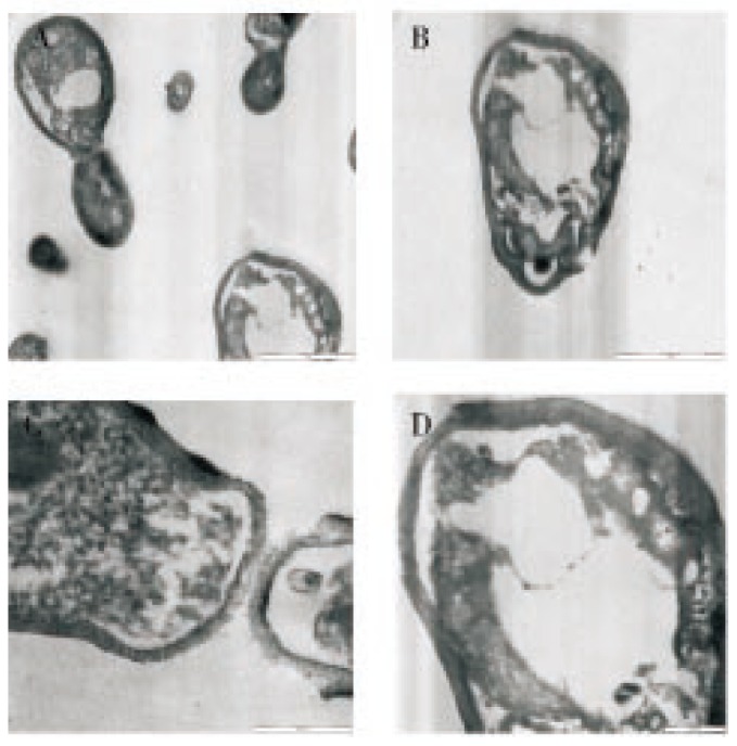
A: 4 000 ×; B: 5 000 ×; C: 10 000 ×; D: 16 000 ×.
Figure 4. TEM images of C. albicans cells after 36 h treatment with E. hirta L. leaf extract.
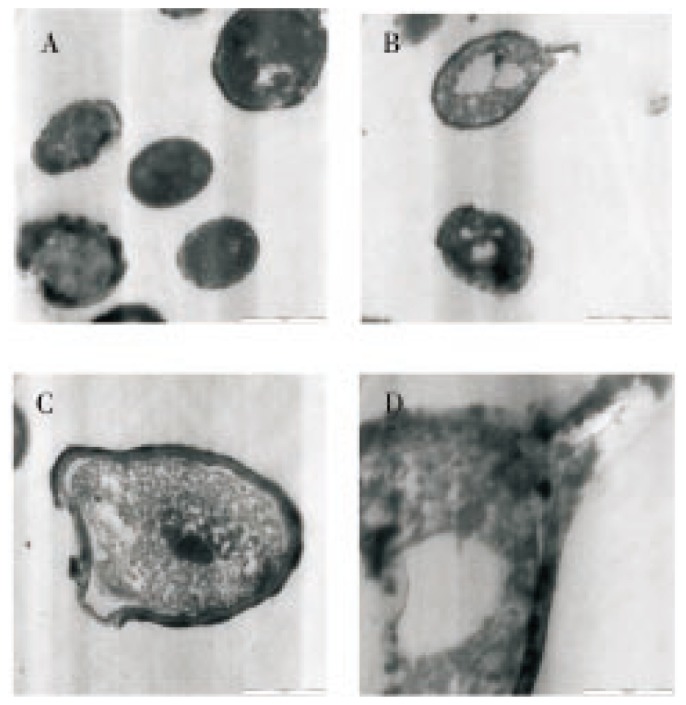
A: 4 000 ×; B: 5 000 ×; C: 10 000 ×; D: 16 000 ×.
4. Discussion
Longer exposure to the extract inhibits the total growth of C. albicans. Seed extract of E. hirta possesses anticandidal activity against C. albicans strain as shown in the electron microscopy observation and further investigations are necessary on mode of action of the extract on C. albicans cells prior to the recommendation for its use as a safe and effective antifungal therapy.
The E. hirta has never been evaluated for anti-Candida activity before, it is the first time to study anti-Candida activity. Therefore, further purification of active compound(s) from E. hirta extract and its individual antimicrobial activity are needed. The significant antimicrobial activities exerted by E. hirta suggest it as a potential source for developing new antimicrobial agents. Further studies of the bioactive compounds and the in vivo evaluation of the antimicrobial activity against C. albicans are therefore suggested.
Acknowledgments
Deep thanks to Islamic Development Bank for the financial support with a master degree scholarship and to the staff of Electron Microscopy Unit, School of Biological Sciences, USM for help and facilities.
Footnotes
Conflict of interest statement: We declare that we have no conflict of interest.
References
- 1.Naglik JR, Challacombe SJ, Hube B. Candida albicans secreted aspartyl proteinases in virulence and pathogenesis. Microbiol Mol Biol Rev. 2003;67(3):400–428. doi: 10.1128/MMBR.67.3.400-428.2003. [DOI] [PMC free article] [PubMed] [Google Scholar]
- 2.Bhavan PS, Rajkumar R, Radhakrishnan S, Seenivasan C, Kannan S. Culture and identification of Candida albicans from vaginal ulcer and separation of enolase on SDS-page. Int J Biol. 2010;2(1):84–93. [Google Scholar]
- 3.O'Dwyer DT, McElduff P, Peterson P, Perbeentupa J, Crack PA. Pituitary autoantibodies in autoimmune polyendocrinopathy-candidiasis-ectodermal dystrophy (APECED) Acta Biomed. 2007;78:248–254. [PubMed] [Google Scholar]
- 4.Terrier B, Degand N, Guilpain P, Servettaz A, Guillevin L, Mouthon L. Alpha-enolase: a target of antibodies in infectious and autoimmune diseases. Autoimmun Rev. 2007;6:176–182. doi: 10.1016/j.autrev.2006.10.004. [DOI] [PubMed] [Google Scholar]
- 5.Sasidharan S, Zuraini Z, Latha LY, Suryani S. Fungicidal effect and oral acute toxicity of Psophocarpus tetragonolobus root extract. Pharm Biol. 2008;46(4):261–265. [Google Scholar]
- 6.Rajeh MA, Zuraini Z, Sasidharan S, Latha LY, Amutha S. Assessment of Euphorbia hirta L. leaf, flower, stem and root extracts for their antibacterial and antifungal activity and brine shrimp lethality. Molecules. 2010;15:6008–6018. doi: 10.3390/molecules15096008. [DOI] [PMC free article] [PubMed] [Google Scholar]
- 7.Sandeep BP, Nilofar SN, Chandrakant SM. Review on phytochemistry and pharmacological aspects of Euphorbia hirta Linn. J Pharma Res Health Care. 2009;1:113–133. [Google Scholar]
- 8.Anonymous Euphorbiahirta L. [Online] Available from: http://florabase.calm.wa.gov.au/browse/profile/4629. (2008). [Access on 31 May 2010]
- 9.Anonymous Euphorbiahirta L. Euphorbia hirta. 2010. [Online] Available from: www.pfaf.org/database/plants.php? [Access on 1 April 2010]
- 10.Sangetha S, Zuraini Z, Suryani S, Sasidharan S. In situ TEM and SEM studies on the antimicrobial activity and prevention of Candida albicans biofilm by Cassia spectabilis extract. Micron. 2009;40:439–443. doi: 10.1016/j.micron.2009.01.003. [DOI] [PubMed] [Google Scholar]