Abstract
Many species have been drastically affected by rapid urbanization. Harris's hawks from their natural habitat of open spaces and a supply of rodents, lizards and other small prey have been forced to change their natural environment adapting to living in open spaces in sub- and peri-urban areas. Specific areas include playgrounds, parks and school courtyards. The migration of this predatory species into these areas poses a risk to individuals, and especially the children are often attacked by claws, talons and beaks intentionally or as collateral damage while attacking rodent prey. In addition, the diverse micro-organisms harbored in the beaks and talons can result in wound infections, presenting a challenge to clinical management. Here we would like to present a case of an 80-year-old man with cellulitis of both hands after sustaining minor injuries from the talons of a Harris's hawk and review the management options. We would also like to draw attention to the matter that, even though previously a rarity, more cases of injuries caused by birds of prey may be seen in hospital settings.
Keywords: Harris hawk, Predatory birds, Deforestation, Urbanization
1. Introduction
Rapid urbanization, increased agricultural and industrial practices leading to destruction of natural forests and habitats have imposed serious environmental pressures on wild life. Many species have been drastically affected and driven to either extinction or near extinction due to displacement from their original habitat. Harris's hawks, known not to migrate from their natural habitat of sparse woodlands, semi-desert and marshes[1], have in the past decade been forced to change their natural environment due to its destruction.
Owing to a preference for open spaces with small trees and a supply of rodents, lizards and other small prey, this predatory species has apparently adapted to living in open spaces in sub-urban and peri-urban areas. Specific areas include playgrounds, parks and school courtyards. These birds are equipped with strong talons and beaks that can cause soft tissue injuries in humans. The migration of this predatory species therefore poses a risk to individuals, and especially the children are often attacked by claws, talons and beaks intentionally or as collateral damage while attacking rodent prey.
Furthermore micro-organisms harbored in the beaks and talons of birds of prey are neither well identified nor reported and such a diverse contaminating flora can result in wound infections in humans which may be difficult to be treated appropriately. This presents a challenge to clinical management. Here we would like to present a case of an 80-year-old man with cellulitis of both hands after sustaining minor injuries from the talons of a Harris's hawk and emphasize that an increasing number of such cases may be seen in hospital settings.
2. Case report
An eighty year old male from a peri-urban area presented to our emergency department (ER) with complaints of swelling and erythema of both hands. He had sustained small puncture wounds and superficial scratches to the dorsum of both hands from the talons of a wild hawk two days ago. Considering his injuries to be trivial, he cleaned the wounds with an antiseptic solution and decided not to seek medical attention. The following day he noticed progressive swelling of both hands complicated with hot flushes and rigors. He presented to the ER 48 hours after the injury on the insistence of his wife.
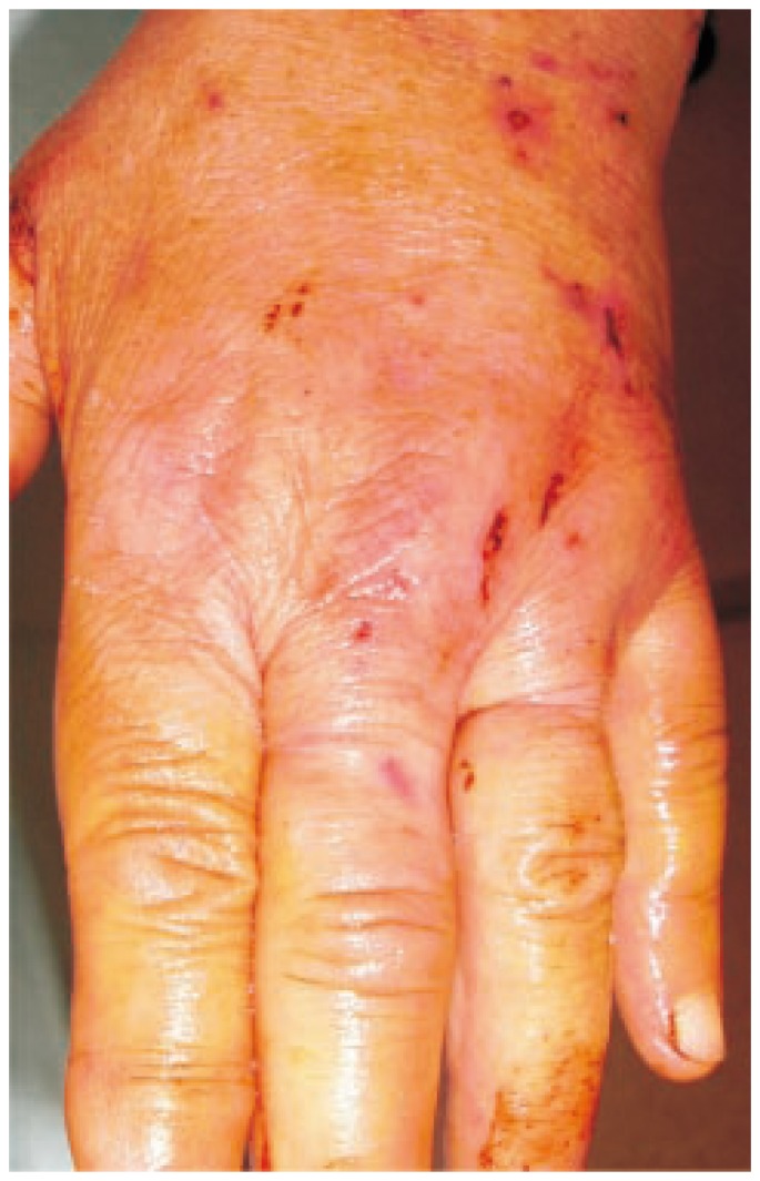
On examination he had tense bilateral swelling and erythema and was unable to make a fist. The puncture wounds and superficial scratches on the dorsum of both hands had since epithelialised and there was no active bleeding or extrusion of pus (Figure 1). Although all digits were swollen, cardinal signs of flexor tenosynovitis or compartment syndrome were absent. A general examination revealed a temperature of 38 °C and clinical signs of sepsis.
Figure 1. The image of the patient's hand shows tense bilateral swelling and erythema, the puncture wounds and scratches on the dorsum of the hand can be noted to have epithelialised and no activebleeding or extrusion of pus was seen.

Complete blood count showed marked leukocytosis with neutrophilia. Wound swabs of the closed puncture sites were taken and sent for culture. Ultrasound imaging of his hands was carried out and no collections were identified in the deep palmar spaces.
The patient was not willing to receive any form of surgical intervention at this stage. The on-call microbiologist was consulted and on his advice a regimen of high dose intravenous co-amoxiclav (2.4 mg TDS) was started in order to offer a broad spectrum cover against gram positive and gram negative anaerobic organisms harbored under the talons of birds of prey.
He was kept under observation and an alternative plan for surgical exploration was kept on the table, in case his situation deteriorated. Within 12 hours the patient improved significantly, his fever resolved and an appreciable difference was noted in his swelling hand.
He was discharged from the hospital 3 days later on oral co-amoxiclav (625 mg TDS) for 5 days. The wound cultures grew non-specific mixed skin organisms and the blood cultures remained negative at day five. On subsequent follow-up he had regained complete functionality of his hand with no permanent disability or deformity.
3. Discussion
There have been no reports of hand injuries inflicted by birds of prey as they are infrequently seen in a high risk cohort of bird handlers, hawk trainers and hunters. However this select group also presents a few cases each year and such injuries are almost never seen in the general urban and suburban population. As pointed out previously the destruction of natural habitats has forced diverse forms of wildlife to migrate to peri- and sub-urban areas, which causes a shift in the disease patterns and health risks of individuals residing in such areas.
Hawks in general, especially the Harris's hawk, seem to have adapted to this environment creating nests in open spaces such as parks, play grounds and school courtyards. Since these predatory birds are equipped with strong talons and beaks they may potentially cause soft tissue injuries in humans in these areas either directly to protect their young or inadvertently while attacking rodent prey.
The micro-organisms which harbored in the beaks and talons of birds of prey are not well defined and this diverse contaminated flora can result in wound infections that can be quite difficult to be treated appropriately. The clinical significance of this flora becomes apparent in patients like ours and the choice of antibiotic becomes an important concern.
In a survey of the choanal and cloacal aerobic bacterial flora in free-living and captive hawks, it has been found that coagulase-negative Staphylococcus/Micrococcus, Corynebacterium and Pasteurella species are the most frequent choanal isolates from both free-living and captive hawks, while the most frequent cloacal isolates of both free-living and captive hawks include coagulase-negative Staphylococcus/Micrococcus, coagulase-positive Staphylococcus, Streptococcus and Escherichia species. Corynebacterium species and Bacillus species have been isolated from the cloaca of free-living birds but not from captive birds. Salmonella species have been isolated from the cloaca of free-living birds of both species but not from captive birds[2].
Moreover, bacteriological examination of the feet especially plantar surfaces, metatarsal interstices and claws of free-living goshawks (Accipiter gentilis) revealed that they were colonized by organisms from many genera. The organisms isolated were mainly associated with the alimentary tract. However, the nestlings also yielded bacteria frequently found in other sites. Isolated species from both older goshawks and nestlings included Staphylococcus epidermis, Staphylococcus aureus, Citrobacter freundii, Pseudomonas aeruginosa, Bacillus, Escherichia coli, Enterobacter, Enterococcus, Klebsiella, Alkaligenes faecalis, Aeromonas and Alpha Hemolytic Streptococci[3].
No specific reason can be given for this, but warmth and the constant availability of prey remnants at nests may provide an environment for the continuity and diversity for bacterial growth, which causes a contamination of hawk feet.
In addition, these birds mostly feed on small animals like rodents by using their talons to grab and shred their victims before devouring them. Therefore, besides the environmental pathogens under the surface of their talons, they can harbor micro-organisms indigenous to rodents, specifically their anaerobic intestinal flora.
Such diverse contaminated flora presents a challenge when the infection involves the closed spaces of the hands which are prone to infection due to the numerous small compartments, which provide the perfect culture environment for anaerobic organisms.
The absence of significant soft tissues separating the skin from bone and joint spaces in hands means that the invaded bacteria can inoculate several spaces (joint, dorsal subcutaneous, dorsal subtendious) and medullary bone simultaneously. With movement of the digits, the tendons retract and carry bacteria to more proximal locations. Furthermore, the epithelialization of the puncture site creates an ideal environment for the inoculated bacteria to flourish[4],[5]. Thus, clinically, trivial puncture wounds can result in significant clinical morbidity.
The level of complication correlates with extent of injury and the time elapsed prior to seeking medical attention[6]. However, patients may initially disregard the injuries as trivial and decide not to seek medical attention as seen in our case. If left untreated, the infection can in severe cases lead to gangrene of the digits or necrotizing fasciitis[7],[8].
Such cases warrant the need for aggressive treatment to prevent further spread of infection. In open injuries, meticulous wound cleaning and irrigation with normal saline is considered the first step, followed by mandatory tetanus prophylaxis[9],[10]. The next step is to administer the appropriate antibiotic treatment. The goal of pharmacotherapy is to eradicate infection and to avoid complications and permanent disability. Broad spectrum antimicrobial therapy should be empirically administered as soon as possible and should cover likely pathogens[7].
Co-amoxilcav appears as a viable empiric therapy owing to its broad spectrum cover against commonly occurring bacterial pathogens. Regardless of the initially chosen antibiotic, subsequent treatment should be guided by wound (in cases of open wounds) or blood cultures. However, as in our case they may result in non-specific or no growth respectively where onwards empirical broad spectrum cover remains the only viable option.
However, surgical debridement with deep incisional tissue biopsy and cultures for anaerobic and aerobic organisms are the gold standard for diagnosis of necrotizing fasciitis[8]. This was not carried out in our case as the patient did not consent to an invasive procedure. Even in the absence of culture results empiric anti-biotic therapy may suffice in treating such injuries.
In light of the case the authors would like to draw attention that, although previously a rarity, more cases of injuries caused by birds of prey may be seen in hospital settings.
Footnotes
Conflict of interest statement: We declare that we have no conflict of interest.
References
- 1.Howell SNG. A Guide to the Birds of Mexico and Northern Central America. Hay-on-Wye: Oxford University Press; 1995. [Google Scholar]
- 2.Lamberski N, Hull A, Fish Allen M, Kimberlee B, Morishita Teresa Y. A survey of the choanal and cloacal aerobic bacterial flora in free-living and captive red-tailed hawks (Buteo jamaicensis) and cooper's hawks (Accipiter cooperii) J Avian Med Surg. 2003;17(3):131–135. [Google Scholar]
- 3.Needham JR, Cooper JE, Kenward RE. A survey of the bacterial flora of the feet of free-living goshawks (Accipiter gentilis) Avian Pathol. 1979;8(3):285–288. doi: 10.1080/03079457908418353. [DOI] [PubMed] [Google Scholar]
- 4.Basadre JO, Parry SW. Indications for surgical debridement in 125 human bites to the hand. Arch Surg. 1991;126(1):65–67. doi: 10.1001/archsurg.1991.01410250071011. [DOI] [PubMed] [Google Scholar]
- 5.Faciszewski T, Coleman DA. Human bite wounds. Hand Clin. 1989;5(4):561–569. [PubMed] [Google Scholar]
- 6.Broder J, Jerrard D, Olshaker J, Witting M. Low risk of infection in selected human bites treated without antibiotics. Am J Emerg Med. 2004;22(1):10–13. doi: 10.1016/j.ajem.2003.09.004. [DOI] [PubMed] [Google Scholar]
- 7.Gibbon KL, Bewley AP. Acquired streptococcal necrotizing fasciitis following excision of malignant melanoma. Br J Dermatol. 1999;141(4):717–719. doi: 10.1046/j.1365-2133.1999.03117.x. [DOI] [PubMed] [Google Scholar]
- 8.Verfaillie GS, Knape, Corne L. A case of fatal necrotizing fasciitis after intramuscular administration of diclofenac. Eur J Emerg Med. 2002;9(3):270–273. doi: 10.1097/00063110-200209000-00013. [DOI] [PubMed] [Google Scholar]
- 9.Goldstein EJ. Management of human and animal bite wounds. J Am Acad Dermatol. 1989;21(6):1275–1279. doi: 10.1016/s0190-9622(89)70343-1. [DOI] [PubMed] [Google Scholar]
- 10.Hagberg C, Radulescu A, Rex JH. Necrotizing fasciitis due to group A Streptococcus after an accidental needle-stick injury. N Engl J Med. 1997;337(23):1699. doi: 10.1056/NEJM199712043372318. [DOI] [PubMed] [Google Scholar]


