

Abstract
Objective
To develop an improved protocol for micropropagation of ethnomedicinally important Scoparia dulcis (S. dulcis) L.
Methods
Explants were inoculated on MS basal medium supplemented with kinetin and 6-benzylaminopurine for shoot bud induction. To enhance the shoot induction, various auxins like 3-indoleacetic acid or 3-indolebutyric acid or α-naphthylacetic acid were tested along with 2.32 M KI and 4.44 µM BAP. The regenerated shoots were rooted in half strength MS medium supplemented with various concentrations of IAA, IBA or NAA. After roots were developed, the plantlets were transplanted to pots filled with vermiculate and sand and kept in growth chamber with 70%–80% humidity under 16 h photoperiod. After acclimatization, the plantlets were transferred to the garden and survival percentage was calculated. Data were statistically analyzed and means were compared using Duncan's multiple range test (P<0.05).
Results
An in vitro method was developed to induce high frequency shoots regeneration from stem, mature leaf and young leaf explants of S. dulcis. Shoot induction on young leaf explants was most successful in MS medium supplemented with combination of two cytokinins (2.32 µM KI and 4.44 µM BAP) 2.85 µM IAA, 10% CM and 1 483.79 µM adenine sulfate. A single young leaf explant was capable of producing 59 shoots after 13 days of culture. Flower was induced in medium supplemented with combination of KI and BAP.
Conclusions
Cytokinins are the key factor to induce the direct shoot regeneration and flowering of S. dulcis.
Keywords: Ethnomedicinal herb, Scoparia dulcis, Shoot induction, Regeneration, Cytokinin, Micropropagation, Explant, Flowering, Auxin, Survival percentage, Bud induction
1. Introduction
Plants are tremendous source for discovering new products with medicinal value for drug development. Scoparia dulcis (S. dulcis) L. is a perennial herb, belongs to the family Scrophulariaceae, and is widely distributed in tropical and subtropical regions. As an ethnomedicinal folklore plant (sweet broom weed), its remedial nature particularly for diabetes mellitus is well documented[1].
S. dulcis is a medicinal plant of growing global interest. A number of the speculated medicinal properties of S. dulcis have been validated by scientific research. These include antiviral activity[2], antitumor promoting activity[3], hypoglycemic activity[4] and antioxidant activity[5]. Traditionally the plant is used to treat kidney stone and urinary troubles[6]–[12]. Though micropropagation has been performed in S. dulcis[13],[14] further studies on factors that may enhance the process are needed. The objective of the present study was to develop an improved protocol for micropropagation of S. dulcis.
2. Materials and methods
Young leaves (0.5-1) cm, mature leaves (1.5-2) cm and young stem (2-3) cm were collected from 3 month old disease-free healthy S. dulcis plants and used as explants. The explants were washed in running tap water for 30 min and then thoroughly rinsed in detergent solution (Tween 20) for 5 min. They were then surface disinfected with HgCl2 (0.1%) for 1 min and thoroughly washed with sterile distilled water. Explants were inoculated on MS[15] basal medium supplemented with kinetin (KI: 2.32, 4.64 and 9.29 µM) and 6-benzylaminopurine (BAP: 4.44, 8.88 and 17.76 µM) for shoot bud induction.
To enhance the shoot induction, various auxins like 3-indoleacetic acid (IAA: 2.85, 5.71, 11.42 and 17.12 µM) or 3-indolebutyric acid (IBA:2.46, 4.92, 9.84 and 14.76 µM) or α-naphthylacetic acid (NAA: 2.68, 5.37, 8.05 and 10.74 µM) were tested along with 2.32 µM KI and 4.44 µM BAP. The effect of coconut milk (CM) and adenine sulphate was also tested for shoot induction.
All media contained 0.8% (w/v) agar. The pH of the medium was adjusted to 5.8 using 0.1 N NaOH or 0.1 N HCl before autoclaving at 121 °C for 15 min. The cultures were incubated at (25±2) °C and 70%-80% relative humidity under a 16-h photoperiod.
The regenerated shoots (2 to 3 cm) were rooted on half strength MS medium supplemented with various concentrations of IAA (2.85, 5.71, 11.42, 17.12 µM), IBA (2.46, 4.92, 9.84, 14.76 µM) or NAA (2.68, 5.37, 8.05, 10.74 µM). After roots were developed, the plantlets were transplanted to pots filled with vermiculate and sand (1:1 ratio) and kept in growth chamber with 70%-80% humidity under 16 h photoperiod. After acclimatization, the plantlets were transferred to the garden and survival percentage was calculated.
Single explant was used per flask. Each experiment was done at least twice using triplicate. Data were statistically analyzed and means were compared using Duncan's multiple range test (P<0.05).
3. Result
In this study, growth regulators in different types or at different concentration and different explants (young leaf, mature leaf and shoot tips) were used to determine optimal culture conditions to regenerate shoots of S. dulcis in vitro. Table 1 showed the growth response of the explants cultured on MS medium supplemented with KI in addition to BAP. Young leaf was found to be an extra prolific over mature leaf and stem. Direct shoots were produced without an intervening callus phase from young leaf explants on MS medium fortified with 2.32 µM KI along with 8.88 µM BAP (Figure 1A). The medium containing 2.32 µM KI along with 4.44 µM BAP was efficient in terms of direct shoot formation after 16 days of culture using young leaf as explants and average of 19.61 shoots were developed. Shoot buds emerged directly from the surface and from the cut end of the explants (Figure 1B).
Table 1. Effect of cytokinins on shoot induction in S. dulcis L.
| Conc. of growth regulators (µM) |
Young leaf |
Mature leaf |
Stem |
|||||||
| KI | BAP | Days req. | Shoot induction (%) | Average number of shoot per explant | Days req. | Shoot induction (%) | Average number of shoot per explant | Days req. | Shoot induction (%) | Average number of shoot per explant |
| 2.32 | 4.44 | 16 | 90.09 | 19.61±2.15 | 25 | 65.08 | 9.81±0.15 | 40 | 45.09 | 4.81±2.15 |
| 8.88* | 19 | 72.56 | 12.45±0.29 | 27 | 63.12 | 7.12±.2.71 | 42 | 35.98 | 3.18±3.12 | |
| 17.76 | 22 | 71.90 | 8.49±1.35 | 30 | 63.56 | 6.15±2.51 | 44 | 35.67 | 2.10±1.21 | |
| 4.64 | 4.44 | 22 | 79.07 | 13.44±0.28 | 25 | 60.12 | 8.89±0.54 | 43 | 42.65 | 3.01±1.32 |
| 8.88 | 25 | 65.32 | 11.45±1.01 | 29 | 59.87 | 5.98±3.45 | 44 | 41.56 | 1.01±0.12 | |
| 17.76 | 27 | 60.12 | 8.28±0.79 | 34 | 56.89 | 3.76±1.37 | 47 | 40.78 | 1.00±0.15 | |
| 9.29 | 4.44 | 30 | 77.13 | 11.97±0.78 | 28 | 59.89 | 5.32±1.46 | 46 | 38.12 | 3.90±1.28 |
| 8.88 | 33 | 55.65 | 9.89±0.91 | 31 | 57.09 | 2.43±0.21 | 49 | 35.34 | 2.01±0.22 | |
| 17.76 | 35 | 51.14 | 5.47±0.56 | 35 | 55.89 | 1.21±0.12 | 50 | 35.12 | 2.08±0.28 | |
* shoot produced without an intervening callus phase.
Figure 1. Growth of S. dulcis.
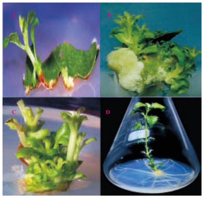
A: Direct shoot initiation from leaf explants of S. dulcis on MS medium with 2.32µM KN and 8.88 µM BAP at 19 days (shoot produced without an intervening callus phase; B: Direct shoot regenerated from leaf explants on medium with 2.32 µM KN and 4.44 µM BAP at 16 days; C: Multiple shoots formation on medium with 2.32µM KI with 4.44 µM BAP and 2.85 µM IAA at 13 days; D: Root regenerated plantlet of S. dulcis on MS medium supplemented with 2.68 µM NAA.
To enhance the shoot formation, young leaves were used as explants with different auxins (IBA, IAA and NAA) combined with 2.32 µM KN + 4.44 µM BAP on culture medium. All auxin showed positive effect in shoot enhancing. Young leaf showed 93% shoot induction on MS medium supplemented with 2.85 µM IAA along with combination of 2.32 µM KN + 4.44 µM BAP after 13 days (Table 2 and Figure 1C).
Table 2. Effect of auxin on shoot induction in S. dulcis L.
| Auxin types | Conc. (µM) | Days required for shoot bud regeneration | Average shoot induction (%) | Average number of shoot per explant |
| IBA | 2.46 | 15 | 80.91 | 40.01±3.18 |
| 4.92 | 16 | 75.32 | 38.34±2.14 | |
| 9.84 | 16 | 70.89 | 34.90±5.19 | |
| 14.76 | 19 | 64.09 | 31.02±2.05 | |
| IAA | 2.85 | 13 | 93.96 | 46.79±2.09 |
| 5.71 | 15 | 90.98 | 42.08±4.12 | |
| 11.42 | 16 | 88.89 | 37.96±3.19 | |
| 17.12 | 16 | 87.78 | 35.08±1.18 | |
| NAA | 2.68 | 16 | 75.67 | 38.90±3.28 |
| 5.37 | 18 | 71.12 | 34.09±1.21 | |
| 8.05 | 18 | 66.12 | 31.09±1.11 | |
| 10.74 | 20 | 64.01 | 25.01±2.14 |
Among coconut milk (CM) or adenine sulfate or in combination of both at different concentration, 10% of CM with 1 481.50 µM adenine sulfate further enhanced the proliferation of shoot buds. About 59 shoots were produced from a single explant (Table 3).
Table 3. Influence of coconut milk and adenine sulfate on shoot induction.
| Coconut milk (% v/v) | Adenine sulfate (µM) | Average shoot induction (%) | Average number of shoot per explant |
| 10 | - | 94.08 | 48.90±2.05 |
| - | 370.94 | 92.21 | 47.09±1.28 |
| 10 | 370.94 | 95.60 | 50..90±5.70 |
| 10 | 741.89 | 97.79 | 51.70±2.50 |
| 10 | 1 112.84 | 100.00 | 55.09±3.50 |
| 10 | 1 483.79 | 100.00 | 59.80±1.70 |
Regenerated shoots were used as explants for flowering induction. Flowering occurred when medium was supplemented with combination of cytokinins alone (BAP and KI) or with combination of auxins, CM and adenine sulfate (Data are not shown here). Flowering was not uniform in each trial. Flowering failed when medium was fortified with any one of the cytokinins at different concentration (KI or BAP) along and auxin alone. In the present study on flowering, the expalnt did not sprout and the base of the explants produced the callus and flowering occurred after 7 days (Figure 2).
Figure 2. In viro flowering of S. dulcis.

Regenerated shoots (2-3 cm long) were transferred to half strength MS medium supplemented with auxin at different concentration for root induction. Hundred percent root induction was achieved (Table 4) when shoots were transferred to half strength MS medium supplemented with 2.68 µM NAA (Figure 1D). The percentage of root induction was less in IAA and IBA (data not shown). The rooted shoots did not require any special treatment in the growth chamber for transplantation. More than 90% of the plants survived in the field.
Table 4. Effect of NAA on rooting response.
| NAA concentration (µM) | Average root (%) | Average No. of root |
| 2.68 | 100.00 | 19.80±1.50 |
| 5.37 | 95.95 | 13.60±3.59 |
| 8.05 | 90.89 | 9.40±0.60 |
4. Discussion
Research in the development and application of in vitro techniques for the large scale multiplication and conservation of the plant germplasm has significantly increased because of its potential for further exploitation of the plant kingdom as a chemical resource and acquired high amount of plant with desired quality in a short period of time[16]. Tissue culture technology offers an alternative method for the conservation of germplasm as well as micropropagation of medicinally important plant resources[17],[18]. Various workers reported the role of cytokinins in shoot bud formation[19]–[21]. In this study, the combination of two cytokinins (BAP and KI) for induction of shoot from explants is reported. KN along with BAP at high concentration had a negative effect on the shoot bud regeneration. Palai et al[22] also reported that the combination of two cytokinins along with auxins increased the rate of shoot multiplication. High concentrations of auxins along with two cytokinins (BAP and KI) inhibit the induction of shoots. The effect of coconut milk and adenine sulfate in shoot induction was reported by Wei et al[23] in Plumbago zeylanica and Sivanesan and Jeong[19] in Sida cordifolia.
Scorz[24] observed that auxin was a major floral inhibitor. Lin et al[25] demonstrated that NAA was a negative regulator of in vitro flowering during Bambusa edulis shoot proliferation. In the present study, combination cytokinins (BAP+KI) alone and along with auxin promote the flowering.
Although in vitro propagation of S. dulcis with leaf explants[14] and with nodal segment[13] were reported, in this report, the total time schedule was compressed as well as produced 59 shoots from a single explants. About three fold increased in direct shoot formation comparing with the previous report.
In conclusion, cytokinins are the key components to promote direct shoot and flowering of S. dulcis. This is an efficient and reproducible protocol for plantlet regeneration from this ethnomedicinally important plant. It provides a hopeful method for in vitro propagation in large scale and conservation of this possible medicinal herb for the requirement of our future generation.
Footnotes
Foundation Project: Supported by a grant from UGC-New Delhi (No. MRP 3011/09).
Conflict of interest statement: We declare that we have no conflict of interest.
References
- 1.Satyanaranan K. Examination of Scoparia dulcis Linn. Indian Chem Soc. 1969;46:765–766. [Google Scholar]
- 2.Hayashi T. Antiviral agents of plants origin 111. Scopaduli: a noval tetracyclic diterpene from Scoparia dulcis L. Chem Pharm Bull. 1990;38(4):945–946. doi: 10.1248/cpb.38.945. [DOI] [PubMed] [Google Scholar]
- 3.Nishing H. Antitumor promoting activity of scopadulic acid B isolated from the medicinal plant Scoparia dulcis L. Oncology. 1993;50(2):100–103. doi: 10.1159/000227156. [DOI] [PubMed] [Google Scholar]
- 4.Venkateswaran PV. Hyperglycemic activity of Scoparia dulcis L. extract in alloxan-induced hyperglycemic rats. Phytochem Res. 2002;7:662–664. doi: 10.1002/ptr.1036. [DOI] [PubMed] [Google Scholar]
- 5.Pari L, Latha M. Effect of Scoparia dulcis (sweet broom weed) plant extract on plasma antioxidant in STZ-induced experiment diabetes in male albino rat. Die Pharmazie. 2004;59:557–560. [PubMed] [Google Scholar]
- 6.Anpin Raja RD, Prakash JW, Jeeva S. Medicinal plants used by Kanis in Aarukani hills of southern Western Ghats. In: Trivedi PC, editor. Indigenous medicinal plants. Jaipur: Pointer Publishers; 2009. pp. 1–48. [Google Scholar]
- 7.Jeeva S, Kingston C, Kiruba S, Kannan D. Sacred forests-treasure trove of medicinal plants: a case study from South Travancore. In: Trivedi PC, editor. Indian folk medicine. Jaipur: Pointer Publishers; 2007. pp. 263–724. [Google Scholar]
- 8.Jeeva S, Kiruba S, Mishra BP, Kingston C, Venugopal N, Laloo RC. Importance of weeds as traditional medicine in Kanyakumari district, southern Western Ghats. J Swamy Bot Club. 2005;22(3&4):71–76. [Google Scholar]
- 9.Jeeva S, Kiruba S, Mishra BP, Venugopal N, Das SSM, Sukumaran S, et al. Weeds of Kanyakumari district and their value in rural life. Indian J Tradit Knowl. 2006;5(4):501–509. [Google Scholar]
- 10.Jeeva S, Kiruba S, Mishra BP, Venugopal N, Regini GS, Das SSM, et al. Diversity of medicinally important plant species under coconut plantation in the coastal region of Cape Comorin. Flora Fauna. 2005;11(2):226–230. [Google Scholar]
- 11.Laila BNR, Sreeja S, Pinky VR, Prakash JW, Jeenath JA. Medicinal plants used by the rural people of Kattathurai, Kanyakumari district, Tamilnadu. J Basic Appl Biol. 2007;1(1):18–22. [Google Scholar]
- 12.Prakash JW, Leena SL, Vidhya DMS, Berin PA, Asbin AN, Veni P, et al. The medicinal plant diversity of Scott Christian College (Autonomous) Campus, Nagercoil, South Tamil Nadu, India. J Nat Conservation. 2006;18(1):81–89. [Google Scholar]
- 13.Hassan S, Afroz F, Bari LS, Munshi JL, Jahan MAA, Khatun R. Callus induction and high frequency regeneration of plantlets of Scoparia dulcis L., a perennial medicinal herb, through auxiliary shoot proliferation. Plant Tissue Cult Biotech. 2008;18(1):75–83. [Google Scholar]
- 14.Ailei M, Kokkirala VR, Kota SR, Umate P, Abbagani S. Efficient in-vitro regeneration from mature leaf explants of Scoparia dulcis L. an ethnomedicinal plant. J Herbs Spices Med Plants. 2008;14(3-4):200–207. [Google Scholar]
- 15.Murashige T, Skoog F. A revised for rapid growth and bioassays with tobacco cultures. Physiol Plant. 1962;15:473–477. [Google Scholar]
- 16.Johnson M, Usha RNA, Babu A. Rapid multiplication of Plumbago auriculata L.-a medicinally important plant. J Basic Appl Biol. 2010;4(1&2):220–226. [Google Scholar]
- 17.John PAL, Ghanthikumar S, Ajitha S. Micropropagation of Mukia maderaspatana (L.) M.J. Roem-a medicinally important plant. J Basic Appl Biol. 2010;4(1&2):5–9. [Google Scholar]
- 18.Indra T, Pandiarajan G, Govindaraj R, Sudarmani DNP. Production of callus from Tinospora cordifolia Miers ex Hook using various plant growth hormones. J Basic Appl Biol. 2010;4(1&2):168–171. [Google Scholar]
- 19.Sivanesan I, Jeong BR. Direct shoot regeneration from nodal explants of Sida cordifolia Linn. In Vitro Cell Dev Biol Plant. 2007;43:436–441. [Google Scholar]
- 20.Sadheeshna KS, Huxley AJ, Sasikala In vitro propagation of medicinally important plant Mimosa invisa. J Basic Appl Biol. 2009;3(3&4):27–32. [Google Scholar]
- 21.Sadheeshna KS, Maybel SN, Huxley AJ. In vitro propagation of Bacopa monnieri (L.)-a wetland medicinal plant. J Basic Appl Biol. 2010;4(3):138–142. [Google Scholar]
- 22.Palai SK, Rout GR, Das P. Micropropagation of giger (Zingiber officinale Rosc.)-interaction of growth regulators and culture conditions. In: Ramana S, Sasikumar KV, Nirmal Babu B, Eapen K, editors. Biotechnology of spices, medicinal and aromatic plants of India. Kerala: Indian Society for Spices; 1997. pp. 20–24. [Google Scholar]
- 23.Wei X, Gou X, Yuan T, Fussell SD. A highly efficient in vitro plant regeneration system and Agrobacterium-mediated transformation in Plumbago zeylanica. Plant Cell Rep. 2006;25:513–521. doi: 10.1007/s00299-006-0114-9. [DOI] [PubMed] [Google Scholar]
- 24.Scorza R. In vitro flowering. Hortic Rev. 1982;4:106–127. [Google Scholar]
- 25.Lin CS, Lin CC, Chang WC. In vitro flowering of Bambusa edulis and subsequent plantlet survival. Plant Cell Tissue Organ Cult. 2003;72:71–78. [Google Scholar]