Abstract
Objectives
To evaluate the radiation dose of the main body CT examinations performed routinely in four regional diagnostic centres, the specific contribution of radiologists and technologists in determining CT dose levels, and the role of radiological staff training in reducing radiation doses.
Methods
We retrospectively evaluated the radiation dose in terms of dose-length product (DLP) values of 2,016 adult CT examinations (chest, abdomen-pelvis, and whole body) collected in four different centres in our region. DLP values for contrast-unenhanced and contrast-enhanced CT examinations performed at each centre were compared for each anatomical area. DLP values for CT examinations performed before and after radiological staff training were also compared.
Results
DLP values for the same CT examinations varied among centres depending on radiologists’ preferences, variable training of technologists, and diversified CT image acquisition protocols. A specific training programme designed for the radiological staff led to a significant overall reduction of DLP values, along with a significant reduction of DLP variability.
Conclusions
Training of both radiologists and technologists plays a key role in optimising CT acquisition procedures and lowering the radiation dose delivered to patients.
Main messages
The effective dose for similar CT examinations varies significantly among radiological centres.
Staff training can significantly reduce and harmonise the radiation dose.
Training of radiologists and technologists is key to optimise CT acquisition protocols.
Keywords: Multidetector computed tomography, Radiation dose, Radiation protection, Staff training
Introduction
The use of computed tomography (CT) has dramatically increased over the last decades, with the number of examinations continuing to grow year by year. Between 2000 and 2007, an estimated 3.1 billion medical procedures involving ionising radiation were performed worldwide, and CT alone contributes almost one half of the total radiation exposure from medical use [1, 2]. This steady increase of the number of CT examinations is undeniably having a beneficial impact on healthcare. However, concerns have been raised about potential cancer induction caused by the increased use of CT and the high radiation dose to patients associated with some multidetector CT (MDCT) protocols [3, 4].
Several epidemiological studies of occupational and atomic bomb survivors have shown that even relatively low doses of ionising radiation can cause cancer, particularly leukaemia and myeloma [5, 6]. The BEIR VII report, published by the National Academies’ Committee in 2006, predicts that there is a 1 % increase in the risk of developing a solid cancer after radiation exposure of 100 mSv, equivalent to approximately 5,000 anteroposterior chest radiographs or 12 abdomen CT examinations. This statistical evaluation is based on a linear no threshold model (currently forming the basis of our radiation protection system), in which the risk is related to the radiation dose and the age of the patient at the time of exposure [7, 8].
Given the increase in the usage of CT and the risks linked to this procedure, it is important to gain full insight into the factors affecting the radiation dose. Until recently, these efforts have been largely concentrated on maximising image quality, while less attention has been paid to the radiation dose. Doses can be expected to differ on the basis of protocol design and the type of CT scanner used, but only few studies have tried to estimate the real dose level of CT examinations performed daily in radiological centres [8–11].
The aim of our study was to record MDCT dose levels associated with some typical diagnostic CT procedures performed in routine clinical practice at four radiological centres in our region by different radiologists and technologists and to evaluate the role of staff training events specifically designed for radiologists and technologists to achieve optimisation of CT protocols.
Materials and methods
Selection of CT examinations and measurement of radiation dose
We collected data concerning the radiation dose absorbed by individual patients undergoing body CT examinations in four different radiological centres in our region between 1 January and 31 December 2010. All the centres involved in the study were general radiology departments of the National Health System with an overall annual rate of around 6,000 CT examinations in 2010. CT examinations were carried out using two 64-row MDCT systems from different manufacturers (centre 1, LightSpeed VCT, General Electric, Milwaukee, WI; centre 2, Somatom Sensation 64, Siemens Healthcare, Forchheim, Germany), one 40-row MDCT system (centre 3, Somatom Sensation 40, Siemens Healthcare), and one 16-row MDCT system (centre 4, Aquilion 16, Toshiba Medical Systems, Tochigi, Japan). All CT systems were equipped with automated tube current modulation algorithms depending on patient’s size, as provided by each manufacturer (smart mA® for centre 1, CareDose® for centres 2 and 3, and SureExposure® for centre 4).
A total of 2,016 CT examinations of male and female outpatients aged 18 years and older were analysed. We elected to differentiate between contrast-unenhanced and contrast-enhanced CT examinations in an attempt to find any difference in dose mainly due to the radiologist’s choice of imaging protocol (e.g. implying the acquisition of one or more contrast-enhanced series for a given diagnostic query) rather than to the selection of acquisition parameters for a single contrast-unenhanced series (which is usually accomplished by technologists). Overall, 862 contrast-unenhanced and 1,154 contrast-enhanced chest, abdomen-pelvis, and whole-body CT studies were evaluated. The distribution of each kind of CT examination is described in Table 1.
Table 1.
Distribution of median and interquartile range of DLP values for each kind of CT examination as observed in the four centres. DLP values are expressed in milliGray∙cm (mGy∙cm)
| Centre 1 | Centre 2 | Centre 3 | Centre 4 | p value | |||||||||
|---|---|---|---|---|---|---|---|---|---|---|---|---|---|
| Median | IQR | No | Median | IQR | No | Median | IQR | No | Median | IQR | No | ||
| Chest (C−) | 667 | 433÷907 | 103 | 580 | 500÷667 | 113 | 527 | 360÷713 | 97 | 680 | 513÷1053 | 127 | <0.001 |
| Chest (C+) | 1647 | 973÷2487 | 37 | 1733 | 1120÷2133 | 35 | 887 | 707÷1133 | 48 | 1253 | 820÷1767 | 35 | <0.001 |
| Abdomen-pelvis (C−) | 1000 | 780÷1133 | 30 | 660 | 647÷733 | 30 | 807 | 573÷993 | 30 | 967 | 633÷1247 | 191 | <0.001 |
| Abdomen-pelvis (C+) | 2940 | 1840÷4727 | 72 | 2453 | 2180÷2913 | 81 | 1960 | 1513÷2700 | 84 | 3227 | 1960÷4547 | 79 | <0.001 |
| Whole body (C−) | 1147 | 807÷1447 | 35 | 893 | 820÷940 | 35 | 747 | 587÷940 | 35 | 1167 | 733÷1600 | 36 | <0.001 |
| Whole body (C+) | 3307 | 2407÷4847 | 49 | 2887 | 2747÷3433 | 54 | 2087 | 1593÷2507 | 71 | 2180 | 1373÷3400 | 509 | <0.001 |
C− indicates contrast unenhanced, C+ contrast enhanced
For each kind of CT examination, at least 30 CT studies were assessed by retrieving data from the Picture Archiving and Communication System (PACS). Radiation dose figures for each patient were collected in terms of dose-length product (DLP) values, which represent an approximation of the total energy absorbed by the body during the entire CT examination. All DLP values were obtained manually from the dose report automatically generated on the CT console at the end of every CT examination.
Evaluation of staff radiation dose training
After collection of DLP values, an intensive training course was organised at centre 4 in an attempt to verify and improve the professional performance of the radiological staff operating in the CT suite. Radiological staff training simultaneously involved radiologists, medical physicists, and technologists and consisted of two consecutive full-day training events in which medical, biological, and technical topics related to CT imaging (i.e. appropriateness criteria, protocol optimisation, assessment of cancer risk due to ionising radiation exposure, risk communication to patients and referring physicians, ethical and legal issues, and continuing education) were tackled from both a theoretical (i.e. through lectures on the principles of CT imaging and updates from the current literature) and practical standpoint (i.e. through hands-on training at the CT console). The training course was held by a group of senior radiologists and technologists with scientific and clinical experience in the fields of radiation protection and CT technology and medical applications. The level of learning was assessed through a test performed at the end of each course. The impact of training on the delivered CT radiation dose was evaluated 6 months thereafter at centre 4 on a total of 406 CT examinations, the distribution of which is reported in Table 2.
Table 2.
Median and interquartile range of DLP values in unenhanced and enhanced CT examinations before and after radiological staff training (involving both technologists and radiologists). DLP values are expressed in mGy∙cm
| Before training (centre 4) | After training (centre 4) | p value | |||||
|---|---|---|---|---|---|---|---|
| Median | IQR | No | Median | IQR | No | ||
| Chest (C−) | 680 | 513÷1053 | 127 | 413 | 323÷577 | 100 | <0.001 |
| Chest (C+) | 1253 | 820÷1767 | 35 | 560 | 380÷760 | 50 | <0.001 |
| Abdomen-pelvis (C−) | 967 | 633÷1247 | 191 | 607 | 460÷793 | 50 | <0.001 |
| Abdomen-pelvis (C+) | 3227 | 1960÷4547 | 79 | 1760 | 1140÷2580 | 50 | <0.001 |
| Whole body (C−) | 1167 | 733÷1600 | 36 | 800 | 547÷1113 | 50 | <0.001 |
| Whole body (C+) | 2180 | 1373÷3400 | 509 | 1479 | 1053÷2180 | 106 | <0.001 |
Statistical analysis
DLP values were expressed as median and interquartile ranges (IQR). Analysis of variance (ANOVA) was performed to compare DLP values for contrast-unenhanced and contrast-enhanced CT examinations of each anatomical area, carried out at each radiological centre. Moreover, in order to assess dose variability among different technologists and radiologists for the same kind of CT examination, DLP values for each kind of CT examination performed at centre 4 were compared among four different technologists with the same radiologist kept constant and among four different radiologists with the same technologist kept constant on a subset of at least 30 CT examinations per each (N = 30÷52, mean 44). The radiologist and technologist kept constant were chosen as the ones with the longest experience in terms of years of radiological practice (both more than 10 years) to ensure that his/her performance could be reasonably assumed to be optimal. Dose distributions were skewed in order to model the log transformation so as to better satisfy the ANOVA assumption of normally distributed outcomes.
Independent t-tests were performed to compare DLP values before and after radiological staff training. In addition, for contrast-enhanced CT examinations, the correlation between the number of acquisitions and DLP was calculated with the Pearson correlation coefficient. A P-value less than 0.05 was considered statistically significant.
Statistical analysis was performed by using software (GraphPad Prism v. 5.0, www.graphpad.com).
Results
Table 1 shows observed doses within each anatomical area (chest, abdomen-pelvis, and whole body) for CT examinations performed in the four different centres. For unenhanced CT, observed doses varied significantly among all centres (P < 0.001), ranging from a median of 527 mGy∙cm to 680 mGy∙cm for chest CT, from 660 mGy∙cm to 1,000 mGy∙cm for abdomen/pelvis CT, and from 747 mGy∙cm to 1,167 mGy∙cm for whole-body CT.
For contrast-enhanced CT examinations, DLP values and differences among centres were markedly increased in all anatomical areas (P < 0.001), rising from 887 mGy∙cm to 1,733 mGy∙cm for chest CT, from 1,960 mGy∙cm to 3,227 mGy∙cm for abdomen/pelvis CT, and from 2,087 mGy∙cm to 3,307 mGy∙cm for whole-body CT.
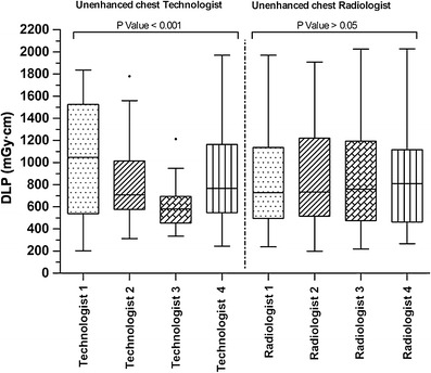
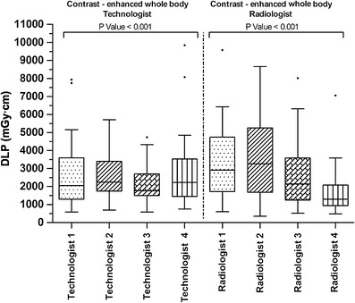
Figures 1 and 2 give the dose values for unenhanced chest CT and contrast-enhanced whole-body CT examinations, observed in the same centre (centre 4) and with the same technical equipment, by dividing the dose contribution for each technologist (with the same radiologist kept constant) and for each radiologist (with the same technologist kept constant). In unenhanced CT, radiologists proved to have no influence on delivered radiation dose, as there were no significant differences in median and IQR (P > 0.05). Conversely, technologists had a great impact on radiation dose, both in terms of median and IQR, with DLP values varying from 580 mGy∙cm to 1,047 mGy∙cm, associated with an approximately ten-fold dose variation for the same kind of CT examination (lowest and highest observed values: 202÷1,970 mGy∙cm, P < 0.001). For all radiologists involved at centre 4, DLP showed a significant linear correlation with the number of acquisitions per CT examination (r = 0.5532, P < 0.0001).
Fig. 1.

DLP values of contrast-unenhanced chest CT examinations sorted by performing technologists and radiologists at centre 4. The box plot diagram shows the distribution of median, interquartile range, minimum and maximum, and outlier observed values. DLP values are expressed in mGy∙cm
Fig. 2.

DLP values of contrast-enhanced whole-body CT examinations sorted by performing technologists and radiologists at centre 4. The box plot diagram shows the distribution of median, interquartile range, minimum and maximum, and outlier observed values. DLP values are expressed in mGy∙cm
In contrast-enhanced whole-body CT studies, the influence of radiologists on the radiation dose was determinant, with median values going from 1,300 mGy∙cm to 3,253 mGy∙cm and a 27-fold dose variation between the lowest and the highest observed values (357÷9,578 mGy∙cm, P < 0.001). Technologists’ influence was significant as well in contrast-enhanced CT examinations, because while median values were comparable, marked differences occurred both in terms of IQR and of minimum and maximum values (580÷9,844 mGy∙cm, P < 0.001).
Table 2 shows the results on the impact of radiological staff training (both technical and medical) on the dose delivered in both unenhanced and enhanced CT examinations. Training led to a significant reduction of the radiation dose associated with both kinds of CT procedures (−39.2 % in unenhanced chest CT examinations and −32.1 % in contrast-enhanced whole-body CT examinations, respectively).
Discussion
Our results showed a significant variability in the radiation dose delivered by CT examinations of several body areas performed in a clinical environment. This finding is consistent with data previously reported in the literature [12–18] and is due to several factors, including usage of different CT equipment with varying dose efficiency for a given image quality, different target image noise (and hence radiation dose) depending on the radiologist’s preferences, variations in training of technologists, and diversified diagnostic scenarios requiring usage of different CT image acquisition protocols. This latter circumstance is likely to account for the significant variation of radiation dose among centres for contrast-enhanced CT examinations. Indeed, all the diagnostic centres involved in the study were general radiology departments dealing with a very wide range of clinical conditions, resulting in the adoption of various CT protocols that can significantly differ in radiation dose for several reasons (i.e. because of a lower target image noise and/or multiple post-contrast acquisitions) [19–22]. In this latter respect, patients with several co-morbidities are often referred to CT with the intent to solve as many diagnostic queries as possible with a single contrast-enhanced CT examination. Such an approach is often seen, on one hand, as a cost- and time-effective solution for the healthcare system (at least in the short term), but on the other hand it contributes to substantially increasing patients' radiation dose [12–18].
The finding of a statistically significant variability in radiation dose for contrast-enhanced CT examinations of several anatomical territories was confirmed on a single-centre basis. In this case, differences due to the CT equipment used are negated, and variation related to the usage of different image acquisition protocols for each anatomical area under investigation should be reduced as well. However, our results reveal that this was not the case, as a significant difference in radiation dose was still detected even for unenhanced CT examinations carried out by different technologists for a given reporting radiologist. To our knowledge, this is the first report of a statistically significant variation within the same diagnostic centre. Small variations could be admitted because of differences in patients’ length or (as in chest CT examinations) breath excursion, but would not likely be significant, as we found in our work. The fact that the dose varied for unenhanced CT of all types (chest, abdomen-pelvis, and whole body) among different technologists indicates that these latter did not operate uniformly. In this context, factors that contributed to increasing the radiation dose were selection of excessively high kV and/or mAs settings for the patient and/or the clinical condition under evaluation, deactivation of tube current modulation algorithms, inaccurate positioning of the patient inside the gantry, and unnecessary prolongation of CT data acquisition beyond the anatomical limits of the body area to be examined [23, 24].
The difference in delivered radiation doses was even more pronounced for contrast-enhanced CT studies, in which the influence of the reporting radiologist with his ability to modify the CT acquisition strategy, depending on the diagnostic query and his own preferences, comes into play. However, several radiologists were tested for a large number of cases, so that any dose variation due to different diagnostic queries among radiologists should have been averaged away. Therefore, it could be expected that dose variability for contrast-enhanced CT examinations depended primarily on radiologist-related factors. Of note, a significant correlation was found between the number of acquisitions per CT examination and radiation dose for all involved radiologists. Indeed, multiphase CT leads to a direct increase in radiation dose per examination and is seldom required in follow-up CT studies, as lesion characterisation can usually be obtained with a single targeted CT examination.
The importance of operators’ choices in determining the radiation dose of CT examinations was emphasised by the significant improvement of dose figures observed after dedicated training of the entire radiological staff. Indeed, training was revealed to have a beneficial effect in reducing and harmonising the overall radiation dose by improving knowledge of CT acquisition protocols on both the technical and medical sides [25, 26].
It should also be noted that the dose values measured in our work were often higher than the reference dose levels recommended by European regulations (EUR 16262 EN) [27]. Besides, such regulations date back more than 10 years ago, so actual doses should be even lower using currently available CT scanners. In this regard, audits of the radiation dose in CT are important because they can provide valuable feedback about the amount of ionising radiation delivered to patients undergoing routine clinical CT examinations and allow gauging any difference between recommended dose levels and real practice.
Our study has some limitations. First, data were obtained retrospectively from patients imaged over a wide time span, who were unclassified by body size and diagnostic query, thus preventing us from finding any correlation between these latter parameters and per-patient radiation exposure. This limitation was also encountered in a large retrospective study by Smith-Bindman et al. in which the radiation dose associated with 11 common types of CT examinations performed on 1,119 adult patients at 4 diagnostic centres was evaluated [9]. However, adult patients of both sexes and no size limitation are routinely imaged at all four diagnostic centres involved in our study, and any patient-related variation in radiation dose such as mentioned above is likely to be dispersed over the large number of enrolled cases, allowing for an expectedly reasonable accuracy of our results. Second, we evaluated CT dose data based on different technologies (from 16- to 64-row MDCT) and from different vendors, potentially resulting in different radiation doses with other parameters kept constant. Third, we collected some data at one single centre (centre 4) because of the ease of systematically observing the behaviour of our own staff technologists and radiologists and having direct access to data. This is partly a limitation, because some types of CT examinations are not performed at our institution, forcing us to restrict our analysis to chest, abdomen-pelvis, and whole body CT studies. However, such examinations are those most frequently performed in a routine clinical setting and account for the highest radiation dose to the population. Moreover, the selection of a single centre allows eliminating the bias due to the presence of different CT systems in different departments, which might introduce discrepancies in the radiation dose burden of equivalent diagnostic protocols. In addition, we elected to discard emergency CT examinations to further reduce dose variability.
In conclusion, the radiation dose delivered to patients for routine body CT examinations shows high variation across different centres and inside the same diagnostic centre. Technologists and radiologists are mainly responsible for such variation, which is most marked for contrast-enhanced CT studies. Several technologies for MDCT dose reduction have been released in the last years, which provide radiologists and technologists with the opportunity to acquire high-quality images while exposing patients to significantly lower radiation doses. In order to become familiar with these tools and innovations, dedicated training can play a key role in reducing and harmonising the radiation dose among all involved technologists and radiologists.
Acknowledgments
We wish to thank Maria Giovanna Neri, BSc., for her valuable support in preparing this manuscript.
References
- 1.Brenner DJ, Hall EJ. CT—an increasing source of radiation exposure. N Engl Med. 2007;22:2277–2284. doi: 10.1056/NEJMra072149. [DOI] [PubMed] [Google Scholar]
- 2.Furlow B. Radiation dose in computed tomography. Radiol Technol. 2010;81:437–450. [PubMed] [Google Scholar]
- 3.Lauer MS. Elements of danger—the case of medical imaging. N Engl J Med. 2009;361:841–843. doi: 10.1056/NEJMp0904735. [DOI] [PubMed] [Google Scholar]
- 4.Smith-Bindman R. Is computed tomography safe? N Engl J Med. 2010;363:1–4. doi: 10.1056/NEJMp1002530. [DOI] [PubMed] [Google Scholar]
- 5.Pearce DA, Preston DL. Radiation-induced cancer risks at low doses among atomic bomb survivors. Radiat Res. 2000;154:178–186. doi: 10.1667/0033-7587(2000)154[0178:RRCRAL]2.0.CO;2. [DOI] [PubMed] [Google Scholar]
- 6.Little MP. Cancer and non-cancer effects in Japanese atomic bomb survivors. J Radiol Prot. 2009;29:43–59. doi: 10.1088/0952-4746/29/2A/S04. [DOI] [PubMed] [Google Scholar]
- 7.Board of Radiation Effects Research Division on Earth and Life Sciences National Research Council of the National Academies . Health risks from exposure to low levels of ionizing radiation: BEIR VII phase 2. Washington, DC: National Academies Press; 2006. [PubMed] [Google Scholar]
- 8.Mettler FA, Huda W, Yoshizumi TT, Mahesh M. Effective doses in radiology and diagnostic nuclear medicine: a catalog. Radiology. 2008;248:254–263. doi: 10.1148/radiol.2481071451. [DOI] [PubMed] [Google Scholar]
- 9.Smith-Bindman R, Lipson J, Marcus R, et al. Radiation dose associated with common computed tomography examinations and the associated lifetime attributable risk of cancer. Arch Intern Med. 2009;169:2078–2086. doi: 10.1001/archinternmed.2009.427. [DOI] [PMC free article] [PubMed] [Google Scholar]
- 10.Griffey RT, Sodickson A. Cumulative radiation exposure and cancer risks estimates in emergency department patients undergoing repeat or multiple CT. AJR Am J Roentgenol. 2009;192:887–892. doi: 10.2214/AJR.08.1351. [DOI] [PubMed] [Google Scholar]
- 11.Foley SJ, McEntee MF, Rainford LA (2012) Establishment of diagnostic reference levels in Ireland. Br J Radiol 2012. doi:10.1259/bjr/15839549 [DOI] [PMC free article] [PubMed]
- 12.Jaffe TA, Yoshizumi TT, Toncheva G, et al. Radiation dose for body CT protocols: variability of scanners at one institution. AJR Am J Roentgenol. 2009;193:1141–1147. doi: 10.2214/AJR.09.2330. [DOI] [PubMed] [Google Scholar]
- 13.Jaffe TA, Hoang JK, Yoshizumi TT, Toncheva G, Lowry C, Ravin C. Radiation dose for routine clinical adult brain CT: variability on different scanners at one institution. AJR Am J Roentgenol. 2010;195:433–438. doi: 10.2214/AJR.09.3957. [DOI] [PubMed] [Google Scholar]
- 14.Hausleiter J, Meyer T, Hermann F, et al. Estimated radiation dose associated with cardiac CT angiography. JAMA. 2009;301:500–507. doi: 10.1001/jama.2009.54. [DOI] [PubMed] [Google Scholar]
- 15.Ngutter LK, Kofler JM, McCollough CH, Vetter RJ. Update on patient radiation doses at a large tertiary care medical center. Health Phys. 2001;81:530–535. doi: 10.1097/00004032-200111000-00007. [DOI] [PubMed] [Google Scholar]
- 16.Street M, Brady Z, Van Every B, Thomson KR. Radiation exposure and the justification of computed tomography scanning in an Australian hospital emergency department. Intern Med J. 2009;39:713–719. doi: 10.1111/j.1445-5994.2009.01956.x. [DOI] [PubMed] [Google Scholar]
- 17.Horowitz JM, Yaghmai V, Miller FH, Russell EJ. Will CT ordering practices change if we educate residents about the potential effects of radiation exposure? Experience at a large academic medical center. Acad Radiol. 2011;18:1447–1452. doi: 10.1016/j.acra.2011.07.006. [DOI] [PubMed] [Google Scholar]
- 18.Shindera ST, Treier R, Allimen JV, et al. An education and training programme for radiological institutes: impact of the reduction of CT radiation dose. Eur Radiol. 2011;21:2039–2045. doi: 10.1007/s00330-011-2168-5. [DOI] [PubMed] [Google Scholar]
- 19.Huda W, Ravenel JG, Scalzetti EM. How do radiographic techniques affect image quality and patient doses in CT? Semin Ultrasound CT MR. 2002;23:411–422. doi: 10.1016/S0887-2171(02)90012-0. [DOI] [PubMed] [Google Scholar]
- 20.Payne JT. CT radiation dose and image quality. Radiol Clin North Am. 2005;43:953–962. doi: 10.1016/j.rcl.2005.07.002. [DOI] [PubMed] [Google Scholar]
- 21.Dougeni E, Faulkner K, Panayiotakis G. A review of patient dose and optimization methods in adult and pediatric CT scanning. Eur J Radiol. 2012;81:e685–683. doi: 10.1016/j.ejrad.2011.05.025. [DOI] [PubMed] [Google Scholar]
- 22.Habibzadeh MA, Ay MR, Asl AR, Ghadiri H, Zaidi H (2011) Impact of miscentering on patient dose and image noise in x-ray CT imaging: phantom and clinical studies. Phys Med. doi:10.1016/j.ejmp.2011.06.002 [DOI] [PubMed]
- 23.McCollough CH, Bruesewitz MR, Kofler JM. CT dose reduction and management tools: overview of available options. Radiographics. 2006;26:503–512. doi: 10.1148/rg.262055138. [DOI] [PubMed] [Google Scholar]
- 24.Zanca F, Demeter M, Oyen R, Bosmans H. Excess radiation and organ dose in chest and abdominal CT due to CT acquisition beyond expected anatomical boundaries. Eur Radiol. 2012;22:779–788. doi: 10.1007/s00330-011-2332-y. [DOI] [PubMed] [Google Scholar]
- 25.Leswick DA, Dumaine CS, Syed NS, Fladeland DA. Computed tomography radiation dose: a primer for administrators. Healthc Q. 2009;12:15–22. doi: 10.12927/hcq.2009.20962. [DOI] [PubMed] [Google Scholar]
- 26.Wallace AB, Goergen SK, Schick D, Soblusky T, Jolley D. Multidetector CT dose: clinical practice improvement strategies from a successful optimization program. J Am Coll Radiol. 2010;7:614–624. doi: 10.1016/j.jacr.2010.03.015. [DOI] [PubMed] [Google Scholar]
- 27.European Guidelines on Quality Criteria for Computed Tomography: Report EUR 16262. Brussels, EC (1999)


