Abstract
Background
The hand and wrist bones are infrequent sites for osteoid osteoma, and its diagnosis can be difficult. This paper reports 25 cases of osteoid osteoma in the hand and wrist.
Methods
Records of the 25 patients who had pathological conditions of osteoid osteoma of the hand and wrist were reviewed and analyzed.
Results
Twenty-five cases of osteoid osteoma of the hand and wrist were treated in 20 years period. The average age was 25.2±7.6 years (range, 16 to 46 years) with men to women and right to left side ratio of 5.25 and 4 respectively. The most common site was in the proximal phalanx (ten cases). The diagnosis was made using x-rays, three- phase Technetium bone scans, CT, and MRI and all the diagnoses were confirmed by histological examination. The average time from the onset of symptom to successful treatment was 16.3±11.1 months, and at a mean follow-up of 36.6±46.9 mouths. Five recurrences of disease took place in which three of them were operated elsewhere. All five patients subsequently were treated and cured by reoperation.
Conclusion
Osteoid osteoma is relatively rare lesions in the hand and wrist that can be a persistent source of hand and wrist pain. Patients under age of 40 who have otherwise unexplained pain should be evaluated.
Keywords: Wrist, Hand, Tumor, Osteoid osteoma
Introduction
Osteoid osteoma usually a symptomatic disease in the second or third decades of life, rarely found in patients over 40 years of age, and approximately twice as many men as women are affected (1–3).
Osteoid osteoma is also a relatively uncommon benign osteoblastic tumor, first described by Jaffe in 1935. It consists of an area of variably calcified osteoid tissue commonly called a nidus within a stroma of relatively loose vascular connective tissue with a rim of sclerotic reactive bone, less than 1 cm in diameters, surroundings the lesion (4).
The most commo n and earliest symptom is gradual increasing pain. The pain is more severe at night and often often relieved by nonsteroidal anti-inflammatory agents (NSAID).
The second most common complaint is swelling, which often occurs in form of painless osteoid osteoma (4–6).
The most frequent location of the osteoid osteoma is the lower extremity. (2, 8) Between 19% and 31% of all osteoid osteoma present in the upper extremity, and of all cases of osteoid osteoma cases, lesions in the hand and wrist comprised 5 to 15% (2, 4, 5).
Methods
Records of the 25 patients (who had pathological reports of osteoid osteoma for hand and wrist) were reviewed. All patients underwent surgery by hand surgeon, in microsurgical unit of Shafa Yahyaian hospital between 1992 and 2011.
All patient data including signs, symptoms, history of trauma, night pain, relief of pain by NSAIDS and duration from onset of symptoms to operation were recorded.
The diagnosis was made using plain X-rays, three- phase technetium bone scans, CT and MRI, and histological examinations confirmed the diagnosis in all cases.
Statistical Analysis
Statistical analyses were conducted using the Microsoft Excel software package. The means and standard deviations (SD) were also derived to analyze the measured data statistically.
Results
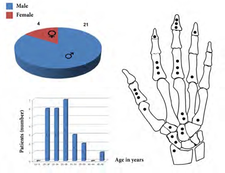
This paper reports on 25 cases of osteoid osteoma in the hand and wrist at 20 years period. There were twenty-one men and four women, with an average age of 25.2 ±7.6 (range: 16-46) years.
Twenty osteoid osteoma were in the right and five in the left side of hands. Fifteen were in the finger phalanges (ten proximal and five distal phalanges), four in the metacarpals, five in the wrist (two in scaphoid, two in capitate and one in trapezium) and one in the styloid of radius.
The most common and earliest symptom was pain. The pain was gradually increased and initially intermittent, but later became more constant and severe. The pain was unaffected by activity and twenty-one of these patients had night pain and in 17 patients partial pain relief obtained by aspirin (five patients had no relief and 3 were not investigated). Blood analyses were normal for all patients, but a history of trauma prior to the onset of symptoms found in five patients.
Physical examination at presentation revealed local swelling and point tenderness. Whenever the osteoid osteoma was close to a joint, restriction of motion and muscle along with atrophy were sometimes present. The physical findings varied with the site of the tumor. In the phalanges, osteoid osteoma induced marked fusiform soft tissue swelling. Proximal phalanx involvement was characterized by a grossly enlarged phalanx with hypertrophy of the soft tissues, and distal phalanx involvement caused finger clubbing, with enlargement of the phalanx, and sometimes hypertrophy of the nail and obliteration of the normal angle between the base of the nail and the skin.
The diagnosis was made using plain x-rays, three-phase Technetium bone scans, CT and MRI.
Only thirteen patients had the characteristic appearances of osteoid osteoma on x-ray and twelve non-specific appearances on initial x-rays.
Three-phase TC- 99 bone scans were done on twenty patients, showed an intense well-defined focal area increased activity during all the three-phase of the scan in eighteen patients. In two cases, the bone scans were non- specific, showing a diffuse increase in isotope uptake. Eighteen CT scans were performed and showed the osteoid osteoma in every case as an obvious lytic lesion with a central granular opacity surrounded by a well-defined sclerotic margin.
The MRI was performed on fifteen cases, and clearly showed an intense soft tissue reaction around the lesion.
The mean delay from the onset of symptoms to definitive treatment was 16.3±11.1 months (range, 3months to 4 years).
Three patients had been treated surgically in another center previously. Twenty-one patients were treated surgically with excisional biopsy and four with curettage and bone grafting.
Fig. 1.
Gender, age, range and number of cases at the wrist and hand for 25 patients with osteoid osteoma.
Histological examinations confirmed the diagnosis in all cases.
At a mean follow up 36.6±46.9 months (range, 3 months to 8 years), twenty patients had no pain or tenderness, and five had recurrence after first surgery in which three of them were initially operated in elsewhere.
Location of the primary osteoid osteoma at recurrence group consisted of one in the metacarpal bone, two in the distal and two in the proximal phalanx.
All patients treated surgically by excisional biopsy (3 patients) or curettage plus bone grafting (2 patients), and patients at follow up of 31.2±11.2 month (recurrence group) were well and had no pain.
Discussion
Osteoid osteomas usually turned into symptomatic disease in the second or third decades of life, and rarely occurred in patients with 40 years of age. Approximately twice as many men as women are affected (3, 5, 9).
The men to women ratio was 5.25 and right to left side hand ratio 4. These ratios were different from other reports.
All studied patients were in second, third or fourth decades and only in one case over 40 years of age. This is similar to other reports.
Ghiam and Bora described 3 carpal tumors in two patients and combined these in the literature to establish a series of 26 cases of carpal osteoid osteomas. The scaphoid was the most commonly involved bone, with the hamate second and the capitate the third most frequently involved carpal bone (10). Marcuzzi & Leti reports of 18 osteoid osteomas of the wrist and hand, two cases was in the scaphoid, two in lunate and one in capitate, and Ambrosia & Wold reported of 19 cases of osteoid osteomas in which included two in capitate, one in hamate and one in the triquetrum (11). We had only five cases in the carpal bone (two in the scaphoid, two in the capitate and one in the trapezium bone).
The most common complaint was pain, often described as being more severe at night. Healey reported improvement of pain after aspirin treatment (3). In the series presented by Bender et al, 86% of patients with an osteoid osteoma of upper extremity also reported pain relief with aspirin use (2).
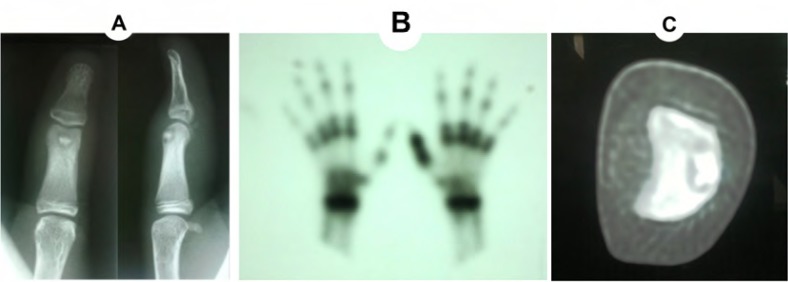
Fig. 2.
Osteoid osteoma of proximal phalanx of thumb. A: Anteroposterior and lateral radiography (nidus with lytic lesion). B: Bone scan. C: CT scan (axial view).
We evaluated relief of pain with NSAID only in 21 patients and found relief of pain in 16 cases (76%).
Physical examination often revealed local tenderness and soft-tissue swelling. When the tumor was located near a joint, it may reduce range of motion and mimic primary arthritis. In the phalanges, typically a fusiform soft-tissue swelling existed with enlargement and clubbing of the phalanges. The mechanism that developed this feature was unclear, but it appeared to be in response to a presumably humoral substance produced by the tumor. This may directly induced proliferation of connective tissue, bone changes and dilation of blood vessels.
Furthermore the edema, produced by local hyperaemia and inflammatory factor release, increases the distance through which oxygen must diffuse before reaching the cells, possibly causing a localized hypoxaemia. This stagnant hypoxia can indirectly induce enlargement of the finger and clubbing of the distal phalanx (4).
Initial x-ray examinations were often normal, as was the case in 12 of our cases, but a three-phase technetium-99m bone scan detected the lesion which was characterized by an intense well-defined focal area of increased uptake, and is readily apparent in all three phases and only in one patient diffuse increase in isotope uptake was seen.
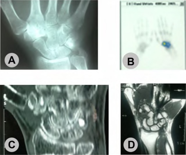
Fig. 3.
Osteoid osteoma of trapezoid bone of 31 years old men with wrist pain since 18 month ago. A: Anteroposterior radiography shows sclerosis at trapezoid bone. B: Bone scan shows increased local uptake. C: CT scan shows nidus at trapezoid (incidental bone iland at triquetrum). D: The MRI shows diffuse edema at wrist.
When the bone scan was positive or equivocal with normal radiographs, computed tomograms employed. This was superior to conventional radiography and MRI, not only for diagnosis, but also for surgical planning and follow-up of osteoid osteoma of the small bones in hand. The wrist MRI showed the tissue reaction around the tumor (1, 2, 11, 12).
Specialized imaging techniques may hasten the diagnosis, but only an accurate clinical history with a high index of suspicion allows one to arrange the appropriate investigations.
Treatment of osteoid osteoma consisted of curettage or en bloc excision of the tumor, which resulted in almost immediate relief of pain. Pre-operative CT assessment of the lesion greatly assist the surgeon and increases the likelihood of an adequate surgical excision. Packing the defect after curettage with cancellous bone chip may not be necessary, but incomplete removal may lead to recurrence or continuation of symptoms. Nowadays the operative treatment is not the only modality recommended to the patients and their family but it is also an alternative choice.
Conclusion
Osteoid osteomas, relatively rare lesions in the hand and wrist that can be a persistent source of hand and wrist pain. Patients under age of 40 who have otherwise unexplained pain should be evaluated. Relief of pain with oral NSAID, most notably aspirin, should suggest the possibility of osteoid osteoma. Examination may demonstrate localized swelling or joint effusion. Point tenderness is also an important symptom in hand and wrist osteoid osteoma. Radiographs should be examined for sclerosis in the region of pain. If radiographs are nonconclusive, a bone scan should be considered. Finally, if the nidus cannot be clearly visualized by radiography and bone scan, a CT scan should be recommended.
References
- 1.Assoun J, Richardi G, Railhac JJ, LeGuennec P, Caulier M, Dromer C, et al. Osteoid Osteoma: MR imaging versus CT. Radiology. 1991;91:217–223,4. doi: 10.1148/radiology.191.1.8134575. [DOI] [PubMed] [Google Scholar]
- 2.Cohen JD, Harrington TM, Ginsberg WW. Osteoid Osteoma: 95 cases and a review of the literature. Semin Arthritis Rheum. 1983;12:265–281. doi: 10.1016/0049-0172(83)90010-0. [DOI] [PubMed] [Google Scholar]
- 3.Healey JH, Ghelman B. Osteoid Osteoma and Osteoblastoma: current concepts and recent advances. Clin Orthop. 1986;204:76–85. [PubMed] [Google Scholar]
- 4.Marcuzzi A, LetiAcciaro A, Landi A. Osteoid Osteoma of the hand and wrist. J Hand surg. 2002;27B:440–443. doi: 10.1054/jhsb.2002.0811. [DOI] [PubMed] [Google Scholar]
- 5.Bender MS, McCormake RR, Glasser D, Weilaad AJ. Osteoid Osteoma of the upper extremity. J Hand Surg. 1993;18A:1019–1025. doi: 10.1016/0363-5023(93)90395-J. [DOI] [PubMed] [Google Scholar]
- 6.Wiss DA, Reid BS. Pamless Osteoid Osteoma of the fingers. J Hand Surg. 1983;8:914–917. doi: 10.1016/s0363-5023(83)80094-x. [DOI] [PubMed] [Google Scholar]
- 7.Jackson RP, Reckling FW, Mantz FA. Osteoid Osteoma and Osteoblastoma: similar histologic lesions with different natural histories. Clin Orthop. 1977;128:303–313. [PubMed] [Google Scholar]
- 8.De Wet IS. Osteoid Osteoma: Review of the literature with a report of five cases. S Afr J Surg. 1967;5:13–24. [PubMed] [Google Scholar]
- 9.Wold LE, Mcleod RA, Sim FH, Unni KK. Atlas of Orthopaedic pathology. Philadelphila: WE Saunders; 1990. pp. 90–9. [Google Scholar]
- 10.Ghiam GF, Bora FW. Osteoid Osteoma of the carpal bones. J Bone Joint Surg. 1978;3:280–283. doi: 10.1016/s0363-5023(78)80093-8. [DOI] [PubMed] [Google Scholar]
- 11.Ambrosia JM, Wold LE, Amadio PC. Osteoid Osteoma of the hand and wrist. J Hand Surg Am. 1987 Sep;12:794–800. doi: 10.1016/s0363-5023(87)80072-2. [DOI] [PubMed] [Google Scholar]
- 12.Woods ER, Martel W, Mandell SH, Crabbe JP. Reactive soft tissue muss associated with Osteoid Osteoma: correlation of MR imaging features with pathologic finding. Radiology. 1993;186:221–225. doi: 10.1148/radiology.186.1.8416568. [DOI] [PubMed] [Google Scholar]


